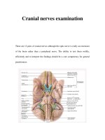
Facial nerve
Bạn đang xem bản rút gọn của tài liệu. Xem và tải ngay bản đầy đủ của tài liệu tại đây (708.01 KB, 10 trang )
<span class="text_page_counter">Trang 1</span><div class="page_container" data-page="1">
1.
<b>FACIAL NERVES</b>
<b>Describe the origin and course of facial nerve.</b><b>Facial nerve is the seventh cranial nerve. It is a mixed (sensory and </b>
motor) nerve.
<b>Origin</b>:Its nuclei lie in the lower part of the pons.
<small>Facial nerve emerges at the lower border of pons as two roots, large medial </small>
<b><small>motor root and a small lateral sensory root (nervus intermedius).</small></b>
<small></small> <b><small>It leaves the cranial cavity by passing through the internal acoustic meatus.</small></b>
<small></small> <b><small>It then enters the facial canal which is divided into three segments viz. </small></b>
<small>labyrinthine, tympanic and mastoid.</small>
<small></small> <b><small>Labyrinthine segment : It lies here above the vestibule of internal ear and </small></b>
<small>passes laterally to reach the medial wall of middle ear where it bends </small>
<b><small>(external genu – location of geniculate ganglion) sharply backwards.</small></b>
<small></small> <b><small>Tympanic segment: The nerve here runs horizontally backward along the </small></b>
<small>medial wall of the middle ear till it reaches the junction of medial and posterior wall of middle ear.</small>
<small></small> <b><small>Mastoid part: It then passes vertically downward in this part along the </small></b>
<small>posterior wall of middle ear to reach the stylomastoid foramen.</small>
</div><span class="text_page_counter">Trang 2</span><div class="page_container" data-page="2"><b>Name the nuclei, functional components of facial nerve </b>
<b>.</b>
Following are the nuclei, functional components of facial nerve:
<b><small>FUNCTIONAL </small></b>
<b><small>SVE (Special visceral efferent)</small></b>
<b><small>Motor nucleus of facial nerve</small></b>
<b><small>All the muscles derived from 2nd branchial arch- Occipitofrontalis,</small></b>
<b><small> Muscles of face, Platysma, Stapedius, Posterior belly of digastric,Stylohyoid</small></b>
<b><small>GVE (General visceral efferent)</small></b>
<b><small>Superior salivatory nucleus</small></b>
<b><small>Secretomotor fibers to submandibular and sublingual gland</small></b>
<b><small>Lacrimatory nucleusSecretomotor fibers to lacrimal glandSVA(Special visceral </small></b>
<b><small>Nucleus of tractus solitarius</small></b>
<b><small>Carry taste sensation from anterior 2/3rd of tongue (except circumvallate papillae)</small></b>
<b><small>GSA (General visceral afferent)</small></b>
<b><small>Spinal nucleus of trigeminal nerve</small></b>
<b><small>Few cutaneous fibers from external ear</small></b>
<b>Name the branches and structures supplied of facial nerve.</b>
<b>Branches in the facial canal</b>
<small></small> <b>Greater petrosal nerve (from geniculate ganglion) – supplies </b>
secretomotor fibers to lacrimal, nasal and palatine glands.<small></small> <b>Nerve to stapedius – supplies stapedius muscle.</b>
<small></small> <b>Chorda tympani nerve – carries taste sensation from anterior </b>
2/3rd of tongue ( except circumvallate papillae) and supplies secretomotor fibers to submandibular and sublingual gland.
<b>Branches below the stylomastoid foramen</b>
<small></small> <b>Posterior auricular nerve– supplies occipital belly of </b>
occipitotemporalis and auricular muscles.
<small></small> <b>Nerve to posterior belly of digastric and stylohyoid muscles.</b>
<b>Branches on face (given within parotid gland)</b>
</div><span class="text_page_counter">Trang 3</span><div class="page_container" data-page="3"><b><small>These five branches supply muscles of facial expression and buccinator muscle.</small></b>
</div><span class="text_page_counter">Trang 4</span><div class="page_container" data-page="4"><b>Infranuclear lesion</b>
Symptoms depend on the site of lesion:
</div><span class="text_page_counter">Trang 5</span><div class="page_container" data-page="5"><b>Lesion at ‘D’: At the stylomastoid foramen (Bell’s palsy)- paralysis of all </b>
the muscles of facial expression on the same side. Symptoms of Bell’s palsy are:
<small></small> Marked facial asymmetry<small></small> Atrophy of facial muscles<small></small> Can’t close eye
<small></small> Tears overflow
<small></small> Smoothing out of forehead and nasolabial folds<small></small> Drooping of corner of mouth
<small></small> Food accumulates in vestibule of mouth
<b>Lesion at ’Ç’: Above the origin of chorda tympani- all the symptoms of </b>
lesion (i) and decreased salivation and loss of taste sensation from anterior 2/3<small>rd</small> of the tongue.
<b>Lesion at ‘B’: Above the origin of nerve to stapedius- all the symptoms of</b>
lesion (i) and (ii) and hyperacusis.
<b>Lesion at ‘Á’: At the geniculate ganglion-: all the symptoms of lesion (i) </b>
and (ii) and(iii) and loss of lacrimation.
</div><span class="text_page_counter">Trang 6</span><div class="page_container" data-page="6"><b>CLINICAL ASPECTS OF FACIAL NERVE</b>
1. clinical significance of the facial nerve
The facial nerve is the seventh cranial nerve. It contains the motor,
sensory, and parasympathetic (secretomotor) nerve fibres, which provide innervation to many areas of the head and neck region. The facial nerve iscomprised of three nuclei: The main motor nucleus.
2. clinical disorders of the facial nerve
<b>Facial Nerve Disorders</b>
<small></small> Bell's Palsy. One of the most common facial nerve disorders is Bell's palsy, which is caused by a viral infection of the facial nerve. ...
<small></small> Facial Spasms. ...
<small></small> Ramsay Hunt Syndrome. ...
<small></small> Neuromas. ...
<small></small> Trauma. ...
3. facial Nerve Paralysis
<b>SYMPTOMS OF FACIAL NERVE PARALYSIS </b>
drooping skin around the brow, eye, cheek, and mouth.
When a muscle loses motor function, it relaxes completely, and the skin above the muscle relaxes as well. Some peoplemay still have partial control over facial muscles or
experience muscle spasms or twitching, while others cannotmove any muscle on the affected side of the face.
Because the condition may prevent you from closing your eyelid, you may develop dry eye and other eye problems. Facial nerve paralysis may also interfere with eating and talking.
</div><span class="text_page_counter">Trang 7</span><div class="page_container" data-page="7"><b> CAUSES OF FACIAL NERVE PARALYSIS</b>
Facial nerve paralysis can be congenital, meaning a person is born with it. But most of the time, the condition occurs in adults as the result of damage to the facial nerves.
One of the most common causes is Bell’s palsy. This type of facial paralysis usually develops suddenly and affects only one side of the face. The cause may be swelling in the facial nerve, which temporarily restricts its blood supply. The
nerve almost always recovers, and facial paralysis typically goes away on its own within a year.
Other causes of sudden one-sided facial nerve paralysis include a traumatic head injury, which may damage the seventh cranial nerve; a stroke, which occurs as a result of aloss of blood supply to the brain stem;
a viral infection, such as herpes simplex or herpes zoster; or,more rarely, Lyme disease.
Sometimes, facial nerve paralysis develops slowly. Muscles on one side of the face gradually lose movement over a period of weeks or months. In this instance, the cause may be a growth on the seventh cranial nerve, such as a facial nerve schwannoma. This slow-growing, noncancerous tumor may press on the nerve and cause increasing paralysis in facial muscles.
Rarely, a cancerous tumor located near the facial nerve or inthe area of the brain that sends signals to facial muscles causes paralysis. In some circumstances, surgery to remove a growth may result in facial nerve paralysis.
Less commonly, a neurological disorder or virus may cause complete paralysis by damaging nerve tissue on both sides of the face. Guillain-Barré syndrome, in which the body’s immune system attacks nerve tissue, is one example.
</div><span class="text_page_counter">Trang 9</span><div class="page_container" data-page="9">as hearing loss or tinnitus, a persistent ringing in the ears. The seventh cranial nerve travels through parts of the skull alongside the eighth cranialnerve, which controls hearing, so these symptoms may occur at the same time.
Tell your doctor if you’ve had any recent insect bites, have spent time in areas known to have ticks, or have been recently diagnosed with a viral infection.
<b>MRI Scan</b>
MRI scans use radio waves and a magnetic field to create computerized, three-dimensional images of soft tissues in the body. MRI is used to examine the entire facial nerve. This imaging test also allows a doctor to identify swelling or a growth on or near the nerve.
MRI uses a contrast agent or dye called gadolinium. When the dye is injected into the bloodstream, it travels to the facial nerve, highlighting areas of inflammation.
<b>Stapedius Reflex Test</b>
The stapedius reflex test is a type of hearing test that audiologists—specialists who study hearing loss, balance problems, and related disorders—use to assess damage to the seventh cranial nerve.During the test an audiologist uses noise to stimulate the stapedius muscle, a tiny muscle in the middle ear. The nerve that controls facial muscles also controls the stapedius muscle. A doctor may be able to pinpoint the location of a problem on the facial nerve based on whether the stapedius muscle responds to the test. If this muscle does not
respond, it may indicate that the area of the facial nerve that controls the stapedius is affected.
Electroneurography is a test used to evaluate the function of peripheral nerves, which include the facial nerve. Ideally, this test is performed within 14 days of the onset of paralysis. The results help doctors determine whether further testing or intervention may be required.
To perform this test, an audiologist places several electrodes on the face and at the base of the ear on both sides of your face. The electrodes are flat, adhesive discs that stick to the skin and are attached to a machine that produces a low electrical current. The sensation may tingle but is not painful.
A doctor stimulates the facial nerves and measures the muscle response to stimulation. The results help doctors determine whether the nerves can
</div><span class="text_page_counter">Trang 10</span><div class="page_container" data-page="10">return to full function without intervention. If the paralyzed muscles
display less than 10 percent of the function that healthy muscles show on the other side, this may suggest that the paralysis may be permanent.Your doctor may conduct this test two or three times during the weeks after diagnosis to assess whether nerve function is improving.
An electromyogram measures the electrical impulses transmitted along nerves and muscle tissue. This test helps doctors evaluate weakness or paralysis in the facial muscles or nerves.
During an electromyogram, a doctor inserts small, thin needle electrodes through the skin and into facial muscles that correspond to specific
nerves. He or she then asks you to move these muscles if you can. The signals recorded when each muscle contracts can indicate which nerves are affected and whether a nerve injury has caused muscle paralysis. The test is also occasionally used after paralysis to determine whether a nerveis recovering.
<b>Blood Test</b>
Occasionally, a doctor may recommend a blood test to determine if a virus or an infection may be the cause of facial nerve paralysis. A specialist draws a small amount of blood and sends the sample to a laboratory for testing. Test results are usually available in a few days.
</div>