PHEOCHROMOCYTOMA – A NEW VIEW OF THE OLD PROBLEM doc
Bạn đang xem bản rút gọn của tài liệu. Xem và tải ngay bản đầy đủ của tài liệu tại đây (19.54 MB, 174 trang )
PHEOCHROMOCYTOMA
– A NEW VIEW
OF THE OLD PROBLEM
Edited by Jose Fernando Martin
Pheochromocytoma – A New View of the Old Problem
Edited by Jose Fernando Martin
Published by InTech
Janeza Trdine 9, 51000 Rijeka, Croatia
Copyright © 2011 InTech
All chapters are Open Access distributed under the Creative Commons Attribution 3.0
license, which allows users to download, copy and build upon published articles even for
commercial purposes, as long as the author and publisher are properly credited, which
ensures maximum dissemination and a wider impact of our publications. After this work
has been published by InTech, authors have the right to republish it, in whole or part, in
any publication of which they are the author, and to make other personal use of the
work. Any republication, referencing or personal use of the work must explicitly identify
the original source.
As for readers, this license allows users to download, copy and build upon published
chapters even for commercial purposes, as long as the author and publisher are properly
credited, which ensures maximum dissemination and a wider impact of our publications.
Notice
Statements and opinions expressed in the chapters are these of the individual contributors
and not necessarily those of the editors or publisher. No responsibility is accepted for the
accuracy of information contained in the published chapters. The publisher assumes no
responsibility for any damage or injury to persons or property arising out of the use of any
materials, instructions, methods or ideas contained in the book.
Publishing Process Manager Masa Vidovic
Technical Editor Teodora Smiljanic
Cover Designer InTech Design Team
Image Copyright ccaetano, 2011. DepositPhotos
First published December, 2011
Printed in Croatia
A free online edition of this book is available at www.intechopen.com
Additional hard copies can be obtained from
Pheochromocytoma – A New View of the Old Problem, Edited by Jose Fernando Martin
p. cm.
ISBN 978-953-307-822-9
free online editions of InTech
Books and Journals can be found at
www.intechopen.com
Contents
Preface IX
Part 1 Pathophysiology 1. Anatomo-Pathological Aspects 1
Chapter 1 Macro and Microscopic Aspects 3
Fernando Candanedo-Gonzalez, Leslie Camacho-Rebollar
and Candelaria Cordova-Uscanga
Chapter 2 Phaechromocytoma with Histopathologic Aspects 15
Servet Guresci, Derun Taner Ertugrul
and Gulcin Guler Simsek
Part 2 Pathophysiology 2. Study Experimental Models 23
Chapter 3 Mouse Models of Human Familial Paraganglioma 25
Louis J. Maher III, Emily H. Smith, Emily M. Rueter,
Nicole A. Becker, John Paul Bida, Molly Nelson-Holte,
José Ignacio Piruat Palomo, Paula García-Flores, José López-Barneo
and Jan van Deursen
Chapter 4 Cell Differentiation Induction
Using Extracellular Stimulation
Controlled by a Micro Device 47
Yuta Nakashima, Katsuya Sato, Takashi Yasuda
and Kazuyuki Minami
Part 3 Pathophysiology 3. Signaling Pathways 63
Chapter 5 Phospholipase A
2
and Signaling
Pathways in Pheochromocytoma PC12 Cells 65
Alexey Osipov and Yuri Utkin
Chapter 6 Programmed Cell Death Mechanisms
and Pheocromocytomas:
Recent Advances in PC12 Cells 85
Davide Cervia and Cristiana Perrotta
VI Contents
Part 4 Clinical Presentation 101
Chapter 7 Headache in Pheochromocytoma 103
Masahiko Watanabe
Chapter 8 Primary Cardiac Pheochromocytoma (Paraganglioma) 111
Iskander Al-Githmi
Part 5 Diagnosis 117
Chapter 9 Diagnosis: Laboratorial
Investigation and Imaging Methods 119
José Fernando Vilela-Martin and Luciana Neves Cosenso-Martin
Part 6 Treatment and Clinical Cases 133
Chapter 10 Undiagnosed Pheochromocytoma
Complicated with Perioperative
Hemodynamic Crisis and Multiple Organ Failure 119
Anis Baraka
Chapter 11 Familial Catecholamine-Secreting Tumors - Three
Distinct Families with Hereditary Pheochromocytoma 149
Shirin Hasani-Ranjbar, Azadeh Ebrahim-Habibi and Bagher Larijani
Preface
Cardiovascular diseases are the major cause of mortality in developed and developing
countries. Hypertension is the most prevalent of all cardiovascular diseases, the major
risk factor for cardio and cerebrovascular injury and the third cause of disability. It is
likely to be involved in 50% of the deaths due to cardiovascular diseases. Genetic and
environmental factors are involved in more than 90% of cases, characterizing essential
hypertension. About 5 to 10% of hypertension cases are represented by cases of
secondary arterial hypertension. In this situation, pheochromocytoma, a catechomine-
secreting tumor that is located in the adrenal medulla (pheochromocytoma) or in the
extra adrenal paraganglionic tissue (paranganglioma) presents prevalence varying
from 0.01% to 0.10% of the hypertensive population, and an incidence of two to eight
cases per million people per year.
When I received the invitation to be editor of a book on pheochromocytoma, a disease
that represents small percentage of cases of secondary hypertension, I was worried
with the development of the book and always wondered what would be the interest
for the medical community. As the chapters were presented and developed by the
authors, this worry has disappeared, because despite its rarity, pheochromocytoma
presents a different clinical picture and several opportunities for clinical and basic
research. Certainly, the level of the authors of this book also did make it an excellent
topic to be discussed, in addition to chapters with new approaches about the clinical
presentation and in the field of experimental research.
The book is divided into 6 sections covering the main aspects of clinical practice and
other issues related to translational research. I hope readers enjoy this book and I
expect it is a reference in the area.
Dr. Jose Fernando Vilela Martin, MD PhD,
Head of Internal Medicine Division,
Coordinator of Hypertension Clinic,
State Medical School of São José do Rio Preto (FAMERP),
Brazil
Part 1
Pathophysiology 1.
Anatomo-Pathological Aspects
1
Macro and Microscopic Aspects
Fernando Candanedo-Gonzalez, Leslie Camacho-Rebollar
and Candelaria Cordova-Uscanga
Department of Pathology, Oncology Hospital,
National Medical Center Century XXI,
Mexico City,
Mexico
1. Introduction
In 1886, Fränkel first described pheochromocytoma at autopsy 1. The term
pheochromocytoma was coined by Poll in 1905 to describe the dusky (pheo) color (chromo)
of the cut surface of the tumour when exposed to dichromate 2. Not until 1926 did Mayo
3 at the Mayo Clinic and Roux 4 in Switzerland successfully remove these adrenal
tumours. Interestingly, neither of these tumours was diagnosed preoperatively.
Pheochromocytomas are rare catecholamine-producing neuroendocrine tumours arising
from the chromaffin cells of the embryonic neural crest mainly of adrenal medulla or the
extra-adrenal chromaffin tissue (paraganglia). Which synthesize, store, metabolize, and
usually but not always secrete catecholamines.
1.1 Incidence
Population studies report an annual incidence of between 0.4 and 9.5 new cases per 100,000
adult persons each year 5,6,7, which constitute a curable form of hypertension in 0.1 to 1%
of hypertension patients 8. Of patients with pheochromocytoma discovered only at
autopsy, 75% died suddenly from either myocardial infartion or a cerebrovascular
catastrophe. Moreover, one third of the sudden deaths occurred during or immediately after
unrelated minor operations 9,10. Referrals for pheochromocytoma have been reported to
be increasing, likely as a result of improved detection.
1.2 Clinical features
The majority of pheochromocytomas are sporadic in origin (80-90%) but may be associated
with other diseases. Classically, pheochromocytomas has been termed a “10% tumour
because roughly 10% of these tumours are malignant, multifocal, and bilateral, arise in
extra-adrenal sites, and occur in children. However, recent evidence suggests the percentage
of familial tumours is considerably higher 11.
1.3 Classic presentation
The classic triad of pheochromocytoma presentation is episodic headache, sweating, and
palpitations. Manifestations of catecholamine excess form a wide spectrum of symptoms in
Pheochromocytoma – A New View of the Old Problem
4
these patients, the foremost being hypertension. Persistent hypertension is frequently
considered part of the presentation. Also is typically found with a diverse set of symptoms,
which may include anxiety, chest and abdominal pain, visual blurring, papilledema, nausea
and vomiting, orthostatic hypotension, transitory electrocardiographic changes, and
psychiatric disorders. As to be expected, these symptoms are not always present and
certainly do not always constitute a diagnosis. Nonfunctioning pheochromocytomas are
distinctly uncommon; nearly all patients with these tumours, at least in retrospect,
demonstrate some characteristic symptom or sign, especially accentuated at the time of
operative tumour manipulation. Diagnosis of pheochromocytoma includes detection of
catecholamines in urine and plasma and radiological tests such as computed axial
tomography, nuclear magnetic resonance imaging and metaiodobenzylguanidine
scintigraphy. Laparoscopic techniques have become standard for treatment of tumours of
the adrenal glands 12.
2. Pathology features
2.1 Macroscopy findings
Nearly 90% of pheochromocytomas are usually confined to the adrenal gland, and may
appear encapsulated. In sporadic pheochromocytomas, even though lobulated, the tumour
is actually a single neoplasm. In contrast, familial tumours are often bilateral and usually
multicentric 13. Pheochromocytomas are of variable size, ranging from 3 cm to 5 cm in
diameter but can be more than 10 cm 14. The weight may range from < 5g to over 3,500g,
the average in patients with hypertension being 100g 15. The cut surface is usually soft,
yellowish white to reddish brown. The larger tumours often have areas of necrosis,
hemorrhage, central degenerative change, cystic change and calcification. The normal gland
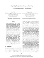
can be seen in most cases but is sometimes attenuated (Fig. 1).
Fig. 1. Adrenal Pheochromocytoma. The round tumour extends torwards the adrenal cortex
but is macroscopically well defined. Focal degenerative change and central hemorrhage is
present. Attached adrenal remnant is also present.
Macro and Microscopic Aspects
5
The other 10 to 15% of cases are found in the neck, mediastinum and heart, or along the
course of the sympathetic chain. The most frequent extra-adrenal site is the aortic
bifurcation, the so-called organ of Zuckerkandl 16.
2.2 Histopathology
Microscopically, the tumour cells are characteristically arranged in well-defined nest
(“Zellballen”) or trabecular pattern bound by a delicate fibrovascular stroma, or a mixture of
the two (Fig. 2A). Diffuse or solid architecture can also be seen. A true capsule does not
usually separate the tumour from the adjacent adrenal but a pseudocapsule may be present,
or the tumour may extend to the adrenal capsule. The border with the adjacent cortex may
be irregular, with intermingling of tumour cells with cortical cells.
The tumour cells vary considerably in size and shape and have a finely granular basophilic
or amphophilic cytoplasm. The nuclei are usually round or oval with prominent nucleoli
and may contain inclusion-like structure resulting from deep cytoplasmic invaginations.
Cellular and nuclear pleomorphism is sometimes prominent (Fig. 2B) 17. Spindle cells are
present in about 2% of cases, usually as a minor component. Haemorrhage and
haemosiderin deposits are common. Mitotic figures are rare, with an average of one per 30
high power fields reported in clinically benign lesions 18.
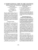
A B
Fig. 2. Benign pheochromocytoma. A) Well-defined nest of cuboidal cells are separated by
highly vascularized fibrous septa (“zellballen”). A granular, basophilic cytoplasm is usually
identified surrounding slightly irregular nuclei; B) nuclear pleomorphisms are sometimes
prominent.
Pheochromocytoma – A New View of the Old Problem
6
2.3 Immunohistochemistry
Specific diagnosis is usually based on morphology and confirmed by immunohisto-
chemistry. Pheochromocytomas are positive for chromogranin A. Other neural markers
such as synaptophysin have been reported to be variably positive in cortical tumours. The
absence of positivity for epithelial membrane antigen helps distinguish pheochromocytoma
from renal cell carcinoma. Immunostaining for S100 protein will demonstrate sustentacular
cells 19 which are usually arranged around the periphery of the cell nests where there is an
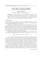
alveolar arrangement (Fig. 3).
A B
Fig. 3. Immunohistochemical staining. A) Positive cytoplasmic immunostain for
chromogranin in the pheochromocytoma; B) Immunostain for S-100 protein shows intense
dark staining of elongated nuclei of sustentacular cells. These are usually located near
vascular channels.
3. Familial pheochromocytoma
Pheochromocytomas are considered to be unique neuroendocrine tumours since they can
occur as part of several familial tumour syndromes. It is now recognized that the frequency
of germline mutations in apparently sporadic presentations is as high as 15%–24% 11,20.
However, the genetic basis of the majority of sporadic pheochromocytomas remains largely
uncharacterized.
Macro and Microscopic Aspects
7
Familial pheochromocytomas are often multifocal or bilateral and generally present at an
earlier age than sporadic pheochromocytoma. Germline mutations in six genes have been
associated with familial pheochromocytoma, namely, the von Hippel-Lindau gene (VHL),
which causes von Hippel-Lindau (VHL) syndrome, the RET gene, leading to multiple
endocrine neoplasia type 2 (MEN 2), the neurofibromatosis type 1 gene (NF1), associated
with neurofibromatosis type 1 (NF1) disease, and the genes encoding subunits B and D (and
also rarely C) of mitochondrial succinate dehydrogenase (SDHB, SDHD, and SDHC), which
are associated with familial paraganglioma/PPC. The recent description of mutations of the
succinate dehydrogenase gene (SDH) has demonstrated a much stronger hereditary
component than formerly thought. Currently, up to 24% of pheochromocytomas may have a
genetic predisposition 11,20.
The genetic susceptibility of malignant and benign pheochromocytomas is similar. However,
advances in molecular genetics continue to underscore the importance of hereditary factors in
the development of pheochromocytoma and propensity to malignancy. Malignant tumours
have been reported in patients with germline mutations of RET, VHL, NF1 and the SDH genes
21,22. On the other hand, malignant pheochromocytomas in the setting of MEN 2 occur less
frequently than sporadic tumours 23,24,25,26. Which suggesting certain groups are
predisposed to malignant disease. For example, patients with SDHB mutations are more likely
to develop malignant disease and nondiploid tumours have also been found to be associated
with malignancy. Gene expression and protein profiling are beginning to identify the genetic
characteristics of malignant pheochromocytoma. However, the genetic changes that induce
malignant disease remain unclear.
4. Malignant disease
Most pheochromocytomas are benign and curable by surgical resection, but some are
clinically malignant 27. The pathologist cannot determine whether a tumour is benign or
malignant based on histological features alone. Although extensive invasion of adjacent
tissues can be considered an indicator of malignant potential, local invasiveness and
malignant disease are not necessarily associated. Currently, there are no prognostic tests
that can reliably predict which patients are at risk of developing metastatic disease. The
World Health Organization tumour definition of a malignant pheochromocytoma is the
presence of metastases, at site distant where chromaffin cells do not normally exist 28.
Metastases occur most frequently to bone, liver, lungs and regional lymph nodes, and can
appear as many as 20 years after initial presentation, which implies that life-long follow-up
of patients (Fig. 4) 29.
Some studies have suggested that the presence of necrosis, vascular invasion, extensive local
invasion, and high rate of mitotic figures may indicate a malignant behavior in
pheochromocytoma. Indeed, a recent study by Thompson used clinical features, histologic
findings, and immunophenotypic studies to indentify parameters that may help distinguish
benign from malignant pheochromocytoma of the Adrenal Gland Scaled Score (PASS) as a
scoring system to differentiate benign from malignant pheochromocytomas. PASS is
weighted for 12 specific histologic features that are more frequently identified in malignant
pheochromocytomas. Factors such as tumour necrosis, high mitotic rate, tumour cell
spindling, and vascular invasion are included in this scoring system (Fig. 5). Thompson
found that tumours with ≥4 were biologically more aggressive than tumours with a PASS
<4, which behaved in a benign fashion (Table 1) 30.
Pheochromocytoma – A New View of the Old Problem
8
Fig. 4. Malignant pheochromocytoma. A and B) Multiple liver and lungs metastatic lesions
were shown by computed tomography; C) Transition from the metastatic
pheochromocytoma component (*) to the liver within the same section; D) By
immunohistochemical was confirmed the presence of a metastatic pheochromocytoma with
the characteristic chromogranin immunoreactivity in the pheochromocytos and the S-100
protein immunoreactivity of the sustentacular cells which contrasted with negative liver
tissue.
A
B
C
D
*
Macro and Microscopic Aspects
9
A
B
C
Fig. 5. Invasive malignant pheochromocytoma. A) A thick fibrous capsule is transgressed by
the neoplastic cells with extension into surrounding addipose connective tissue in malignant
pheochromocytoma; B) Extension into a vascular spaces is noted in a malignant
pheochromocytoma.
Pheochromocytoma – A New View of the Old Problem
10
Microscopic feature Score
Extension into peri-adrenal adipose tissue 2
Presence of large nests or diffuse growth
(>10% of tumour volume)†
2
Central tumour necrosis (in the middle of large nests or confluent necrosis) 2
High cellularity 2
Tumour cell spindling even when focal 2
Cellular monotony 2
Mitotic figures >3/10 high-power field 2
Atypical mitotic figures 2
Vascular invasion* 1
Capsular invasion 1
Profound nuclear pleomorphism 1
Nuclear hyperchromasia 1
Total 20
*Defined by direct extension into vessel lumen, intravascular attached tumour thrombi, and/ or tumour
nests convered by endothelium identified in a capsular or extracapsular vessel.
†Defined as 3-4 times the size of a zellballen or the normal size of the medullary paraganglia nest.
Table 1. Pheochromocytoma of the Adrenal Gland Scoring Scale (PASS) 30.
Additional markers that might be useful prognostic indicators in the pathological
assessment of these tumours are sought. However, some studies with markers for important
events in the cell cycle showed that less p21
/WAF1
expression and aneuploidy correlated with
malignant pheochromocytomas 31,32,33.
4.1 Prognosis and predictive factors
The rarity of this tumours and the resulting fragmented nature of studies, typically
involving small numbers of patients, represent limiting factors to the development of
effective treatments and diagnostic or prognostic markers for malignant disease. The
prognosis for patients with benign pheochromocytoma is primarily dependent upon a
successful surgical resection and extend of preoperative complications related to
hypertension. The usual prognosis of malignant pheochromocytoma is poor, with a 45-55%
5-year survival 30,34,35,36,37,38. However, some patients may have indolent disease, with
life expectancy of more than 20 years 39. Until further studies identify precise biological
markers that can accurately predict the clinical behaviour of catecholamine-secreting
tumours, it may be advisable for all pheochromocytoma patients to undergo lifelong
hormonal monitoring and imaging studies to detect recurrence and metastases 40.
5. Composite pheochromocytoma
Ordinary pheochromocytoma is composed of polygonal to spindled cells arranged in an
alveolar, trabecular, or solid pattern, often with a typical Zellballen appearance. Composite
pheochromocytomas account for only 3% of both adrenal and extra-adrenal
pheochromocytomas and can be associated with MEN 2A and phakomatoses 41,42.
Composite pheochromocytoma is a rare tumour composed of typical pheochromocytoma
and other components, most often neuroblastoma 43, ganglioneuroblastoma, or
Macro and Microscopic Aspects
11
ganglioneuroma in adult cases, and pediatric were very rare. Rare cases have displayed
pheochromocytoma with other coexisting neural or neural crest–derived tumours such as
malignant peripheral nerve sheath tumour. Little is known about the biologic potential,
outcome, or molecular genetic profile.
Because composite pheochromocytoma clinically resembles a typical pheochromocytoma,
diagnosis is frequently made by the pathologist. The median age is 16 yr (9 to 24 yr) 43.
The pathologic diagnosis of composite pheochromocytomas creates a clinical dilemma
because it is not known whether the neuroblastic component results in therapeutic and
prognostic implications different from those in ordinary pheochromocytoma.
Neuroblastoma is the most immature of the neuroblastic tumours; the others are
ganglioneuroblastoma and ganglioglioma (Table 2). These tumours are differentiated based
on the amount of schwannian stroma and the presence or absence of ganglion cell
differentiation. This dual phenotype is supported by light microscopy and corroborated by
immunohistochemistry and ultrastructural findings. Prognosis of coexistence with
pheochromocytoma and ganglioneuroblastoma or neuroblastoma is variable.
Coexistence with No. of cases %
Ganglioneuroma 41 70
Ganglioneuroblastoma 7 11
Neuroblastoma 4 9
Schwannoma 4 7
Neuroendocrine carcinoma 1 2
Total 57 100
Table 2. Cases of composite pheochromocytoma of adrenal gland 43.
6. New insights on pheochromocytoma
The molecular events involved in the malignant transformation of pheochromocytoma are
poorly understood. There are also no reliable and uniformly accepted histopathologic
criteria to distinguish benign from malignant pheochromocytoma. Unsupervised cluster
analysis showed 3 main clusters of tumors that did not have complete concordance with the
clinical and pathologic groupings of pheochromocytomas. Supervised cluster analysis
showed almost completely separate clustering between benign and malignant tumours. The
differentially expressed genes with known function belonged to 8 biologic process
categories; signal transduction, transcription, protein transport, protein synthesis, smooth
muscle contraction, ion transport, chemotaxis, and electron transport. Gene set enrichment
analysis revealed significant correlation between the microarray profiles of malignant
pheochromocytomas and several known molecular pathways associated with
carcinogenesis and dedifferentiation. Ten differentially expressed genes had high diagnostic
accuracy, and 5 of these genes (CFC1, FAM62B, HOMER1, LRRN3, TBX3, ADAMTS) in
combination distinguishing benign versus malignant tumours. Differentially expressed
genes between benign and malignant pheochromocytomas distinguish between these
tumours with high diagnostic accuracy. These findings provide new insight into the genes
and molecular pathways that may be involved in malignant pheochromocytomas 44.
Pheochromocytoma – A New View of the Old Problem
12
7. Future directions
Much attention has recently been devoted to pheochromocytoma as the understanding of
this disease continues to improve. If it becomes widely available, it would greatly aid in the
staging and management of malignant disease. Continually improving detection methods,
especially screening of high-risk populations, will only contribute to the treatment and
knowledge of these conditions in the future. It has become clear that many apparently
sporadic pheochromocytomas have a genetic component. Not only has there been a great
deal of attention directed toward the hereditary components, but better predictive molecular
factors have been identified for malignant pheochromocytoma, which could lead to more
effective genetic testing. In addition, microarray studies have identified a set of genes
preferentially expressed in malignant pheochromocytoma. The combination of an
identifiable hereditary component along with an understanding of the genetic and
molecular defects in sporadic pheochromocytoma makes this a promising model and
approach for insights into other cancers. The future is wide open for improvements in the
understanding and treatment of this disease.
8. References
[1] Fränkel F. Ein fall von doppelseitigen vollig latent verlaufen nebennier entumor und
gleichseitiger nephritis mit veranderungen am circulation sappart und retinitis.
Virchow Arch A 1886;103:244.
[2] Poll H. Die vergleichende Entwicklung der nebennierensysteme. In: Hertwig O, ed.
Handbuch der Entwicklungsgeschichte des Menschen und der Wirbeltiere. Jena:
Gustave Fishcer, 1905:443-448.
[3] Mayo CH. Paroxystmal hypertension with tumor of retroperitoneal nerve. JAMA
1927;89:1047.
[4] Roux C. Thesis Lausanne. Cited by Welbourne RB. Early surgical history of
pheochromocytoma. Br J Surg 1987;74:594.
[5] De Graeff J, Horak BJV. The incidence of phaeochromocytoma in the Netherlands. Acta
Med Scand 1964;176:583-593.
[6] Beard CM, Sheps SG, Kurland LT, Carney JA, Lie JT. Ocurrence of pheochromocytoma in
Rochester, Minnesota, 1950 through 1979. Mayo Clin Proc 1983;58:802-804.
[7] Sheps SG, Jiang NS, Klee GG. Diagnostic evaluation of pheochromocytoma. Endocrinol
Metab Clin North Am 1988;17:397-414.
[8] Samaan NA, Hickey RC, Shutts PE. Diagnosis, localization, and management of
pheochromocytoma: Pitfalls and follow-up in 41 patients. Cancer 1988;62:2451-2460.
[9] Graham JB. Phaeochromocytoma and hypertension; an analysis of 207 cases. Int Abstr
Surg 1951;92:105-121.
[10] Sutton MG, Sheps SG, Lie JT. Prevalence of clinically unsuspected pheochromocytoma:
review of a 50-year autopsy series. Mayo Clin Proc 1981;56:354-360.
[11] Neumann HP, Bausch B, McWhinney SR, Bender BU, Gimm O, Franke G, Schipper J,
Klisch J, Altehoefer C, Zerres K, Januszewicz A, Eng C, Smith WM, Munk R, Manz
T, Glaesker S, Apel TW, Treier M, Reineke M, Walz MK, Hoang-Vu C, Brauckhoff
M, Klein-Franke A, Klose P, Schmidt H, Maier-Woelfle M, Peczkowska M,
Szmigielski C. Germ-line mutations in nonsyndromic pheochromocytoma. N Engl J
Med 2002;346:1459-1466.
Macro and Microscopic Aspects
13
[12] Gil-Cárdenas A, Cordón C, Gamino R, Rull JA, Gómez-Pérez F, Pantoja JP, Herrera MF.
Laparoscopic adrenalectomy: lessons learned from and initial series of 100 patients.
Surg Endosc 2008;22:991-994.
[13] Webb TA, Sheps SG, Carney JA. Differences between sporadic pheochromocytoma and
pheochromocytoma in multiple endocrine neoplasia type 2. Am J Surg Pathol
1980;4:121-126.
[14] Page DL, DeLellis RA, Hough AJJ. Tumors of the Adrenal. 2
nd
ed. Armed Forces
Institute of Pathology: Washington, D.C.
[15] ReMine WH, Chong GC, van Heerden JA, Sheps SG, Harrison EGJr. Current
management of pheochromocytoma. Ann Surg 1974;179:740-748.
[16] van Heerden JA, Sheps SG, Hamberger B, Sheedy PF 2
nd
, Poston JG, ReMine WH.
Pheochromocytoma: Current status and changing trends. Surgery 1982;91:367-373.
[17] DeLellis RA, Suchow E, Wolfe HJ. Ultrastructure of nuclear “inclusions” in
pheochromocytoma and paraganglioma. Hum Pathol 1980;11:205-207.
[18] Linnoila RI, Keiser HR, Steinberg SM, Lack EE. Histopathology of benign versus
malignant sympathoadrenal paragangliomas: clinicopathologic study of 120 cases
including unusual histologic features. Hum Pathol 1990;21:1168-1180.
[19] Lloyd RV, Blaivas M, Wilson BS. Distribution of chromogranin and S100 protein in
normal and abnormal adrenal medullary tissues. Arch Pathol Lab Med
1985;109:633-635.
[20] Bryant J, Farmer J, Kessler LJ, Townsend RR, Nathanson KL. Pheochromocytoma: the
expanding genetic differential diagnosis. J Nat Cancer Ints 2003;1196-1204.
[21] Koch CA, Vortmeyer AO, Huang SC, Alesci S, Zhuang Z, Pacak K. Genetic aspects of
pheochromocytoma. Endocr Regul 2001;35:43-52.
[22] Neumann HP, Berger DP, Sigmund G, Blum U, Schmidt D, Parmer RJ, Volk B, Kriste G.
Pheochromocytomas, multiple endocrine neoplasia type 2, and von Hippel-Lindau
disease. N Engl J Med 1993;329:1531-1538.
[23] Casanova S, Rosenberg-Bourgin M, Farkas D, Calmettes C, Feingold N, Heshmati HM,
Cohen R, Conte-Devolx B, Guillausseau PJ, Houdent C, Bigogne JC, Boiteau V,
Caron J, Modigliani E. Phaeochromocytoma in multiple endocrine neoplasia type 2
A: survey of 100 cases. Clin Endocrinol (Oxf) 1993;38:531-537.
[24] Medeiros LJ, Wolf BC, Balogh K, Federman M. Adrenal pheochromocytoma: a
clinicopathologic review of 60 cases. Hum Pathol 1985;16:580-589.
[25] Modigliani E, Vasen HM, Raue K, Dralle H, Frilling A, Gheri RG, Brandi ML, Limbert E,
Niederle B, Forgas L, Rosenberg-Bourgin M, Calmettes C. Pheochromocytoma in
multiple endocrine neoplasia type 2: European study. The Euromen Study Group. J
Intern Med 1995;238:363-367.
[26] Scopsi L, Catellani MR, Gullo M, Cusumato F, Camerini E, Pasini B, Orefice S.
Malignant pheochromocytoma in multiple endocrine neoplasia type 2B syndrome.
Case report and review of the literature. Tumori 1996;82:480-484.
[27] Lehnert H, Mundschenk J, Hahn K. Malignant pheochromocytoma. Front Horm Res
2004;31:155-162.
[28] DeLellis RA, Lloyd RV, Heitz PU, Eng C. Eds 2004. Tumours of Endocrine Organs.
IARC Press. Lyon.
[29] Strong VE, Kennedy T, Al-Ahmadie H, Tang L, Coleman J, Fong Y, Brennan M,
Ghossein RA. Prognostic indicators of malignancy in adrenal pheochromocytomas:
Pheochromocytoma – A New View of the Old Problem
14
clinical, histopathologic, and cell cycle/apoptosis gene expression analysis. Surgery
2008;143:759-768.
[30] Thompson LDR. Pheochromocytoma of the Adrenal Gland Scaled Score (PASS) to
separate benign from malignant neoplasms. A clinicopathologic and
immunophenotypic study of 100 cases. Am J Surg Pathol 2002;26:551-566.
[31] Candanedo-Gonzalez F, Barraza IB, Cerbulo VA, Saqui SM, Gamboa DA. Aneuplody
and low p21/WAF1 expression in malignant paragangliomas. Virchow Archiv
2005;447:430.
[32] Nativ O, Grant CS, Sheps SG, O’Fallon JR, Farrow GM, van Heerden JA, Lieber MM.
The clinical significance of nuclear DNA ploidy pattern in 184 patients with
pheochromocytoma. Cancer 1992;69:2683-2687.
[33] Carisen E, Abdullan Z, Kazmi SM, Kousparos G. Pheochromocytomas, PASS, and
immunohistochemistry. Horm Metab Res 2009;41: 715-719.
[34] Modlin IM, Farndon JR, Shepherd A, Johnston ID, Kennedy TL, Montgomery DA,
Welbourn RB. Phaeochromocytomas in 72 patients: clinical and diagnostic features,
treatment and long term results. Br J Surg 1979;66:456-465.
[35] Pommier RF, Vetto JT, Bilingsly K, Woltering EA, Brennan MF. Comparison of adrenal
and extra-adrenal pheochromocytomas. Surgery 1993;114:1160-1165.
[36] Scott HWJr, Halter SA. Oncologic aspects of pheochromocytoma: importance of follow-
up. Surgery 1984:96:1061-1066.
[37] Reynolds V, Green N, Page D, Oates JA, Robertson D, Roberts S. Clinical experience
with malignant pheochromocytomas. Surg Gynecol Obstet 1982;154:801-818.
[38] Shapiro B, Sisson JC, Lloyd R, Nakajo M, Satterlee W, Beierwaltes WH. Malignant
phaeochromocytoma: clinical, biochemical and scintigraphic characterization. Clin
Endocrinol (Oxf) 1984;20:189-203.
[39] Young AL, Baysal BE, Deb A, Young WF Jr. Familial malignant catecholamine-secreting
parganglioma with prolonged survival associated with mutation in the succinate
dehydrogenase B gene. J Clin Endocrinol Metab 2002;87:4101-4105.
[40] Tang SH, Chen A, Lee CT, Yu DS, Chang SY, Sun GH. Remote recurrence of malignant
pheochromocytoma 14 years after primary operation. J Urol 2003;169:269.
[41] Jansson S, Dahlstrom A, Hansson G, Tisell LE, Ahlman H. Concomitant occurrence of
an adrenal ganglioneuroma and a contralateral pheochromocytoma in a patient
with von Recklinghausen’s neurofibromatosis. An immunocytochemical study.
Cancer 1989;63:324-329.
[42] Tischler AS. Divergent differentiation in neuroendocrine tumors of the adrenal gland.
Semin Diagn Pathol 2000;17:120-126.
[43] Candanedo Gonzalez F, Alvarado Cabrero I, Gamboa Dominguez A, Cerbulo Vazquez
A, Lopez Romero R, Bornstein Quevedo L, Salcedo Vargas M. Sporadic type
composite pheochromocytoma with neuroblastoma: clinicomorphologic, DNA
content, and ret gene analysis. Endocrine Pathol 2001;12:343-350.
[44] Suh I, Shribru D, Eisenhofer G, Pacak K, Duh QY, Crack OH, Kebebew E. Candidate
genes associated with malignant pheochromocytomas by genome-wide expression
profiling. Ann Surg 2009;983-990.
2
Phaechromocytoma with
Histopathologic Aspects
Servet Guresci, Derun Taner Ertugrul and Gulcin Guler Simsek
Kecioren Training and Research Hospital
Turkey
1. Introduction
Phaeochromocytoma is a term used for catecholamine secreting tumors that arise from
chromaffin cells of sympathetic paraganglia. The new World Health Organisation (WHO)
classification of endocrine tumors has recommended to reserve the term
phaeochromocytoma for intraadrenal tumors only and the others are defined as sympathetic
or parasympathetic paragangliomas, further categorised by site. Although it was the first
adrenal tumor to be recognised, the term phaeochromocytoma was introduced many years
later by Pick in 1912. The name is based on the fact that the tumors get dark brown after
exposure to potassium dichromate because of chromaffin reaction.
2. The usual adrenal medulla
2.1 Anatomy
The human adrenal glands are located in retroperitoneum superomedial to kidneys. They
are composite endocrine organs made up of cortex and medulla which have different
embriyonic origin, function and histology. On fresh or formalin fixated cut surface the two
portions, a relatively thick outer yellow cortex and inner, pearly gray medulla, is readily
visible. The medulla is mainly situated in head and partly body of the organ . It may
variably extend to tail and focally to alae. It’s weight comprises about 8%-10% of the total.
Medulla is of neuroectodermal origin and secretes and stores catecholamines, especially
epinephrine.
2.2 Histology
On histological examination the cortex-medulla junction is sharp with no intervening
connective tissue but the border is irregular. The medulla is mainly composed of chromaffin
cells (phaeochromocytes, medullary cells) that are arranged in tight clusters and trabeculae
seperated by a reticular fiber network. Embriyologically, they are modified sympathetic
postganglionic neurons which have lost their axons. They are all innervated by cholinergic
endings of preganglionic symptathetic neurons. There are sustentacular cells at the
periphery of clusters which can only be demonstrated by immunostaining for S-100 protein.
The chromaffin cells are polygonal to columnar and larger than cortical cells. They have
basophilic cytoplasm which have fine secretory granules and/or vacuoles. These granules
contain catecholamines and derivates of tyrosine which transform to colored polymers by