GLAUCOMA – CURRENT CLINICAL AND RESEARCH ASPECTS potx
Bạn đang xem bản rút gọn của tài liệu. Xem và tải ngay bản đầy đủ của tài liệu tại đây (25.46 MB, 388 trang )
GLAUCOMA –
CURRENT CLINICAL
AND RESEARCH ASPECTS
Edited by Pinakin Gunvant
Glaucoma – Current Clinical and Research Aspects
Edited by Pinakin Gunvant
Published by InTech
Janeza Trdine 9, 51000 Rijeka, Croatia
Copyright © 2011 InTech
All chapters are Open Access distributed under the Creative Commons Attribution 3.0
license, which permits to copy, distribute, transmit, and adapt the work in any medium,
so long as the original work is properly cited. After this work has been published by
InTech, authors have the right to republish it, in whole or part, in any publication of
which they are the author, and to make other personal use of the work. Any republication,
referencing or personal use of the work must explicitly identify the original source.
As for readers, this license allows users to download, copy and build upon published
chapters even for commercial purposes, as long as the author and publisher are properly
credited, which ensures maximum dissemination and a wider impact of our publications.
Notice
Statements and opinions expressed in the chapters are these of the individual contributors
and not necessarily those of the editors or publisher. No responsibility is accepted for the
accuracy of information contained in the published chapters. The publisher assumes no
responsibility for any damage or injury to persons or property arising out of the use of any
materials, instructions, methods or ideas contained in the book.
Publishing Process Manager Petra Zobic
Technical Editor Teodora Smiljanic
Cover Designer Jan Hyrat
Image Copyright Yuri Arcurs, 2011. Used under license from Shutterstock.com
First published October, 2011
Printed in Croatia
A free online edition of this book is available at www.intechopen.com
Additional hard copies can be obtained from
Glaucoma – Current Clinical and Research Aspects, Edited by Pinakin Gunvant
p. cm.
ISBN 978-953-307-263-0
free online editions of InTech
Books and Journals can be found at
www.intechopen.com
Contents
Preface IX
Part 1 Basic Science Research in
Understanding Pathogenesis in Glaucoma 1
Chapter 1 Oxidative Stress in Anterior
Segment of Primary Open Angle Glaucoma 3
Sergio C. Saccà and Alberto Izzotti
Chapter 2 Manipulating Glia to Protect
Retinal Ganglion Cells in Glaucoma 25
Denise M. Inman, Caroline B. Lupien
and Philip J. Horner
Chapter 3 Role of the Matrix Metallo-Proteinases in the Cellular
Re-Modeling in a Glaucoma Model System in Rat 51
Claudio De Seta, Nicola Calandrella, Valerio Berardi,
Antonio Mazzarelli, Gianfranco Scarsella
and Gianfranco Risuleo
Chapter 4 Systemic C-Reactive Protein Levels
in Normal-Tension Glaucoma and
Primary Open-Angle Glaucoma 63
Wei-Wen Su and Shih-Tsung Cheng
Chapter 5 Molecular Analysis of Italian
Patients with Congenital Glaucoma 71
Italo Giuffre’
Part 2 Review of Evidence in
Clinical Management of Glaucoma 83
Chapter 6 Tonometry – Past, Present and Future 85
Elliot M. Kirstein, Ahmed Elsheikh
and Pinakin Gunvant
VI Contents
Chapter 7 Clustered Trend-Type Analysis to Detect
Progression of Visual Field Defects in
Patients with Open-Angle Glaucoma 109
Takeo Fukuchi, Takaiko Yoshino, Masaaki Seki,
Tetsuya Togano, Hideko Sawada and Haruki Abe
Chapter 8 Neovascular Glaucoma 127
Kurt Spiteri Cornish
Chapter 9 Current Diagnosis and Management
of Angle-Closure Glaucoma 145
Rafael Castañeda-Díez, Mariana Mayorquín-Ruiz,
Cynthia Esponda-Lamoglia and Oscar Albis-Donado
Part 3 Various Issues Related to Medical Therapy in Glaucoma 169
Chapter 10 Topical Pressure Lowering Drug:
Racial Respond Variation and Pharmacogenetics 171
Ahmad Tajudin Liza-Sharmini
Chapter 11 Pharmacogenomics of Open-Angle Glaucoma 187
Stephen G. Schwartz and Tomomi Higashide
Chapter 12 Drops, Drops, and More Drops 197
John Walt and Fern Alexander
Chapter 13 Pressure Lowering Medications 223
Liza-Sharmini Ahmad Tajudin
and Yaakub Azhany
Part 4 Review of Evidence in Using Lasers
in Management of Glaucoma 255
Chapter 14 Selective Laser Trabeculoplasty 257
Silvia Pignatto, Daniele Veritti,
Andrea Gabai and Paolo Lanzetta
Part 5 Various Surgical Aspects in Glaucoma 273
Chapter 15 Combined Approach to Coexisting Glaucoma
and Cataract: Choice of Surgical Techniques 275
Brig JKS Parihar, Lt Col Jaya Kaushik,
Surg Lt Cdr AS Parihar and Ashwini KS Parihar
Chapter 16 A Surgical Technique for Difficult Glaucoma Cases:
Combined Cyclectomy/Trabeculectomy 325
Kaya N. Engin and Günay Engin
Contents VII
Chapter 17 Glaucoma Surgery with Fugo Blade 337
Daljit Singh and Richard Fugo
Chapter 18 Secondary Glaucoma After Vitreoretinal Procedures 361
Lizette Mowatt
Preface
It is difficult to imagine how intellectually gifted individuals, like Leonardo da Vinci,
could demonstrate such mastery of so many subjects: art, mathematics and medicine.
Such comprehensive academic proficiency was uncommon in the 15
th
century, and
perhaps is even more rare today. It would be an understatement to say that we live in
an era where information is exploding at a rate faster than what can be assimilated.
This means that it is nearly impossible to be specialists in all areas and has, in turn,
produced “super specialists” in all fields––including medicine.
This book features a collection of articles authored by various researchers who are
super specialists in the pathogenesis, diagnosis and treatment of glaucoma. With such a
heterogeneous nature, you may even argue that glaucoma is not one disease, but a
group of diseases. A lot is already known about glaucoma, and a lot more has yet to be
determined. Likewise, what information we deem to be "fact" today may be
questioned and deconstructed by the thought leaders of tomorrow. But, that is the
beauty of science and research.
This book summarizes current literature about research and clinical science in
glaucoma. By no means is this a comprehensive guide to the subject; rather, it is more
of a synopsis and translation of the research conducted by individuals who are known
in each of their respective areas.
The book can be divided into two broad sections: basic science and clinical science.
The basic science section examines bench- and animal-modeling research in an attempt
to understand the pathogenesis of glaucoma. The clinical science section addresses
various diagnostic issues and the medical, laser and surgical techniques used in
glaucoma management. We hope that both clinicians and scientists find this work
useful and stimulating.
The e-book and open-access model provided by the publishers ensure that the
information in this book will be widely circulated and available to everyone who
wants to learn. This model echoes the words of Nobel laureate Rabindranath Tagore
who wrote the poem given below in his book Gitanjali about 100 years ago:
“Where the mind is without fear and the head is held high;
Where knowledge is free;
X Preface
Where the world has not been broken up into fragments by narrow domestic walls;
Where words come out from the depth of truth;
Where tireless striving stretches its arms towards perfection;
Where the clear stream of reason has not lost its way into the dreary desert sand of dead habit;
Where the mind is led forward by thee into ever-widening thought and action
Into that heaven of freedom, my Father, let my country awake.”
My sincere gratitude goes to Subba Gollamudi MD, Eye Specialty Group, Memphis
Tennessee; Shelly Gupta MD, faculty The Ohio State University, Columbus, Ohio; and
Jasmine Yumori OD, Western University of Health Sciences, Pomona, California. Their
expert opinion and assistance in reviewing parts of the book was invaluable.
Additionally, I would like to thank the managing editor, Ms. Petra Zobic and editorial
team manager Ms. Anna Nikolic for their assistance throughout the duration of this
project. Most importantly, thanks to all the contributing authors whose dedication to
research and the art of scientific knowledge dissemination has made this book a
reality.
Dr. Pinakin Gunvant
College of Optometry,
Western University of Health Sciences,
Pomona California
This book is dedicated to
My dear parents Minaben and Gunvant Davey
for all that they have done so I am here today.
My darling wife Payal for her care and
continued support to my professional career
My beloved Professors Daniel O’Leary and Edward Essock,
whose teaching laid the foundation of my research and understanding glaucoma
Part 1
Basic Science Research in
Understanding Pathogenesis in Glaucoma
1
Oxidative Stress in Anterior Segment
of Primary Open Angle Glaucoma
Sergio C. Saccà
1
and Alberto Izzotti
2
1
Department of Head/Neck Pathologies, St Martino Hospital, Ophthalmology Unit, Genoa,
2
Department of Health Sciences, Faculty of Medicine, University of Genoa,
Italy
1. Introduction
The glaucomas is a group of complex and heterogeneous ocular diseases representing the
second leading cause of blindness, and almost 75 million people are affected worldwide
(Quigley 1999) being a major issue for public health. The prevalence of glaucoma increases
with age (Friedman et al. 2006). The reported prevalence among whites in their 80s varies
widely across these studies, with estimates as low as 1.9%3 to as high as 8.8%. Glaucoma is a
syndrome characterized by a progressive optical atrophy resulting from the apoptosis of the
retinal ganglion cells (RGCs). Growing evidence obtained from clinical and experimental
studies over the past decade strongly suggests the involvement of the reactive oxygen
species (ROS) in glaucoma. Free radicals can directly induce neuronal death by a protease
and phosphatase-gated mechanism distinct from apoptosis (Sée and Loeffler 2001). In
glaucoma free radicals may damage the trabecular meshwork (TM) (Saccà et al. 2005) while
in the posterior segment of the eye the process of apoptotic retinal ganglion cell death starts
with exposure of glial cells to elevated concentrations of free radicals (Nakazawa et al. 2006).
The final neurological damage results in progressive RGCs death, axon atrophy and
degeneration also extending to the brain cortex (visual areas) finally leading to the
characteristic optical-cup neuropathy and irreversible visual loss (Weber and Harman 2005)
(Yucel et al. 2000). In addition to the loss of the ganglion cells, the most of glaucoma types is
characterized by having a high intraocular pressure (IOP). This is the most important risk
factor for this disease, even if it is not yet clear what are the pathogenic events connecting
IOP to glaucoma phenotype. In any case the TM damage has a key role in the increasing of
IOP.
1.1 Trabecular meshwork: Functional anatomy
The chambers of the eye are filled with
aqueous humour, a fluid with an ionic composition
very similar
to the blood plasma and with two main functions: to provide nutrients
to the
structures of the eye: cornea, iris and lens and
to maintain intraocular pressure. Therefore
the anterior chamber of the eye can be regarded as a highly specialized vascular
compartment whose inner walls are composed of the endothelia of iris, cornea, and
trabecular meshwork (Brandt and O’Donnell 1999). Aqueous humor is secreted by the
ciliary body into the posterior chamber of the eye. Aqueous humor cannot traverse the intact
iris and thus it passes through the pupil to reach the anterior chamber of the eye. At the iris-
Glaucoma – Current Clinical and Research Aspects
4
corneal angle, the main part of this flow enters a pathway composed of the trabecular
meshwork (TM), the juxtacanalicular connective tissue (JCT), the endothelial lining of the
inner wall of Schlemm’s canal, Schlemm’s canal itself, and the collecting channels that lead
to the episcleral veins and episcleral vessels. This outflow pathway is called the
“conventional way” to distinguish it from the non-conventional outflow called the
uveoscleral way. The posterior way or uveoscleral outflow pass through the iris root and the
anterior face of ciliary muscle, passing in the connective tissue interposed between the
bundles of ciliary muscle to sovracoroideal space. This pathway carries less than 10% of the
total flow in the older adult human eye (Gabelt and Kaufman 1989). The TM resides in the
ocular limbus between the cornea and the sclera and comprises perforated, interlacing
collagenous lamellae, called the TM beams. These have a core of collagenous and elastic
fibers, and are covered by flat cells which rest on a basal lamina. The space between the
beam is filled with extracellular matrix where the AH filters through (Chen and Kadlubar
2003). The beams are encapsulated by a single layer of endothelial-like cells (Polansky and
Alvarado 1994) (Figure 1). The outermost juxtacanalicular or cribriform region has no
collagenous beams, but rather several cell layers which some authors claim to be immersed
in loose extracellular material/matrix (Tian et al., 2000). Histologic studies of POAG do not
find a specific ‘‘plug’’ of the outflow pathways, suggesting instead that derangement of a
cellular physiologic function may be involved (Johnson 2005). The functional aspects and
morphology of the aqueous outflow pathways is still not clearly understood (Epstein and
Rohen 1991). Some authors think that aqueous humor (AH) flow through TM structures in a
passive way (Freddo and Johnson 2008; Tamm 2009) relegating the role of TM to a passive
filter. Still others believe the TM is a tissue that is actively crossed from an active flow (Saccà
et al. 2005; Alvarado et al. 2005a and b). Anyway, the locus of aqueous humor outflow
resistance in the normal eye has not yet been unequivocally determined. Nevertheless
experimental evidence supports the conclusion that the source of normal outflow resistance
as well as the source of increased outflow resistance in glaucoma is attributable to the inner
wall endothelium, its basement membrane, JCT, or some combination of all three of these
tissues.
1.2 The juxtacanalicular tissue
The JCT, is the region of the meshwork positioned between the beams of the corneoscleral
meshwork and the basal lamina of the inner wall of Schlemm’s canal. Its small flow
pathways would suggest a significant outflow resistance, but it is not supported by
hydrodynamic studies (Ethier et al. 1986; Seiler and Wollensak 1985). Rather it manifests a
decrease in extracellular matrix (ECM) components in hyaluronan (Knepper et al. 1996a)
or an increase in outflow resistance to excess accumulation of glycosaminoglycans (Knepper
et al. 1996b). It is possible that other extracellular matrix components have a major role in
contributing to outflow resistance in human eyes. Several ECM proteins may contribute to
homeostatic modifications of AH outflow resistance, being up- or downregulated (Vittal et
al., 2005) and lower concentrations of oxidized low-density lipids stimulate ECM
remodeling (Bachem et al., 1999). Interestingly, an increased fibronectin synthesis could
result in concomitant increase of IOP (Fleenor et al., 2006). Transforming growth factors
(TGFs) are a family of cytokines that control the production of a wide variety of ECM genes,
including elastin, collagens, fibrillin, laminin, and fibulin. One of its isofom the TGF-b2
levels are elevated in glaucomatous human AH (Tripathi et al., 1994) and alter ECM
Oxidative Stress in Anterior Segment of Primary Open Angle Glaucoma
5
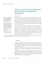
Fig. 1. Scanning electron microscope photograph of the human sclerocorneal trabecular
meshwork (magnification 2000 X).The conventional outflow pathway consists of trabecular
lamellae covered with human trabecular meshwork (HTM) cells, in front of a resistor
consisting of juxtacanalicular HTM cells and the inner wall of Schlemm’s canal. This tissue
has unique morphologic and functional properties involved in the regulation of AH
outflow. Endothelial cells of TM seem to have a leading role in outflow: probably, their
tridimensional architecture and allocation on the trabecular beams considerably increases
the filtration surface whose degeneration, resulting in the decay of HTM cellularity, causes
IOP increase and triggers glaucoma (Saccà and Izzotti 2008).
Glaucoma – Current Clinical and Research Aspects
6
metabolism (Wordinger et al., 2007). TGF in the AH is also responsible for anterior chamber-
associated immune deviation, a mechanism that protects the eye from inflammation and
immune-related tissue damage (Wilbanks et al., 1992). Indeed, TGF-b2 is one of the most
important immunosuppressive cytokines in the anterior chamber of the eye and has a
fibrogenic effect in trabecular cells (Alexander et al., 1998). Finally ECM production in the
TM may be mediated by vitamin C (Epstein et al., 1990; Sawaguchi et al., 1992). Ascorbic
acid is reported to stimulate increased hyaluronic acid synthesis in glaucomatous TM cells
compared with normal human TM cells (Schachtschabel and Binninger, 1993). Also,
ascorbate reduces the viscosity of hyaluronic acid, thus increasing outflow through the
trabeculum (McCarty, 1998). Indeed, Virno already in 1966 discovered that high doses of
vitamin C decreases IOP (Virno 1966). Other molecules that seem to play a very important role
on collagen remodeling are the metalloproteinases (MMPs). MMPs are a family of calcium-
and zinc-dependent extracellular endoproteases that degrade ECM proteins (Nagase and
Woessner, 1999). Matrix metalloproteinases (MMPs) comprise a family of at least 25 secreted
zinc proteases, which are of eminent importance not only for the ECM turnover, but also for
interactions between cells and their surrounding structures (Sternlicht and Werb 2001). Indeed,
increased MMP activity decreases collagen deposition, and AH outflow facility is increased by
stimulating MMP activity (Saccà and Izzotti 2008). Anyway, it remains unclear what fraction
of total resistance is attributable to the JCT and how ECM or specific ECM molecules might be
involved in generation of this resistance (Overby et al. 2009). Anyway, ECM turnover is
required to maintain the appropriate outflow resistance. (Bradley et al. 1998).
By analogy to other basement membranes in the body, the inner wall basement membrane
has the potential to generate a significant portion of outflow resistance. This is
discontinuous (Gong et al. 1996) and this characteristic may be related to the flow of
aqueous humor into Schlemm’s canal (Buller and Johnson, 1994). The resistance by this
tissue seems to be substantially limited (Overby et al. 2009).
On the basis of electron microscopy studies, it has been proposed that aqueous humor
mainly crosses the inner endothelium wall of Schlemm’s canal by two different mechanisms:
a paracellular route through the junctions formed between the endothelial cells (Epstein and
Rohen 1991) and a transcellular pathway through intracellular pores of the same cells
(Johnson and Erickson 2000).
Nevertheless trabecular meshwork pores contribute only 10% of the aqueous outflow
resistance (Sit et al. 1997). Furthermore characteristics of inner wall pores depend on fixation
conditions. In particular, the density of inner wall pores increases with the volume of
fixative perfused through the outflow pathway (Johnson et al. 2002). Scott et al. (2009)
provided by confirmation that the inner wall and underlying juxtacanalicular connective
tissue work together to regulate outflow resistance .
1.3 Meshwork endothelial cells
According to Alvarado, we know that in conventional aqueous outflow pathway there are
two endothelial cell barriers separating the venous circulation from the aqueous humor,
which are specialized and positioned in series: the trabecular meshwork endothelial (TME)
cells and then, subsequently, the endothelial cells that line the lumen of Schlemm’s canal
(SCE) cells. Between these two barriers, there is the juxtacanalicular tissue, which contains a
loose extracellular matrix through which the AH flows (Alvarado et al. 2004). The TME cells
release factors into the AH, and these ligands flow downstream from TMEs to bind and
Oxidative Stress in Anterior Segment of Primary Open Angle Glaucoma
7
actively regulate the permeability properties of the SCEs. These factors, upon binding to
SCE cells, increase the permeability of the SCE barrier (Alvarado et al. 2005a) inducing a
400% enhance in SCE conductivity by means of the activation of specific TME genes
(Alvarado et al. 2005b). In particular interleukin-1α and 1β and tumor necrosis factor-α
released by TME cells induce cell division and migration (Bradley et al. 2000) in those cells
near Schwalbe’s line, while inducing the release of matrix metalloproteinases (Kelley et al.
2007) and an increase of fluid flow across extracellular matrix tissues near JCT (Alvarado et
al. 2005b). In a recent research (Izzotti et al. 2010a) for the first time we have provided
evidence that aqueous humour molecular alterations reflect glaucoma pathogenesis. The
expression of 1,264 proteins was analysed detecting remarkable changing in the aqueous
humour proteins of glaucomatous patients as compared to matched controls. Among the
others AH proteins we have observed that in patients with glaucoma those cytokines
referred by Alvarado are expressed in significantly greater amount compared with controls.
This finding is likely related to the fact that these cytokines are produced to improve the TM
working but in the case of glaucoma TM does not respond properly because malfunctioning
and therefore we are seeing an over-expression of these cytokines. Therefore, the cytokines
released by TME cells regulate the permeability of the SCE barrier in active way (Alvarado
et al. 2005b) . Regulatory volume responses of TM cells influence the tissue permeability too;
indeed, hyperosmotic solutions increased and hyposmotic solutions decreased outflow
facility, respectively (Al-Aswad et al. 1999; Gual et al. 1997 ). The molecular mechanisms for
regulating water balance in many tissues are unknown, but TM cells express aquaporin-1, a
multiple water channel protein transporting water through membranes that can modulate
cell volume (Stamer et al. 2001). Aquaporins also facilitate cell migration, (Verkman 2005 ),
cell proliferation, neuroexcitation, fat metabolism, hydration, and others cell functions
(Tradtrantip et al. 2009 ). Aquaporin may be implicated in the pathogenesis of glaucomatous
optical neuropathy, indeed in animal model elevated IOP reduce its expression (Naka et al.
2010). Anyway, chronic sublethal injury due to cellular stress is a common theme in the
pathogenesis of diverse diseases including atherosclerosis, glomerulonephritis and
pulmonary fibrosis (Dunn 1991; Ross 1995). During glaucoma course, sublethal damage to
the outflow pathways is developing, being the result of accumulated oxidative stress arising
from the environment, vascular dysregulation, aging and/or the pathogenic processes
(Flammer et al. 1999). Molecular changes in the surviving cells determine the expression of
new genes (Dunn 1991; Ross 1995) dependent on the nature of the damaging stimulus and
on tissue type (Mercurio and Manning 1999; Itoh and Nakao 1999). Glaucomatous eyes
exhibit a high level of TM cell loss, above and beyond that of age-matched controls
(Alvarado et al. 1984). The decline of human TM cellularity is linearly related to age
(Alvarado et al., 1984). Grierson has calculated that at 20 years of age the estimated TM cell
number for the whole meshwork is 763,000 (Grierson and Howes, 1987) and the cells
number decreases to 403,000 by the age of 80 years, with a loss rate of 6000 per year
(Grierson et al., 1982).
The mechanism of cell loss and the environmental factors contributing to it are not yet
known. However, this phenomenon may be brought about by cell death caused by noxious
insult, such as free radical attack (Yan et al., 1991; Padgaonkar et al., 1994).
In the anterior chamber (AC), oxidized lipoproteins and free radicals are considered to be
major causes of tissue stress and serve as local triggers for tissue inflammation (Xu et al.
2009). The up-regulation of a large number of inflammatory genes, including genes involved
Glaucoma – Current Clinical and Research Aspects
8
in complement activation and inflammatory cytokine/chemokine production, which in turn
cause abnormal leukocyte-endothelial interactions and ultimately vascular damage (Xu et al.
2009). Furthermore, the innate immune system in general and monocytes in particular play
an important role in aqueous outflow homeostasis: presumably under the influence of
chemotactic signals, the monocytes circulate through the trabecular meshwork in the normal
state and cytokines regulate the permeability of Schlemm's canal endothelial cells (Shifera et
al.2010) and monocytes increase aqueous outflow (Alvarado et al. 2010).
This last mechanism is most easily understood if we think AC as a vase and its endothelium
as that of specialized vase in which flows AH; the endothelia of this vase is the TM whose
complex structure represent a system to increase the area of contact between the TME cells
and the AH. Indeed, the TM has been shown to be composed of contractile elements, which
helps to regulate the outflow facility (Wiederholt et al. 2000). Therefore, opening or
fastening its slots, TM can change the quantity of cells involved in the passage of AH from
AC to SC. Its malfunction thus leads to the intraocular pressure increase. Finally, it is to
remember that fluid flow is not equal within the trabecular meshwork and that preferential
pathways or flow to areas of lower resistance exist in the trabecular meshwork. (De Kater et
al. 1989; Tripathi 1971). One factor contributing to preferential flow may be changes in
extracellular matrix interactions with Schlemm's canal cells in collector channel regions. As
collector channels become altered with age or disease, other collector channels are available
to assume the functional burden (Hann and Fautsch 2009).
2. Oxidative stress and Trabecular Meshwork
The interaction of many risk factors converge on intracellular signaling pathways, affecting
the balance between protein synthesis and breakdown, inducing mitochondrial damage and
apoptosis, which cause the primary glaucoma pathology through a significant loss of TM
endothelial cells. It is not known the exact sequence in which this disease starts, but it is a
fact that glaucoma is not a condition relating to IOP alone. Oxidative stress (Izzotti et al.
2006), vascular abnormalities (vasospasm, systemic hypotension, reduced vascular
perfusion in the optic nerve head and/or retina) (Flammer et al. 2002), glial activity (Kirwan
et al. 2005), immune system (Tezel 2007), inflammatory stimulus (Rönkkö et al. 2007) are
involved in glaucoma injury.
The “trait de union” of all these components is the oxidative stress (Kumar and Agarwal
2007). Oxidative DNA damage is an inevitable consequence of cellular metabolism and it is
secondary to free-radical formation (Cooke et al., 2003).
The oxidative stress and related molecular damages occur in a cell, tissue, and organ in
response to the exposure to oxidizing agents. The mayor types of free radicals are reactive
oxygen species (ROS) and reactive nitrogen species. The free radicals are molecule
fragments equipped with an unpaired electron (odd number of electrons in the last orbital,
when normally the electrons are coupled). Under normal physiological conditions, a small
fraction of the oxygen consumed by mitochondria is constantly converted to superoxide
anions, hydrogen peroxide, hydroxyl radicals, and other ROS. To cope with the ROS, human
cells express an array of antioxidant enzymes, including Mn2+-dependent superoxide
dismutase (SOD), copper/zinc SOD, glutathione peroxidase (GP), glutathione reductase
(GR), and catalase (CAT). SOD convert superoxide anions to hydrogen peroxide, which is
then transformed to water by CAT. The NO radical is produced in organisms by the
oxidation of one of the terminal guanido- nitrogen atoms of L-arginine (Palmer et al. 1988).
Oxidative Stress in Anterior Segment of Primary Open Angle Glaucoma
9
This process is catalyzed by the enzyme NOS. Depending on the microenvironment,
superoxide and NO are readily converted by enzymes or nonenzymic chemical reactions
into reactive non radical species such as singlet oxygen, hydrogen peroxide, or peroxynitrite
, i.e., species which can in turn give rise to new radicals. At moderate concentrations,
however, nitric oxide (NO), superoxide anion, and related reactive oxygen species (ROS)
play an important role as regulatory mediators in signaling processes. Many of the ROS-
mediated responses actually protect the cells against oxidative stress and reestablish “redox
homeostasis.” (Dröge 2002 ). Anyway, there is an age dependent increase in the fraction of
ROS and free radicals that may escape these cellular defense mechanisms and exert damage
to cellular constituents, including DNA, RNA, lipid, and proteins. Any signal or stimulus
that triggers overproduction of ROS may induce the opening of the membrane permeability
transition pore in mitochondria and release of cytochrome c and other apoptogenic factors,
which ultimately lead the cell into apoptosis (Tatton and Olanow 1999).
The anterior chamber (AC) is a highly specialized structure of the eye. It is composed of
several tissues and structures, including the posterior surface of the cornea, the anterior
surface of the iris, the pupil, the pupillary portion of the lens, and peripherally, the
sclerocorneal angle, where the trabecular meshwork (TM), the scleral spur, the ciliary body,
and the iris root are located.
Both vitamin C and glutathione operate in fluid outside the cell and within the cell (Cardoso
et al., 1998); the ascorbate content is higher in diurnal than in nocturnal aqueous humor
(Reiss et al. 1986). Aqueous humor could act as a liquid ultraviolet-light filter for the lens by
virtue of the ascorbic acid in the anterior chamber (Ringvold 1980). Also in vitreous vitamin
C has a very important role; indeed human vitreous gel consumes oxygen by an ascorbate
dependent mechanism. Anyway, the concentration of ascorbate in human vitreous is
remarkably high (Shui et al. 2009). Hence, the vitamin might protect against oxidative or
photo-oxidative damage (Garland 1991; Rose et al. 1998) in both the central corneal
epithelium and aqueous humour (Giblin et al., 1984) and reacts with O
2
to form H
2
O
2
, which
in turn is neutralized by SOD and CAT. Continuous exposure of HTM cells to oxidative
stress via H
2
O
2
results in ROS generation in mitochondria. This, in turn, stimulated NF-kB
activation and subsequent production of interleukins and the induction of inflammatory
mediators. (Li et al., 2007). A high level of ascorbic acid is necessary to maintain oxidative
balance in the AH (Izzotti et al. 2009). A synergism between vitamin E and C has been
envisaged, because vitamin C reduces oxidized vitamin E, which is crucial for protecting cell
membranes from lipid peroxidation; thus, this synergism may have a role in the
pathogenesis of glaucoma (Varma 1991; Kang et al. 2003). Indeed, resistance to AH outflow
increases in the presence of high levels of H
2
O
2
in eyes with a glutathione (GSH)-depleted
TM (Kahn et al. 1983). GSH plays a critical role as an intracellular defense system providing
detoxification of a broad spectrum of reactive species and allowing their excretion as water-
soluble conjugates (Meister, 1989). Glaucomatous patients exhibit low levels of circulating
glutathione (Gherghel et al. 2005) and glutathione participates directly in the neutralization
of free radicals and reactive oxygen species, and maintains exogenous antioxidants such as
vitamins C and E in their reduced (active) forms (Saccà et al. 2007). Insufficient glutathione
combined with exogenous H
2
O
2
may induce collagen matrix remodeling and TM cell
apoptosis independently of mitochondria (Veach 2004). Therefore in glaucoma course
occurs a decline in total antioxidant defenses and in particular in AH (Ferreira et al. 2004)
that have a great impact on the TM endothelium: because this is not more protected
Glaucoma – Current Clinical and Research Aspects
10
properly and because TM is the most sensitive tissue to oxidative radicals in the AC (Izzotti
et al. 2009). This leads to the reduction in the TM cellularity, to TM failure and subsequently
to IOP increase. Of course, oxidative DNA damage in the TM of patients with primary open-
angle glaucoma is significantly higher than in controls (Izzotti et al. 2003). Furthermore,
oxidative DNA damage in patients with glaucoma correlate significantly with intraocular
pressure and with visual field defects (Saccà et al. 2005, Izzotti et al. 2003; Fernández-
Durango et al. 2008). Therefore it is possible that the process that occurs in AC and at the
level of the optic nerve head is the same, and that in both districts the oxidative stress play
an important role. Indeed, in glaucoma animal model, through the induction of oxidative
damage, mechanical and vascular factors working synergistically lead to the same final
pathologic consequence, i.e. glaucoma (Prasanna et al., 2005).
3. Mitochondrial dysfunction
The most important and frequent ocular degenerative diseases including cataract, glaucoma
and age-related macular degeneration are caused by multiple interacting factors. A large
number of environmental and genetic factors play a role in common eye pathologies. Many
of these risk factors are genotoxic therefore being emerging evidence that DNA damage play
a role in the development of the degenerative diseases of the eye (Saccà et al. 2009). ROS
production has been principally implicated in the pathogenesis of eye disease. In particular,
it is possible to believe that the ROS are responsible for the decline of cells that occurs in TM
during aging and glaucoma that is the basis of its bad functioning and failure (Alvarado et
al. 1984 and 2005a; Saccà et al. 2005).
Nowadays it is established that POAG patients bear a spectrum of mitochondrial
abnormalities (Abu-Amero et al. 2006). Mitochondrial theory of ageing, a variant of free
radical theory of ageing, proposes that the accumulation of damage in mitochondria, and in
particular in mitochondrial DNA (mtDNA), leads to human and animal ageing. Oxidative
modification and mutation of mtDNA easily and frequently occur, and the extent of such
alterations of mitochondrial DNA increases exponentially with age. Oxidative modification
in mtDNA is much more extensive than that in nuclear DNA (Ames et al.1993; Yakes and
Van Houten 1997). Age-related alterations in the respiratory enzymes not only decrease ATP
synthesis but also enhance the production of reactive oxygen species (ROS) through
increasing electron leakage in the respiratory chain. With the accumulation of genetic
defects in mechanisms of mitochondrial energy production, the issue of neuronal
susceptibility to damage as a function of ageing becomes important (Parihar and Brewer
2007). Damage to mtDNA induces alterations to the polypeptides encoded by mtDNA in the
respiratory complexes, with a consequent decrease in electron transfer efficiency, leading to
further production of ROS, thus establishing a vicious circle of oxidative stress and energy
decline. This deficiency in mitochondrial energy capacity is regarded as the cause of ageing
and age-related degenerative diseases (Genova et al. 2004). On the basis of the fact that
mitochondria are the major intracellular source and vulnerable target of ROS (Linnane et al.
1989) accumulation of somatic mutations in mtDNA is a major contributor to human aging
and degenerative diseases. Hence, a vicious cycle contributes to the progression of
degenerative process. In this cycle, first a primary mitochondrial mutations induces a
mitochondrial respiratory defect, which increases the leakage of ROS from the respiratory
chain. Then the ROS would trigger accumulation of secondary mtDNA mutations in
Oxidative Stress in Anterior Segment of Primary Open Angle Glaucoma
11
postmitotic cells, leading to further aggravation of mitochondrial respiratory defects and
increased production of ROS and lipid peroxides from mitochondria, and thus resulting in
degeneration of cellular components (Tanaka et al. 1996). This is the basis of mitochondrial
dysfunction that occurs in glaucoma. In the anterior chamber of POAG patients the
relationship between mitochondria and TM is still rather obscure. From a morphological
point of view, we know that in the cribriform layer often contained small mitochondria
(Rohen et al.1993). Besides using an in vitro culture system of bovine trabecular meshwork
cells Dexamethasone-treatment developed an increased number of mitochondria (Sibayan et
al. 1998). TM cells from patients with POAG cells have high endogenous reactive oxygen
species, low ATP, and that mitochondrial complex I defect is associated with the
degeneration of TM cells (He et al. 2008). Therefore other information is taken not “in vivo”,
but only from the study of cell cultures. Recently, it has been demonstrated in vitro that
expression of mutant myocilin, a mitochondrial protein whose role in the arising of early
glaucoma is established, sensitizes Cells to Oxidative Stress-Induced Apoptosis (Myung
Kuket al., 2010).
Recently we have demonstrated that mtDNA damage occurs in the target tissue of POAG:
the TM, but not in other anterior chamber districts, e.g, the iris (Izzotti et al. 2010b).
Furthermore genetic polymorphisms for antioxidant and apoptosis related genes affect the
amount of mtDNA damage in TM (Izzotti et al. 2010b). The remarkable interindividual
variability observed in this study could be due to differences in diseases status because all
patients were carriers of advanced unbalanced POAG or related to mitochondrial
haplotypes, which are characterized by different sensitivity to oxidative damage (Kofler et
al.2009). The lack of mtDNA damage in tissues different from TM observed in this study in
iris is in agreement with the negative findings reported by other studies in blood
lymphocytes of patients with POAG (Abu-Amero et al.: 2007). These findings indicate that
glaucoma may be envisaged as a mitochondriopathy specifically occurring in TM.
Mitochondrial damage and loss occurring in TM trigger both degenerative and apoptotic
phenomena resulting in cell loss, as specifically occurring only in primary open-angle
glaucoma and in pseudoexfoliative glaucoma and not in other glaucoma types ( Izzotti et al.
2011). Decreased cellularity of the TM appears to be a particular characteristic of POAG, but
other authors reported that it does not seem to play a role in the pathogenesis of PEX
glaucoma (Schlotzer-Schrehardt and Naumann 1995). Anyway, in all types of glaucoma in
which IOP is high, TM malfunction resulting from cellularity decrease is the key to the
development of the disease, in acute and chronic angle closure glaucoma too (Sihota et al.
2001).
Furthermore, glaucoma itself could also produce apoptosis of TM cells through mechanical
stress (Grierson 1987) or through trabecular hypoperfusion (Rohen et al. 1993). An increase
in oxidative stress may also contribute to cell loss or alterations in the functioning of TM
cells (Izzotti et al. 2003; Saccà et al. 2005; Dela Paz et al. 1996). Mitochondrial damage has
been detected only in primary open angle and pseudo-exfoliation glaucoma, as the outflow
dysfunction in the other glaucomas studied may have a different underlying bases ( Izzotti
et al. 2011). A further confirmation of this type of pathogenesis is given by the study of AH.
4. Aqueous Humour proteome reflects glaucoma pathogenesis
The AC endothelium (ACE) is not only a group of cells that act as a barrier between AH and
the surrounding tissues, but like in vessels, is a real organ with the function of modulating
Glaucoma – Current Clinical and Research Aspects
12
the tone and the flow rate in response to humoral, nervous and mechanics stimuli. In
physiological conditions the ACE plays an active role cellular interchange, being able to
adapt functionally and structurally to changes in the environment (Verma et al. 2003). The
normal endothelial function depends on both the continuity of cellular anatomical
monolayer and by its functional integrity (Furchgott and Zawadzki 1980). In vessels, the
endothelial dysfunction is characterized by vasoconstriction, platelet aggregation,
proliferation of smooth muscle cells, and is related to a reduced bioavailability of nitric
oxide (NO), to an excess oxidative burden, and to an increased action of endothelin ET-1
(Monnink et al. 2002; Landmesser et al. 2002).
NO is one of the more important substances produced by endothelium; it is a powerful
vasodilatator, an inhibitor of endothelial cell growth and inflammation. NO is a major
intracellular and extracellular effective agent against oxidative stress, and it has a beneficial
antioxidant effects against reactive oxygen species, such as H
2
O
2
, whose detrimental effects
on aqueous humor outflow are established (Lutjen-Drecoll 2000). A prominent role in
endothelial dysfunction must be assigned to NO inactivation by ROS. ROS react with NO
producing peroxy-nitrites, which are cytotoxic agents, thus decreasing NO bioavailability
or directly inactivating NO (Aslan et al. 2008).
The Endothelin-1(ET-1), a potent vasoconstricting peptide of endothelial production acts on
specific receptors present only on smooth muscle cells and cause vasoconstriction and cell
growth. ET-1 determine stimulates the NO production, which acts as negative feedback by
inhibiting further ET-1production (Heitzer et al. 2001). In case of reduced NO bioavailability
this negative feedback mechanism is compromised, and consequently ET-1 vasoconstricting
effect is increased (Haynes and Webb 1998).
A balance between vasoconstrictors and vasodilators is necessary for the maintenance of the
physiological structure and function of endothelia (Gibbons 1997). Whenever the balance
between vasoconstriction and vasodilatation is disrupted, as in glaucoma, the outcome is
endothelial dysfunction and injury that has as consequence cellular proteins loss in AH.
Anterior chamber (AC) is a lumen of a vessel constituted by the cornea and iris and joint
together by means of TM. The AC contains the aqueous humor (AH). The volume of the AC
is approximately 0.25 mL, whereas the volume of the posterior chamber is 0.06 mL. AH is
needed to guarantee optical transparency, structural integrity, and nutrition in the absence
of blood vessels (Izzotti et al. 2009). Furthermore, this liquid has the task of protecting and
supplying nutrients to the cornea, lens, and TM (Izzotti et al. 2006; Fuchshofer and Tamm
2009). Other functions ascribed to AH inflow have been less clearly defined (Krupin and
Civan 1996) and include the delivery of antioxidant, such as ascorbate, and participation in
local immune responses. The ciliary epithelium concentrates ascorbate in AH rendering its
concentrations 40-fold higher in AH than in blood plasma (Krupin and Civan 1996). It is
possible that ascorbate is not only a scavenger of ROS, but may be also a regulator of ion
channel activity functioning as an endogenous modulator of neuronal excitability (Nelson
et al. 2007). In any case, cells in the outflow pathway are subjected to chronic oxidative
stress and go towards an impaired proteasome activity in TM (Govindarajan et al. 2008).
Therefore, any alteration in proteasome function due to oxidative stress or aging would be
also expected to increase the rate of accumulation of misfolded mutant myocilin in the
endoplasmic reticulum and contribute to the pathogenic effect of this mutant protein in the
mitochondrial functions in human TM cells (He et al. 2009). AH represents a protein-
containing biological fluid fundamental for eye pathophysiology (Civan 2008). However,
Oxidative Stress in Anterior Segment of Primary Open Angle Glaucoma
13
the relationship between TM and AH proteins as related to POAG pathogenesis has not yet
been explored. Many proteins expressed at high levels in healthy patients are reduced in
POAG patients, while other proteins detected at low levels in normal subjects are increased
in POAG patients.
Recently we have discovered 6 classes of protein specifically present in the AH of advanced
glaucomatous patients (Izzotti et al. 2010a).
5. Proteins in glaucomatous Aqueous Humour
The first class of proteins were mitochondrial proteins involved in the electron transport
chain, trans-membrane transport, protein repair, and mitochondrial integrity maintenance.
The second class were proteins involved in apoptosis induction, through the intrinsic, i.e.,
mitochondrial-dependent, pathway.
The third class were proteins connecting cells. These include catenins, junctional plaque
protein, dynein, and cadherins.
The fourth class is composed by neuronal proteins like optineurin or growth and
differentiation factors involved in neurogenesis and neuron survival.
The fifth class includes the protein kinase involved in apotosis activation and signal
transduction.
Finally, the sixth class concerns the oxidative stress and includes: nitric oxide synthetase,
superoxide dismutase and microsomal glutathione S-transferase 1.
All these proteins testify that the AH composition is affected and reflects the mechanisms of
glaucoma pathogenesis. Mitochondrial proteins presence, segregated into intact
mitochondrial under normal conditions, reflects that TM endothelium cells is affected by
mitochondria loss and dysfunction, as specifically occurring only in TM and not other AC
tissues during glaucoma. The presence of mitochondrial proteins in AH indicates that TM
undergoes structural alterations and that in particular its endothelial cells loss
mitochondrial proteins as a consequence of mitochondrial DNA deletion that leads to
mitochondrial TM malfunction and destruction leading to apoptosis. Cellular loss is
determined not only by proapoptotic proteins but also by other mechanisms involved and
revealed by the presence of other protein: inflammation, vascular dysregulation, and
hypoxia (Choi and Benveniste 2004; Li et al. 2006). Indeed, proteins of these functional
groups are expressed in the AH of glaucomatous patients. This is the situation for BIK
protein, normally located inside mitochondria, which activates apoptosis process through
the intrinsic pathway, and for FAS protein, that is responsible for the activation of apoptosis
through the extrinsic pathway in response to inflammation and/or oxidative stress.
Furthermore, FAS has been demonstrated to provoke apoptosis increasing myocilin release
from mitochondria to cytosolic compartments of TM cells (Sakai et al. 2007).
The presence of “proteins connecting cells” in glaucomatous AH, reflects the impairment of
cytoskeleton organization, cell-cell adhesion and migration (Lee and Tomarev 2007).
Calnexin presence in AH can be linked with the presence of mutant myocilin. Myocilin, the
first protein genetically associated with the development of glaucoma, is a constituent of
human AH and is expressed in many ocular tissues, with highest expression observed in
cells of the trabecular meshwork (Adam et al., 1997;). While calnexin is a calcium-binding
protein playing a major role in the quality control apparatus of the endoplasmic reticulum
by the retention of incorrectly folded proteins. Normally, located in melanosomes, calnexin