BREAST CANCER – CURRENT AND ALTERNATIVE THERAPEUTIC MODALITIES pptx
Bạn đang xem bản rút gọn của tài liệu. Xem và tải ngay bản đầy đủ của tài liệu tại đây (22.81 MB, 550 trang )
BREAST CANCER –
CURRENT AND
ALTERNATIVE
THERAPEUTIC MODALITIES
Edited by Esra Gunduz and Mehmet Gunduz
Breast Cancer – Current and Alternative Therapeutic Modalities
Edited by Esra Gunduz and Mehmet Gunduz
Published by InTech
Janeza Trdine 9, 51000 Rijeka, Croatia
Copyright © 2011 InTech
All chapters are Open Access articles distributed under the Creative Commons
Non Commercial Share Alike Attribution 3.0 license, which permits to copy,
distribute, transmit, and adapt the work in any medium, so long as the original
work is properly cited. After this work has been published by InTech, authors
have the right to republish it, in whole or part, in any publication of which they
are the author, and to make other personal use of the work. Any republication,
referencing or personal use of the work must explicitly identify the original source.
Statements and opinions expressed in the chapters are these of the individual contributors
and not necessarily those of the editors or publisher. No responsibility is accepted
for the accuracy of information contained in the published articles. The publisher
assumes no responsibility for any damage or injury to persons or property arising out
of the use of any materials, instructions, methods or ideas contained in the book.
Publishing Process Manager Silvia Vlase
Technical Editor Teodora Smiljanic
Cover Designer Jan Hyrat
Image Copyright Serdar Tibet, 2011. Used under license from Shutterstock.com
First published October, 2011
Printed in Croatia
A free online edition of this book is available at www.intechopen.com
Additional hard copies can be obtained from
Breast Cancer – Current and Alternative Therapeutic Modalities,
Edited by Esra Gunduz and Mehmet Gunduz
p. cm.
ISBN 978-953-307-776-5
free online editions of InTech
Books and Journals can be found at
www.intechopen.com
Contents
Preface IX
Part 1 Targeting Signaling Pathways and Extracellular Matrix 1
Chapter 1 Novel Therapeutic Strategies and
Combinations for HER2-Overexpressing Breast Cancer 3
Sylvia Shabaya and Rita Nahta
Chapter 2 Therapeutic Targeting of
Osteopontin in Breast Cancer Cells 23
Gopal C. Kundu, Supriya Saraswati, Megha Sanyal,
Anuradha Bulbule, Anuja Ramdasi, Dhiraj Kumar, Reeti Behera,
Mansoor Ahmed, Goutam Chakraborty, Vinit Kumar,
Shalini Jain, Gowrishankar S. and Pompom Ghosh
Chapter 3 Targeting Cas Family Proteins as
a Novel Treatment for Breast Cancer 37
Joerg Kumbrink and Kathrin H. Kirsch
Chapter 4 Breast Cancer and
Current Therapeutic Approaches:
From Radiation to Photodynamic Therapy 63
Peter Ferenc, Peter Solár, Jaromír Mikeš,
Ján Kovaľ and Peter Fedoročko
Part 2 Anti-Tumor Compounds 89
Chapter 5 Boron Compounds in the Breast Cancer
Cells Chemoprevention and Chemotherapy 91
Ion Romulus Scorei
Chapter 6 Benzo-Fused Seven- and Six-Membered Derivatives
Linked to Pyrimidines or Purines Induce Apoptosis of
Human Metastatic Breast Cancer MCF-7 Cells In Vitro 115
Joaquín M. Campos, M. Carmen Núñez,
Ana Conejo-García and Olga Cruz-López
VI Contents
Chapter 7 The Analogues of DNA Minor-Groove
Binders as Antineoplastic Compounds 133
Danuta Drozdowska
Chapter 8 Fractionation and Characterization
of Bioactive Components in Kefir Mother
Culture that Inhibit Proliferation
of Cultured MCF-7 Human Breast-Cancer Cells 149
Chujian Chen, Hing Man Chan and Stan Kubow
Part 3 Targeting Coagulation Factor VII 173
Chapter 9 Factor VII-Targeted Photodynamic
Therapy for Breast Cancer and Its Therapeutic
Potential for Other Solid Cancers and Leukemia 175
Zhiwei Hu
Chapter 10 Ectopic Synthesis of Coagulation Factor VII
in Breast Cancer Cells: Mechanisms, Functional
Correlates, and Potential for a New Therapeutic Target 197
Shiro Koizume and Yohei Miyagi
Part 4 Use of Herbal Medicine and Derivatives 213
Chapter 11 Lunasin, a New Breast Cancer
Chemopreventive Seed Peptide 215
Chia-Chien Hsieh, Blanca Hernández-Ledesma and
Ben O. de Lumen
Chapter 12 Experimental Therapeutics in Breast Cancer Cells 243
Weena Jiratchariyakul and Tanawan Kummalue
Chapter 13 Red American Ginseng and Breast Cancer 269
Chong-Zhi Wang, Guang-Jian Du and Chun-Su Yuan
Chapter 14 Synthesis and In Vitro Screening of Novel Heterocyclic
Compounds as Potential Breast Cancer Agents 283
Narsimha Reddy Penthala, Thirupathi Reddy Yerramreddy,
Nikhil Reddy Madadi, Vijayakumar Sonar and Peter A. Crooks
Chapter 15 The Beneficial Effects of Nutritional
Compounds on Breast Cancer Metastasis 295
Jeffrey D. Altenburg and Rafat A. Siddiqui
Chapter 16 Legume-Derived Bioactive Compounds for
the Prevention and Treatment of Breast Cancer 319
Graziella Joanitti, Sonia Freitas and Ricardo Azevedo
Contents VII
Part 5 Novel Therapeutics: Gene Therapy, Nanoparticles,
Experimental Therapeutics 345
Chapter 17 Nanobody, New Agent for
Combating Against Breast Cancer Cells 347
Fatemeh Rahbarizadeh, Fatemeh Rahimi Jamnani and
Farnoush Jafari Iri-Sofla
Chapter 18 Experimental Therapeutics for
the Treatment of Triple Negative Breast Cancer 371
Julian Dzeyk, Babasaheb Yadav and Rhonda J. Rosengren
Chapter 19 New Experimental Therapies
Targetting Breast Cancer Cell 395
Di Benedetto Melanie
Chapter 20 Future Therapeutic Strategies:
Implications for Brk Targeting 413
Amanda Harvey and Rajpal Burmi
Chapter 21 Immunoliposomes: A Multipurpose
Strategy in Breast Cancer Targeted Therapy 435
Enrique Barrajón-Catalán, María P. Menéndez-Gutiérrez,
Alberto Falcó, Miguel Saceda, Angela Catania and Vicente Micol
Chapter 22 Treatment of Breast Cancer Lytic
Skeletal Metastasis Using a Model in Nude Rats 453
Michael Zepp, Tobias J. Bäuerle, Victoria Elazar,
Jenny Peterschmitt, Rinat Lifshitz-Shovali, Hassan Adwan,
Franz P. Armbruster, Gershon Golomb and Martin R. Berger
Chapter 23 Inhibition of Tumor Growth
and Metastasis by a Combination of
Anti-VEGF-C and Enhanced IL-12 Therapy in
an Immunocompetent Mouse Mammary Cancer Model 489
Masa-Aki Shibata, Junji Morimoto, Eiko Shibata,
Mariko Harada-Shiba and Shigekazu Fujioka
Part 6 Drug Resistance 503
Chapter 24 Roles and Mechanisms of Estrogen and Estrogen
Receptors in Breast Cancer Resistant to Chemotherapy 505
Weimin Fan and Meihua Sui
Chapter 25 Tamoxifen Resistant Breast
Cancer and Autophagy 523
Grey A. Wilkinson, Adam N. Elwi and Sung-Woo Kim
Preface
Cancer is the leading cause of death in most countries and continues to increase
mainly because of the aging and growth of the world population as well as habitation
of cancer-causing behaviors such as smoking and alcohol. Based on statistics of the
GLOBOCAN 2008, about 12.7 million cancer cases and 7.6 million cancer deaths are
estimated to have occurred in 2008 (Siegel et al. Ca Cancer J Clin 61:212-236, 2011).
Breast cancer is the most frequently diagnosed cancer and the leading cause of cancer
death among females, accounting for 23% of the total cancer cases and 14% of the
cancer deaths. Thus cancer researches, especially breast cancer, are important to
overcome both economical and physiological burden. The current book on breast
cancer aims at providing information about recent clinical and basic researches in the
field. The book includes chapters written by well-known authors, who are worldwide
experts in their research areas and mainly covers therapeutic applications in breast
cancer. Other topics covered in this book are: therapeutic modalities targeting
signaling pathways, coagulation factor VII as well as extracellular matrix, use of anti-
tumor compounds, use of herbal medicine and derivatives as well as application of
alternative medicine, and recent novel therapies including gene therapy, nanoparticles
as well as other experimental methods, and finally, the issue of chemoresistance is also
discussed. We hope that the book will serve as a good guide for the scientists,
researchers and educators in the field.
Assoc. Prof. Dr. Esra Gunduz
Prof. Dr. Mehmet Gunduz
Fatih University Medical School
Turkey
Part 1
Targeting Signaling Pathways and
Extracellular Matrix
1
Novel Therapeutic Strategies and Combinations
for HER2-Overexpressing Breast Cancer
Sylvia Shabaya and Rita Nahta
Emory University,
USA
1. Introduction
Approximately 20-30% of breast cancers show increased expression of the HER2 receptor
tyrosine kinase. Elevated levels of HER2 are associated with aggressive disease, high
metastatic potential, and reduced survival versus other breast cancer subtypes (Slamon,
1987). Trastuzumab (Herceptin) is a monoclonal antibody targeted against an extracellular
region of HER2 (Carter, 1992). Clinical trials have shown that 15-30% of patients with HER2-
overexpressing metastatic breast cancer respond to single-agent trastuzumab for a median
duration of approximately 10 months (Baselga, 1996; Cobleigh, 1999). Response rates
improve when trastuzumab is combined with chemotherapy in patients with HER2-
overexpressing metastatic breast cancer (Esteva, 2002; Slamon, 2001). A subset of
trastuzumab-resistant breast cancers respond to the dual EGFR/HER2 kinase inhibitor
lapatinib, although the majority (70% or more) show primary resistance (Geyer, 2006).
Similar to trastuzumab treatment, clinical trials with lapatinib indicated that the median
duration of response to lapatinib in a heavily pre-treated, trastuzumab-refractory
population was less than one year (Geyer, 2006). Hence, resistance to clinically available
HER2-targeted agents is a major concern in the treatment of patients with HER2-
overexpressing metastatic breast cancer.
2. HER2 and breast cancer
The human epidermal growth factor receptor 2 (HER2) is overexpressed in approximately
25% of invasive breast carcinomas. HER2 is a member of the epidermal growth factor
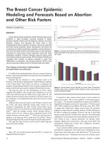
receptor (EGFR) family, which also contains two other receptors, HER3 and HER4 (Fig. 1).
Each of these cell surface receptors has an extracellular ligand-binding domain and a
transmembrane-spanning domain (Nielsen, 2008). All HER family receptors except HER2
bind specific ligands that induce conformational changes and receptor homo- or hetero-
dimerization. Several HER family ligands have been identified including transforming
growth factor alpha (TGFa), epidermal growth factor (EGF), and the heregulins (Nielsen,
2008). In addition, all except HER3 contain an intracellular tyrosine kinase domain. Receptor
dimerization activates the kinase function of receptors, leading to receptor auto- or trans-
phosphorylation. The phosphorylated tyrosine residues serve as docking sites for SH2 and
PTB-domain containing proteins, which links the receptors to multiple cell survival and
proliferation pathways including the phosphatidylinositol-3 kinase (PI3K) and mitogen-
Breast Cancer – Current and Alternative Therapeutic Modalities
4
activated protein kinase (MAPK) cascades (Spector, 2009; Graus-Porta, 1997). HER2 is the
preferred dimerization partner for the other HER family members, as HER2 heterodimers
have increased ligand binding affinity and increased catalytic activity relative to other
heterodimer complexes (Spector, 2009; Graus-Porta, 1997). In particular, the HER2-HER3
heterodimer has the strongest kinase activity and transforming ability, as HER3 possesses
multiple PI3K docking sites in its cytoplasmic tail.
Fig. 1. HER/erbB family of growth factor receptors. The four members of the EGFR family
are illustrated. The inactive ligand-binding domains of HER2 and the inactive kinase
domain of HER3 are denoted with an X. Trastuzumab binds to domain IV of the
extracellular region of HER2.
2.1 Targeting HER2 in breast cancer
Patients who are diagnosed with HER2-overexpressing breast cancer have a poor prognosis,
and shorter progression-free and overall survival compared to patients with other subtypes
of breast cancer (Eccles, 2001). HER2-overexpressing tumors have been found to be larger in
size, and higher in nuclear grade, S phase fraction, and aneuploidy (Nielsen, 2008).
Traditional cancer treatments have targeted DNA replication or cell division, leading to
nonspecific cytotoxicity (Oakman, 2010). The identification of abnormal signaling from
HER2 led to the development of trastuzumab (Herceptin) (Genentech, San Francisco, CA,
USA), which is the first drug to target the genetic lesion or oncogenic addiction found in
patients with HER2-overexpressing breast cancer. Clinically, trastuzumab was found to
significantly enhance the effectiveness of conventional chemotherapies. However, the
median duration of response was less than one year, indicating rapid development of
Novel Therapeutic Strategies and Combinations for HER2-Overexpressing Breast Cancer
5
resistance. The precise mechanism of action of trastuzumab is unclear, but it is thought to
involve HER2 downregulation (Cuello, 2001; Gajria, 2011), selective inhibition of HER2-
HER3 heterodimerization (Junttila, 2009; Gajria, 2011), prevention of HER2 extracellular
domain proteolytic cleavage (Molina, 2001; Gajria, 2011), and activation of an immune
response including antibody-dependent cellular cytotoxicity (Sliwkowski, 1999). As a single
agent, trastuzumab achieved an overall response rate for a median duration of about nine
months (Baselga, 1996; Cobleigh, 1999; Nielsen, 2008; Slamon, 2001). The low response rate
indicates that many patients with HER2-overexpressing breast cancer have primary resistance
to trastuzumab, while the short duration of response indicates rapid development of acquired
resistance. Multiple mechanisms contributing to trastuzumab resistance have been proposed,
resulting in multiple approaches to potentially treat resistant cancers (Table 1).
Target Role in trastuzumab resistance
PI3K Increased PI3K signaling due to PIK3CA mutations or PTEN loss was reported
in trastuzumab-resistant cancers
mTOR As a downstream molecule of PI3K, mTOR has become a target of inhibition in
resistant cancers; multiple mTOR inhibitors are in advanced phases of clinical
development
IGF-
IR
Increased expression of IGF-IR has been shown to reduce response to
trastuzumab; increased IGF-IR overexpression was associated with lower
response to neoadjuvant trastuzumab; IGF-IR/HER2 interaction and crosstalk
were associated with acquired resistance
Src Trastuzumab-mediated inhibition of Src activity appears to be important to its
anti-cancer activity; resistance to trastuzumab was associated with PTEN loss
and increased Src activity; targeting Src with dasatinib or genetic knockdown
blocked growth of resistant cancers
Cdk2 Reduced p27kip1 levels or amplification of cyclin E gene have been reported to
result in increased cdk2 activity in trastuzumab-resistant cancers
Table 1. Potential pharmacologic targets in trastuzumab-resistant HER2-positive breast
cancers.
3. Targeting PI3K/mTOR signaling in HER2-overexpressing breast cancer
HER2 signaling is initiated upon receptor dimerization, which induces phosphorylation of
tyrosine residues within the receptor cytoplasmic domain. The phosphorylated residues
serve as docking sites for adaptor proteins and link the receptor to downstream survival
pathways including the PI3K/Akt/mTOR axis (Spector, 2009). The PI3K pathway is
frequently hyper-activated in many cancers. An association between oncogenic PI3K
mutations and trastuzumab resistance was found in a study examining HER2-
overexpressing tumors from patients with trastuzumab-refractory disease (Berns, 2007).
About 25% of tumors analyzed had PIK3CA mutations, and reduced phosphatase and
tensin homolog (PTEN) expression was present in 22% of the tumors.
Immunohistochemistry studies performed in a retrospective analysis of HER2-amplified
breast tumors treated with trastuzumab plus taxanes showed a postive correlation between
PTEN down-regulation and tumor response (Nagata, 2004). To evaluate the role of PI3K
Breast Cancer – Current and Alternative Therapeutic Modalities
6
post-trastuzumab exposure, tumors that had progressed on trastuzumab were analyzed for
changes in PI3K signaling. The findings demonstrated that PI3K mutations and PTEN loss
were identified in patients who had initially responded to trastuzumab; reduced PTEN
expression was identified in tumors that had developed trastuzumab resistance, but had not
been identified before trastuzumab treatment. This finding indicates that PI3K mutations
can occur as a result of trastuzumab treatment in some tumors (Kalinsky, 2009; Sakr, 2010;
Gajria, 2011). Thus, there is ample rationale for co-targeting PI3K and HER2 in breast cancer.
Activated Akt regulates several downstream signaling molecules including mTOR, a highly
conserved 289-kDa serine/threonine kinase that plays roles in cell proliferation, survival,
and motility (Lang, 2010). mTOR activation is initiated when phosphorylated PI3K/Akt
inhibits the TSC1/TSC2 complexes, thereby preventing Rheb from inhibiting mTOR.
mTORC1 (mTOR, Raptor, mLST8/GBL and PRAS40) and mTORC2 (mTOR, RICTOR,
mLST8/GBL, SIN1, and PROTOR/PRR5) are the two distinct complexes through which
mTOR exerts cellular effects. The complexes have different functional roles, with mTORC1
having been implicated in cell cycle progression, motility, and protein biosynthesis, while
mTORC2 regulates cytoskeleton organization, and regulates cell growth and survival
(Wullschleger, 2005; Van der Heijen, 2011).
Preclinical in vivo studies in which mice were treated with single agent trastuzumab, the
mTOR inhibitor rapamycin, or a combination of trastuzumab plus rapamycin showed that
the combination was more effective at inducing tumor regression than either of the single
agent treatments (Miller, 2009). In cell culture experiments using the rapamycin analogue
RAD001, a greater amount of growth inhibition was observed with combination mTOR
inhibition plus HER2-targeting than with either drug alone. Trastuzumab partially
decreased PI3K activity, but not mTOR activity (Miller, 2009). Increased PI3K signaling is a
validated mechanism of trastuzumab resistance, but its association with lapatinib resistance
is yet to be determined due to conflicting data (Eichhorn, 2008; O’Brien, 2010). Patients with
HER2-overexpressing breast cancer who have developed resistance to trastuzumab may be
given the dual EGFR/HER2 tyrosine kinase inhibitor lapatinib. Response to single agent
lapatinib is less than 25%, indicating cross-resistance between trastuzumab and lapatinib
(Blackwell, 2010; Eichhorn, 2008). As with trastuzumab treatment, the small subset of
patients who initially responded to lapatinib eventually developed resistance, at which
point there is no standard therapeutic approach available. Phase I trials have indicated that
in patients with trastuzumab-resistant, heavily pretreated breast cancer, combined
everolimus plus trastuzumab could be a promising treatment (Jerusalem, 2011). It is thought
that the inability of trastuzumab to completely inhibit PI3K/Akt/mTOR signaling may
permit escape from growth inhibition; mTOR inhibitors would thus synergize with
trastuzumab to prevent the continued growth of HER2-dependent cancer cells.
In contrast to PI3K, very little has been published regarding the role of MAPK signaling in
trastuzumab resistance. Our data suggests that phosphorylation of Erk1/2, which is a
marker of MAPK activity, is not increased in resistant cells (Fig. 2A). Inhibition of MEK
(upstream of Erk1/2) using a small molecule MEK kinase inhibitor called PD0325901
reduces p-Erk1/2 levels in parental HER2-overexpressing breast cancer cells and in acquired
trastuzumab-resistant and primary trastuzumab-resistant cells (Fig. 2B). However,
trastuzumab-naïve and trastuzumab-resistant cells are relatively resistant to PD0325901, in
that doses up to 10 uM do not block proliferation of HER2-overexpressing trastuzumab-
naïve or resistant cells (Fig. 2C). Thus, our data indicate that MAPK signaling may not be a
major mechanism of trastuzumab resistance.
Novel Therapeutic Strategies and Combinations for HER2-Overexpressing Breast Cancer
7
Fig. 2. Role of MAPK signaling in trastuzumab-resistant cells. (A) SKBR3 parental,
trastuzumab-resistant pool 2, and BT474 parental, and trastuzumab-resistant clone 2 and
clone 3 cells were Western blotted for phosphorylated and total Erk1/2. (B) BT-parental, BT-
c2 (resistant clone 2), and MDA-MB-361 primary trastuzumab-resistant cells were treated
with MEK inhibitor PD0325901 at 10, 100, or 1000nM for 6 hours or with DMSO control (C)
corresponding to the volume found in the highest dose of PD0325901. Total protein lysates
were Western blotted for phosphorylated and total Erk1/2. (C) BT-parental, resistant clone 2
and 3, MDA361, and MDA453 cells were treated with MEK inhibitor PD0325901 at 1, 10,
100, 1000, or 10, 000nM for 48 hours with six replicates per treatment group. Control cells
were treated with DMSO corresponding to the volume found in the highest dose of
PD0325901. Proliferation was assessed by MTS assay, and is shown as a percentage of
control group per line.
4. Targeting IGF-IR signaling in HER2-overexpressing breast cancer
The insulin-like growth factor receptor I (IGF-IR) is a heterotrimeric transmembrane
tyrosine kinase receptor that regulates cell metabolism and growth (Chaves, 2010), and has
Breast Cancer – Current and Alternative Therapeutic Modalities
8
been associated with increased risk and maintenance of multiple cancers including HER2-
overexpressing breast cancer (Esparis-Ogando, 2008; Hankinson, 1998; Surmacz, 2000).
Circulating ligands of the insulin-like growth factor (IGF) system include IGF-I and IGF-II,
with IGF-I having the highest affinity for IGF-IR. Upon binding to IGF-IR, a receptor
conformational change is induced that leads to tyrosine phosphorylation and activation of
several downstream survival signaling pathways such as the Ras/Raf/mitogen activated
protein kinase pathway (MAPK), and the PI3K/Akt/mTOR pathway. Activation of these
pathways results in cell cycle progression and resistance to apoptosis (Chaves, 2011; Adams,
2000). The IGF binding proteins (IGFBPs) modulate IGF-IR activity by binding to the IGF
ligands thereby sequestering them and preventing ligand-induced receptor activation
(Adams, 2000). Higher levels of circulating IGF-I have been linked to trastuzumab resistance
in HER2-overexpressing breast cancer, with the addition of IGFBP3 decreasing IGF-IR
activity, and subsequently resulting in an increased response to trastuzumab (Lu, 2001;
Jerome, 2006).
We found by gene microarray analysis that IGFBP3 and IGFBP5 were down-regulated in
resistant versus sensitive cells (Table 2). However, ELISA of secreted IGFBP3 (Fig. 3A) or
real-time PCR analysis of endogenous IGFBP3 or IGFBP5 transcript level (Fig. 3B) failed to
show any differences in IGFBP3 or IGFBP5 level in resistant versus parental cells. Thus, our
data do not support down-regulation of IGFBP3 or IGFBP5 as a mechanism of increased
IGF-IR signaling in trastuzumab resistance.
Gene
Name Fold Change ILMN_GENE DEFINITION
IGFBP5 -20. 55848937 IGFBP5
Homo sapiens insulin-like
g
rowth factor bindin
g
protein
5 (IGFBP5), mRNA.
IGFBP5 -20. 0185274 IGFBP5
Homo sapiens insulin-like
g
rowth factor bindin
g
protein
5 (IGFBP5), mRNA.
IGFBP3 -7. 77282369 IGFBP3
Homo sapiens insulin-like
g
rowth factor bindin
g
protein
3 (IGFBP3), transcript variant 2, mRNA.
PKIA -6. 484521044 PKIA
Homo sapiens protein kinase (cAMP-dependent,
catalytic) inhibitor alpha (PKIA), transcript variant 7,
mRNA.
IGFBP3 -6. 193624741 IGFBP3
Homo sapiens insulin-like
g
rowth factor bindin
g
protein
3 (IGFBP3), transcript variant 1, mRNA.
PKIA -5. 371909749 PKIA
Homo sapiens protein kinase (cAMP-dependent,
catalytic) inhibitor alpha (PKIA), transcript variant 6,
mRNA.
BASP1 -4. 444496135 BASP1
Homo sapiens brain abundant, membrane attached
signal protein 1 (BASP1), mRNA.
HERC6 -4. 048474978 HERC6 Homo sapiens hect domain and RLD 6 (HERC6), mRNA.
FRAS1 -3. 988854857 FRAS1 Homo sapiens Fraser s
y
ndrome 1 (FRAS1), mRNA.
THBS1 -3. 966312615 THBS1 Homo sapiens thrombospondin 1 (THBS1), mRNA.
Table 2. Genes that are down-regulated in SKBR3- and BT474-derived acquired
trastuzumab-resistant cells versus parental SKBR3 and BT474 cells by 4-fold or more.
Novel Therapeutic Strategies and Combinations for HER2-Overexpressing Breast Cancer
9
Fig. 3. IGFBP3 and IGFBP5 in resistant and sensitive cells. (A) Secreted IGFBP3 was assessed
by ELISA in SKBR3 parental, resistant pool 2, BT474 parental, resistant clone 2 and clone 3
cells. IGFBP3 is shown in pg/mL and was measured in triplicate with reproducible results
per line. (B) Real-time PCR analysis of IGFBP3 and IGFBP5 was examined in triplicate per
line, with error bars representing standard deviation between replicates. Housekeeping
gene RPLPO was measured as an internal control; IGFBP3 and IGFBP5 values are
normalized to RPLPO.
A subset of HER2-/ IGF-IR-overexpressing cells were found to be less sensitive to the
growth inhibitory effects of trastuzumab when compared to HER2-overexpressing cells that
do not overexpress IGF-IR (Lu, 2001). Flow cytometry revealed that after trastuzumab
Breast Cancer – Current and Alternative Therapeutic Modalities
10
treatment, HER2 overexpressing cells were less likely to progress through the cell cycle and
stopped at the G1 phase, while a greater number of HER2/IGF-IR overexpressing cells
passed the restriction point and completed the cell cycle. These results demonstrate that
IGF-IR interferes with the growth inhibitory actions of trastuzumab, supporting therapeutic
strategies that co-target HER2 and IGF-IR. Further, we discovered that signaling interactions
exist between IGF-IR and HER2 in trastuzumab-resistant cancers (Nahta, 2005; Jin, 2008).
Immunoprecipitation and immunoblotting experiments revealed that IGF-I stimulation
results in an increase in IGF-IR phosphorylation more rapidly in trastuzumab-resistant cells
than in trastuzumab-sensitive cells. Furthermore, IGF-IR heterodimerization with HER2
results in HER2 activation in trastuzumab-resistant cells, but not in trastuzumab-sensitive
cells, indicating crosstalk between the two receptors. Kinase inhibition or antibody blockade
of IGF-IR restores trastuzumab sensitivity. Treatment of trastuzumab-resistant breast cancer
cells with the highly specific IGF-IR antibody alpha IR3 disrupted the IGF-IR/HER2
heterodimer and increased trastuzumab sensitivity. These results suggest that IGF-IR-
targeted treatments may be useful in combination with trastuzumab.
The association of increased IGF-IR activity with the development of trastuzumab resistance
in HER2-overexpressing breast cancer makes IGF-IR an important target. Researchers have
been working toward the goal of developing agents that target IGF-IR for the past several
years with each generation of agents aimed at producing a greater benefit for the patient
while decreasing adverse effects. IGF-IR and the insulin receptor (IR) are 60% homologous,
with one of the adverse effects of IGF-IR antibody treatment being downregulation of the IR,
leading to hyperglycemia (Sachdev, 2006). In an effort to remedy this problem,
pharmacological agents like the small molecule tyrosine kinase inhibitor NVP-AEW541
(Novartis Pharma, Basel Switzerland) are specific for IGF-IR and less likely to interfere with
glucose metabolism. Combination treatment with NVP-AEW541 and trastuzumab showed
synergistic growth inhibitory effects, indicating that inhibiting IGF-IR plus HER2 could
benefit patients whose tumors overexpress both receptors (Esparis-Ogando, 2008).
IGF-IR overexpression and crosstalk with HER2 suggests that IGF-IR plays a crucial role in
conferring trastuzumab resistance. The molecular signaling pathways by which IGF-IR
confers resistance to trastuzumab is not clear, although downstream focal adhesion kinase
(FAK) and PI3K/Akt pathway signaling likely play a role (Yang, 2010). This data linking
IGF-IR to the development of trastuzumab resistance, along with the increased sensitivity to
trastuzumab upon IGF-IR inhibition provides a rational for the development of
combinatorial HER2 and IGF-IR targeting.
5. Targeting Src in HER2-overexpressing breast cancer
Trastuzumab treatment of HER2-overexpressing breast cancer cells results in inhibition of
Src non-receptor tyrosine kinase (Nagata, 2004). Src inhibition appears to be important to
trastuzumab-mediated anti-cancer activity, as increased Src signaling is associated with
trastuzumab resistance (Mitra, 2009; Liang, 2010; Zhang, 2011). One mechanism leading to
increased Src activity appears to be a variant of HER2 called HER2 delta 16 (Mitra, 2009),
which shows increased oncogenic activity. Local disease progression involved HER2Delta16
in 89% of breast cancer patients with HER2-positive tumors (Mitra, 2009). Transfection of
MCF7 or NIH3T3 cells with HER2 delta 16 promoted receptor dimerization, invasion, and
trastuzumab resistance (Mitra, 2009). The oncogenic properties of HER2Delta16 were
mediated through direct interaction of HER2Delta16 with Src kinase. Activated Src kinase
Novel Therapeutic Strategies and Combinations for HER2-Overexpressing Breast Cancer
11
was found in 44% of HER2Delta16-positive breast carcinomas (Mitra, 2009). Dual targeting
of HER2Delta16 plus Src with dasatinib resulted in Src inactivation, destabilization of
HER2Delta16, and decreased tumorigenicity (Mitra, 2009). In addition, Src activation via
Jak2 has been shown to reduce trastuzumab activity (Liang, 2010). Recombinant human
erythropoietin activated Jak2-Src signaling and inactivated PTEN in HER2-positive cells
(Liang, 2010). Combined treatment with recombinant human erythropoietin plus
trastuzumab reduced response to trastuzumab in cell culture and in vivo models. Further,
shorter progression-free and overall survival was found in patients with HER2-positive
breast cancer treated concurrently with erythropoietin and trastuzumab (Liang, 2010). Src
was also shown to be activated in primary and acquired trastuzumab resistance as a
consequence of PTEN loss (Zhang, 2011). Src-targeted therapy blocked growth of
trastuzumab-resistant tumors in vivo (Zhang, 2011). Thus, Src activation may occur via
multiple mechanisms, ultimately abrogating sensitivity to trastuzumab. Combining Src-
targeted therapy with trastuzumab may offer benefit to patients with HER2-overexpressing
breast cancer.
6. Role of p27 and cdk2 in HER2-overexpressing breast cancer
Trastuzumab induces G1 arrest by several mechanisms including increased expression of
cyclin-dependent kinase inhibitor p27kip1, which inhibits cyclin E/cdk2 and cyclin
A/cdk2 complexes and blocks cell cycle progression through S phase (Lane, 2001; Le,
2003). Trastuzumab induces p27kip1expression by suppressing expression of proteins that
sequester p27kip1, which also results in increased interaction between p27kip1 and cdk2
leading to cdk2 inactivation (Lane, 2001). We previously reported (Nahta, 2004b) that cells
with acquired trastuzumab resistance showed increased proliferation, reduced p27kip1
expression, reduced p27kip1-cdk2 interaction, and increased cdk2 activity relative to
parental, trastuzumab-sensitive cells. Transfection of wild-type p27kip1 increased
trastuzumab sensitivity in cells with acquired resistance (Nahta, 2004b). Yakes et al.
(Yakes, 2002) showed that knockdown of p27kip1 reduced trastuzumab sensitivity in
HER2-overexpressing breast cancer cell lines, further supporting a requirement of p27kip1
expression for optimal response to trastuzumab. Post-translational modification of
p27kip1 occurs primarily by phosphorylation, with subsequent protein ubiquitination and
degradation. Preliminary data supporting ubiquitin-proteasome degradation of p27kip1
as a mechanism of p27kip1 down-regulation in trastuzumab resistance includes our
finding that proteasome inhibitor MG132 induced p27 expression and reduced viability of
resistant cells (Nahta, 2004b). Further, Cardoso et al. (Cardoso, 2006) showed that
proteasome inhibitor bortezomib induced p27kip1 and increased the efficacy of
trastuzumab in HER2-overexpressing breast cancer cells. PI3K inhibition has been shown
to induce p27kip1 expression, and is believed to contribute to p27kip1 down-regulation
and acquired trastuzumab resistance. In addition to observing reduced p27kip1 levels in
models of acquired resistance, our data indicates that p27kip1 expression is down-
regulated post-transcriptionally in cells with primary trastuzumab resistance (Fig. 4).
Cyclin E expression has been shown to be regulated by HER2 expression status, in that
HER2 knockdown resulted in reduced cyclin E level and reduced cyclin E-associated
kinase activity (Mittendorf, 2010). In addition, HER2-overexpressing breast cancers that
also show increased cyclin E expression have lower 5 year disease-free survival versus
those that have lower cyclin E levels (Mittendorf, 2010). Recently, cyclin E overexpression
Breast Cancer – Current and Alternative Therapeutic Modalities
12
in HER2-overexpressing breast cancer cells that have acquired trastuzumab resistance was
shown to be due to amplification of the cyclin E gene (Scaltriti, 2011). Amongst 34 patients
with HER2-overexpressing breast cancer, cyclin E amplification was associated with
worse response to trastuzumab (Scaltriti, 2011). Knockdown of cyclin E or cdk2 inhibition
reduced proliferation and induced apoptosis of trastuzumab-resistant tumors (Scaltriti,
2011). Thus, cdk2 inhibition is a potential pharmacologic strategy for treating
trastuzumab-resistant HER2-overexpressing breast cancers that show reduced p27kip1 or
increased cyclin E levels.
Fig. 4. p27 down-regulation in models of intrinsic (primary resistance). (A) SKBR3 and
BT474 trastuzumab-sensitive cells and trastuzumab-resistant HCC1419, HCC1954, and
JIMT-1 cells were examined by Western blotting for p27 and actin internal control. (B) BT474
and acquired resistant clone BT-HRc1 and primary resistant HCC1954 and JIMT-1 cells were
examined by real-time PCR for p27 transcript which was normalized to RPLPO
housekeeping gene.
7. Combining multiple HER2-targeted agents in HER2-overexpressing breast
cancer
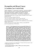
Two HER2-targeted agents are currently approved for use in the setting of metastatic HER2-
positive breast cancer, trastuzumab and lapatinib. These agents target HER2 via distinct
mechanisms (Fig. 5). Trastuzumab is a monoclonal antibody that specifically recognizes and
binds to an extracellular part of HER2. Since antibodies are large, bulky molecules,
trastuzumab is unable to cross the blood-brain barrier and thus cannot combat brain
metastases. In contrast, lapatinib is a small molecule kinase inhibitor targeted against the
EGFR and HER2 active sites. Since it is a small molecule, it is believed that lapatinib has the
potential to enter the brain and target metastatic cells that overexpress HER2. A phase II
trial of lapatinib in patients with trastuzumab-refractory disease and CNS metastases
showed some volumetric changes in brain lesions and improved neurologic symptoms (Lin,
2008; Lin, 2009). Amongst 50 patients who were terated with lapatinib plus capecitabine,
20% showed a CNS objective response and 40% experienced 20% or greater volumetric
reduction in their CNS lesions (Lin, 2009), suggesting that lapatinib may have some utility in
limiting CNS metastases of primary HER2-overexpressing breast cancers.
Novel Therapeutic Strategies and Combinations for HER2-Overexpressing Breast Cancer
13
Fig. 5. Novel targeted agents in trastuzumab-resistant HER2-positive breast cancer. T-DM1,
Trastuzumab-DM1; TRAST, Trastuzumab; PERT, Pertuzumab; IGFR, insulin growth factor
receptor; EGFR, epidermal growth factor receptor; LAP, lapatinib; NER, neratinib.
7.1 Combining trastuzumab with lapatinib
Combination of trastuzumab plus lapatinib has been shown to induce apoptosis in part via
down-regulation of survivin in cell culture and animal models (Xia, 2005). Initial phase I
data suggested that the combination is well-tolerated and elicits partial or complete
responses in a subset of patients who have progressed on prior trastuzumab therapy
(Storniolo, 2008). The combination has been tested clinically in advanced phase trials in
patients who have progressed on trastuzumab-based regimens. Progression-free survival
and quality of life were improved in patients treated with the combination versus lapatinib
alone (Wu, 2011). EGF104900 showed that the combination was superior to lapatinib alone
in the trastuzumab-resistant setting, with a clonical benefit rate of 24. 7% versus 12. 4%
(Blackwell, 2010). A potentially important mechanism of action of this drug combination is
that lapatinib has been shown to induce accumulation of inactive HER2 dimers via reduced
receptor ubiquitination, providing increased pharmacologic target for trastuzumab-
mediated antibody-dependent cellular cytotoxicity (Scaltriti, 2009). Combining trastuzumab
with lapatinib offers a chemotherapy-free option for treating HER2-positive trastuzumab-
resistant disease.
7.2 Combining trastuzumab with pertuzumab
Pertuzumab is an anti-HER2 monoclonal antibody that targets an extracellular epitope
distinct from what is targeted by trastuzumab. Pertuzumab binds to HER2 near the center of
Breast Cancer – Current and Alternative Therapeutic Modalities
14
domain II, sterically blocking a binding pocket necessary for receptor dimerization and
signaling (Franklin, 2004). In contrast, trastuzumab does not significantly inhibit HER2
interaction with other erbB receptors. We were the first to show that combining pertuzumab
with trastuzumab results in synergistic inhibition of proliferation of HER2-overexpressing
breast cancer cells (Nahta, 2004a). Trastuzumab increased pertuzumab-mediated disruption
of HER2 dimerization with EGFR and HER3, and further reduced pertuzumab-mediated
inhibition of PI3K signaling (Nahta, 2004a). Phase II data shows that combining trastuzumab
with pertuzumab in patients who have progressed on prior trastuzumab regimens achieves
clinical benefit rate of 50%, objective response rates of 24%, and median progression-free
survival of 5. 5 months (Baselga, 2010a). A potential mechanism of synergy is non-
overlapping mechanisms by single agents, trastuzumab-mediated inhibition of p95HER2
cleavage and pertuzumab-mediated disruption of dimerization (Scheuer, 2009). Clinical
evaluation of pertuzumab and trastuzumab (CLEOPATRA) is an international, randomized,
double-blind, placebo-controlled phase III trial. Patients with HER2-positive breast cancer
with locally recurrent or metastatic disease will be randomized to receive docetaxel,
trastuzumab, and pertuzumab or docetaxel, trastuzumab, and placebo. Progresion-free
survival will be assessed to determine efficacy of combination pertuzumab plus
trastuzumab in the trastuzumab-refractory setting (Baselga, 2010b).
8. Novel HER2-targeted agents in clinical development
8.1 Trastuzumab-DM1
One novel preparation of trastuzumab is a drug conjugate called trastuzumab-DM1, which
is trastuzumab conjugated to a microtubule-depolymerizing drug called maytansinoid
(Lewis Phillips, 2008). Trastuzumab-DM1 blocks growth of trastuzumab-naive and
trastuzumab-refractory HER2-overexpressing breast tumors in vivo (Lewis Phillips, 2008),
and retains the mechanistic activity of unconjugated trastuzumab (Junttila, 2010). Antibody-
dependent cellular cytotoxicity was induced by trastuzumab-DM1, and tumor growth of
trastuzumab-resistant cells was blocked by trastuzumab-DM1 due to induction of apoptosis
and mitotic catastrophe (Barok, 2011). A phase I dose-escalation study in patients who had
progressed on trastuzumab showed clinical benefit of 73% in 15 of 24 patients, including
objective responses in 5 patients (Krop, 2010). A phase II study of trastuzumab-DM1 in
patients with trastuzumab-refractory HER2-positive breast cancer showed objective
response of 25. 9% and median progression-free survival of 4. 6 months (Burris, 2011). Thus,
trastuzumab-DM1 HER2 antibody-chemotherapy conjugate is a promising treatment for
HER2-positive breast cancer that has progressed on prior HER2-directed therapies.
8.2 Irreversible pan-HER kinase inhibitors
In contrast to lapatinib, which is a reversible EGFR/HER2 kinase inhibitor, irreversible pan-
HER inhibitors are being developed for use against HER2-dependent breast cancers (Ocana,
2009). Neratinib, an irreversible EGFR/HER2 inhibitor, achieved a response rate of 26% in
trastuzumab-pretreated patients and 55% in trastuzumab-naïve patients (Burstein, 2009).
Progression-free survival at 16 weeks was 60% and 77%, respectively, for trastuzumab-
pretreated and naïve patients (Burstein, 2009). Finally, the median time to progression was
23 weeks and 40 weeks, respectively, for trastuzumab-pretreated and naïve patients
(Burstein, 2009). Canertinib (CI-1033) is an irreversible inhibitor of all HER proteins.
Response to canertinib was higher in patients with HER2-positive breast cancer, although
toxicity at the most effective dose was limiting and unacceptable (Rixe, 2009).
Novel Therapeutic Strategies and Combinations for HER2-Overexpressing Breast Cancer
15
9. Conclusion
In conclusion, several major mechanisms of trastuzumab resistance have been proposed,
including increased signaling from PI3K/mTOR, Src, and IGF-IR, as well as reduced
p27kip1 and increased cdk2 activity. These mechanisms have uncovered new therapeutic
targets for which multiple pharmacologic agents have been developed. Some of the most
promising include mTOR-targeted agents derived from rapamycin and trastuzumab-DM1.
Combining multiple HER2-targeted agents appears to be beneficial due to different
mechanisms of action. Future studies should more clearly address the role of IGF-IR in
acquired versus primary resistance, and test IGF-IR-targeted agents in combination with
trastuzumab and/or lapatinib in a trastuzumab-refractory setting. In addition, studies
examining the role of estrogen receptor (ER) signaling in trastuzumab resistant HER2-
positive ER-positive disease should be performed. Finally, biological predictors of response
or resistance need to be developed to determine which patients are most likely to benefit
from trastuzumab therapy, thus allowing for more specific individualization of targeted
therapy in patients with HER2-overexpressing breast cancer.
10. References
Adams TE, Epa VC, Garrett TP, & Ward CW. (2000). Structure and function of the type 1
insulin-like growth factor receptor. Cell Mol Life Sci, Vol. 57, No. 7, (July 2000), pp.
1050-93
Barok M, Tanner M, Koninki K, & Isola J. (2011). Trastuzumab-DM1 causes tumor growth
inhibition by mitotic catastrophe in trastuzumab-resistant breast cancer cells in
vivo. Breast Cancer Res Vol. 13, No. 2, (2011 Apr 21), pp. R46
Baselga J, Tripathy D, Mendelsohn J, Baughman S, Benz CC, Dantis L, Sklarin NT, Seidman
AD, Hudis CA, Moore J, Rosen PP, Twaddell T, Henderson IC, & Norton L. (1996).
Phase II study of weekly intravenous recombinant humanized anti-p185HER2
monoclonal antibody in patients with HER2/neu-overexpressing metastatic breast
cancer. J Clin Oncol, Vol. 14, No. 3, (March 1996), pp. 737-44
Baselga J, Gelmon KA, Verma S, Wardley A, Conte P, Miles D, Bianchi G, Cortes J, McNally
VA, Ross GA, Fumoleau P, & Gianni L. (2010a). Phase II trial of pertuzumab and
trastuzumab in patients with human epidermal growth factor receptor 2-positive
metastatic breast cancer that progressed during prior trastuzumab therapy. J Clin
Oncol Vol. 28, No. 7, (2010 Mar 1), pp. 1138-44
Baselga J & Swain SM. (2010b). CLEOPATRA: a phase III evaluation of pertuzumab and
trastuzumab for HER2-positive metastatic breast cancer. Clin Breast Cancer Vol. 10,
No. 6, (2010 Dec 1), pp. 489-91
Berns K, Horlings HM, Hennessy BT, Madiredjo M, Hijmans EM, Beelen K, Linn SC,
Gonzalez-Angulo AM, Stemke-Hale K, Hauptmann M, Beijersbergen RL, Mills GB,
van de Vijver MJ, & Bernards R. (2007). A functional genetic approach identifies the
PI3K pathway as a major determinant of trastuzumab resistance in breast cancer.
Cancer Cell Vol. 12, No. 4, (2007 Oct), pp. 395-402
Blackwell KL, Burstein HJ, Storniolo AM, Rugo H, Sledge G, Koehler M, Ellis C, Casey M,
Vukelja S, Bischoff J, Baselga J, O'Shaughnessy J. (2010). Randomized study of
Lapatinib alone or in combination with trastuzumab in women with ErbB2-