EPILEPSY IN CHILDREN – CLINICAL AND SOCIAL ASPECTS pptx
Bạn đang xem bản rút gọn của tài liệu. Xem và tải ngay bản đầy đủ của tài liệu tại đây (9.4 MB, 248 trang )
EPILEPSY IN
CHILDREN – CLINICAL
AND SOCIAL ASPECTS
Edited by Željka Petelin Gadže
Epilepsy in Children – Clinical and Social Aspects
Edited by Željka Petelin Gadže
Published by InTech
Janeza Trdine 9, 51000 Rijeka, Croatia
Copyright © 2011 InTech
All chapters are Open Access articles distributed under the Creative Commons
Non Commercial Share Alike Attribution 3.0 license, which permits to copy,
distribute, transmit, and adapt the work in any medium, so long as the original
work is properly cited. After this work has been published by InTech, authors
have the right to republish it, in whole or part, in any publication of which they
are the author, and to make other personal use of the work. Any republication,
referencing or personal use of the work must explicitly identify the original source.
Statements and opinions expressed in the chapters are these of the individual contributors
and not necessarily those of the editors or publisher. No responsibility is accepted
for the accuracy of information contained in the published articles. The publisher
assumes no responsibility for any damage or injury to persons or property arising out
of the use of any materials, instructions, methods or ideas contained in the book.
Publishing Process Manager Dragana Manestar
Technical Editor Teodora Smiljanic
Cover Designer Jan Hyrat
Image Copyright dusan964, 2011. Used under license from Shutterstock.com
First published August, 2011
Printed in Croatia
A free online edition of this book is available at www.intechopen.com
Additional hard copies can be obtained from
Epilepsy in Children – Clinical and Social Aspects, Edited by Željka Petelin Gadže
p. cm.
ISBN 978-953-307-681-2
free online editions of InTech
Books and Journals can be found at
www.intechopen.com
Contents
Preface IX
Part 1 Ethiology and Pathogenesis
of Epilepsy: Data from Research 1
Chapter 1 Polymicrogyria: A Clinical and
Experimental Approach to Epilepsy 3
Tomoyuki Takano
Chapter 2 Sequential Prefrontal Lobe Volume
Changes in Epileptic Patients with
Continuous Spikes and Waves During Slow Sleep 13
Hideaki Kanemura and Masao Aihara
Part 2 Clinical Presentation of Epilepsy
and Epileptic Syndromes of Childhood 25
Chapter 3 Neonatal Seizures 27
Isam AL-Zwaini
Chapter 4 Epileptic Encephalopathy Syndromes in Infancy 47
Raidah Albaradie
Chapter 5 The Lessons from Angelman
Syndrome for Research and Management 59
Karine Pelc, Guy Cheron and Bernard Dan
Part 3 Therapy of Epilepsy:
Medicamentous and Surgical Approach 77
Chapter 6 Novel Neuroprotective Strategies
and Targets of Intervention in Epilepsy 79
Ryan D. Readnower, Laurie M. Davis
and Patrick G. Sullivan
VI Contents
Chapter 7 Zonisamide – An Overview 93
Sowmini Raman, Lakshmi Narasimhan Ranganathan
and Sridharan Ramaratnam
Chapter 8 Practical Use of the
Ketogenic Diet in Childhood Epilepsy 105
Da Eun Jung and Heung Dong Kim
Chapter 9 Epilepsy Surgery in Children 115
Vera Cristina Terra,
Américo C Sakamoto and Hélio Rubens Machado
Chapter 10 Corpus Callosotomy in Pediatric Intractable Epilepsy:
Microsurgical Technique Implication and Variation 133
Eun Kyung Park and Dong-Seok Kim
Part 4 Social Aspects of Epilepsy 145
Chapter 11 Childhood Age Epilepsy and Family 147
Cicek Hocaoglu and Ayse Koroglu
Chapter 12 Health-Related Quality of Life in Children
and Adolescents with Epilepsy: A Systematic Review 161
Dejan Stevanovic, Ivana Tadic and Tanja Novakovic
Chapter 13 Frontal Lobe Epilepsies: Neuropsychological
and Behavioral Consequences in Children 187
Chiara Vago, Sara Bulgheroni,
Silvana Franceschetti and Daria Riva
Chapter 14 Physiotherapy for Children with Cerebral Palsy 213
Mintaze Kerem Günel
Preface
Epilepsy is a neurological condition that accompanies mankind probably since its
inception. About 400 years before Christ, the disease was already known by
Hippocrates, who wrote the book “On The Sacred Disease”, in which he refuted the
idea that the upheaval was the work of spirits and wisely related it to the brain. This
concept was not fully accepted until modern era (John Hughlings Jackson, 1873).
Classically, epilepsy is defined as a chronic condition characterized by an enduring
propensity to generate seizures, which are paroxysmal occurring episodes of abnormal
excessive or synchronous neuronal activity in the brain. According to WHO epilepsy
accounts for about 1% of the total burden of disease worldwide, about the same as
breast cancer in women and lung cancer in men.
Out of all brain disorders, epilepsy is the one that offers a unique opportunity to
understand normal brain functions as derived from excessive dysfunction of neuronal
circuits, because the symptoms of epileptic seizures are not the result of usual loss of
function that accompanies many disease that affect the brain. I am therefore extremely
honoured to present this book. The 15 very interesting chapters of the book cover
various fields in epileptology – they encompass the etiology and pathogenesis of the
disease, clinical presentation with special attention to the epileptic syndromes of
childhood, principles of medical management, surgical approaches, as well as social
aspects of the disease.
Author Takao dedicated the chapter to the clinical and experimental investigations in
polymicrogyria, that were reviewed with special reference to the epileptogenicity of
this malformation. The cortical hyperexcitability in polymicrogyria may be reduced by
the inhibitory neuronal network constructed by a population of aberrantly migrating
inhibitory interneurons, which are mobilized from the ganglionic eminence during the
development of polymicrogyria. Authors Kanemura and Aihara wrote about epileptic
patients with continuous spikes and waves during slow sleep, in which mentioned
electroencephalographic findings were associated with frontal lobe growth
disturbance. They state that seizures and paroxysmal anomaly durations may be
influenced by prefrontal lobe growth, which relates to neuropsychological problems.
Authors Isam et al wrote the chapter about neonatal seizures. Neonatal seizures are
common and the incidence is variable according to age and maturity of the neonate,
X Preface
weight and the severity of the underlying condition. It has been estimated that the
incidence rate of clinical seizures varies from 1.1 to 8.6 per 1000 live births. No period
carries the danger of seizures to the individual person like the first four weeks of life,
because of immaturity of the brain cells that are more vulnerable to injury and because
of wide range of factors that might cause seizures in this period. Neonatal seizures
tend to be brief, because immature neurons are unable to sustain repetitive activity for
a long period of time, and to be focal or multifocal. It requires immediate evaluation
because of the variable conditions that might insult developing and vulnerable
neurons of neonate, some of which might endanger the life of neonate. Some time
seizures might be the first and probably the only manifestations of underlying
significant dysfunction of the central nervous system of the newborn infant.
Furthermore, these seizures are sometimes difficult to be diagnosed clinically,
resulting in delaying treatment and worsening of short and long term prognosis. There
is still a great debate about pathophysiology, clinical classification,
electroencephalographic (EEG) significance and treatment of neonatal seizures.
Chapter by authors Dan, Pelc et al is dedicated to patients with Angelman syndrome,
that, compared to many other neurodevelopmental disorders, has the remarkably high
risk for epilepsy. In particular, early-childhood onset of refractory epilepsy with
atypical absences and myoclonic seizures with predisposition to developing non-
convulsive status epilepticus is a common presentation. This may be due to propensity
to hypersynchronous neuronal activity, which might be related to abnormal GABA-
mediated transmission due to lack of UBE3A expression, or other factors. On the one
hand, non-epileptic stereotyped or paroxysmal events (including motor or behavioural
manifestations) may lead to overdiagnosis. On the other hand, the epileptic nature of
relatively subtle manifestations such as absences, myoclonias or non-convulsive status
epilepticus may be under-recognised in the context of behavioural and motor features.
The neurocognitive effects of seizures are difficult to evaluate. There is a major need
for evidence on which to base rational treatment.
A diagnostic scheme for patients with epileptic seizures and with epilepsy proposed
by ILAE Commission (2001) newly adopted the concept of “epileptic encephalopathy”
as one of the new key terms. It is defined as a condition in which epileptiform
abnormalities are believed to contribute to the progressive disturbance in cerebral
function, but this definition may be ambiguous. Authors Raidah et al state that the
proposal include 8 syndromes: early myoclonic encephalopathy, Ohtahara syndrome,
West syndrome, Dravet syndrome, myoclonic status in non-progressive
encephalopathies, Lennox-Gastaut syndrome, Landau-Kleffner syndrome, and
epilepsy with continuous spike-waves during slow-wave sleep. To these syndromes,
the migrating partial seizures in infancy and severe epilepsy with multiple
independent spike foci may be reasonably added. In the chapter authors concentrate
on the epileptic encephalopathies that occur only in infancy.
The frontal lobes of the brain constitute more than a third of the human cerebral cortex
and are characterized by a complex functional organization supporting higher level
Preface XI
integration circuits. The complexity of the frontal lobe, in terms of its neuroanatomy
and connections, determines a marked variability in the epileptic manifestations with
fast and inter- and intra-hemispheric propagation. Vago et al discuss about the
epilepsies involving the frontal lobe – they describe the characteristic EEG discharges,
neuropsychological and behavioral consequences, in the light of the complexity of
frontal regions, and they also focus on the interactions between EEG features,
demographic variables and neuropsychological outcome.
Authors Readnower et al discuss about the novel neuroprotective strategies and
targets of intervention in epilepsy. Development of new anticonvulsive therapies
designed as both an anticonvulsive as well as a neuroprotectant would be the best way
to treat acute seizure conditions and to possibly prevent the development of chronic
epilepsy. One of the newer broad spectrum antiepileptic drug, widely used in the
management of epilepsy, is zonisamide (ZNS). Narasimhan et al state that zonisamide
is effective as adjunctive therapy for refractory partial seizures, and as monotherapy
for newly diagnosed or refractory partial seizures. It can also be administrated in
patients with post-operative seizures, may be useful in the treatment of patients with
progressive myoclonic epilepsy (studies have found it to be useful in Unverricht-
Lundborg disease), West syndrome, and brain tumour related epilepsy.
Chapter by Jung et al will provide practical recommendations to guide the
management of the ketogenic diet in childhood epilepsy and give a review on the
current state of ketogenic diet. Special chapter written by Günel et al is dedicated to
the physiotherapy for children with cerebral palsy.
Epilepsy affects 1-2% of children. In childhood, epilepsy is more common in the first
year of life, and its incidence decreases progressively with increasing age, affecting
approximately 100 children per 100,000 births in the first year of life, 40 children for
every 100,000 births in subsequent years, and approximately 20 individuals per
100,000 adolescents. In 75% of these cases, seizures are well controlled with
antiepileptic drugs and in the remaining 25% epilepsy is refractory to pharmacological
treatment and surgical approach should be considered. Terra et al state that surgery
for epilepsy in childhood has become an effective method in treating this condition,
and should be indicated as early as possible. Peculiarities of epilepsy in children
should be considered to achieve optimal results. Although a reduction of seizures is
the primary goal of surgery, the maintenance of cognitive and motor development
milestones is essential to allow the child have a quite normal life in adulthood.
Extratemporal epilepsy in children closes more cases compared to those observed in
adults, but still dominates the temporal lobe as the site of ictal onset, and surgical
results are very encouraging. Surgical option should take in account several factors
such as child´s age, underlying pathology and lesion extension. Neuronal plasticity
can be an ally for the development of minor post-operative neurological deficits.
Authors Park and Kim state that callosotomy in pediatric epilepsy is a valuable tool to
control seizures early on, in order to protect the developing brain from further damage
and to give chance to recover neuropsychological function from damage done by
XII Preface
seizure itself as well as seizure medication. They advocate that one stage total
callosotomy in young patients with medically intractable epilepsy without localizing
lesions is especially effective in drop attacks and secondary generalized epilepsy. With
improvement in microsurgical techniques, excellent seizure outcome as well as
functional outcome may be reached without previously known high rate of morbidity
and mortality.
Authors Hocaoglu and Koroglu state that childhood epilepsy has a significant effect
on the child himself and the family because of its psychological and social results. In
the studies the increasing economical responsibility of the families whose children
undergo chronic diseases is distinctively described. Still, epilepsy in childhood is
different from the other chronic diseases due to the fact that its sudden symptoms and
early unpredictable effects are all specific for itself. In many studies about epilepsy,
despite the fact that the patient’s quality of life and relationship with the family are
examined, in few ones problems belonging to family members that result from
epilepsy are pointed. Clinicians should consider both neurological and psychosocial
factors, including the family system, when treating psychopathology in children with
epilepsy. The chapter by Stevanovic et al systematically reviewed synthesizing
different studies that evaluated health-related quality of life (HRQOL) in children and
adolescents with epilepsy over 12 past years. The affected domains, predictors, and
impacts on HRQOL of specific and non-specific treatments were reviewed. Previous
reviews evaluated methodological issues in HRQOL assessment, components of
theoretical model, and determinants of HRQOL in pediatric epilepsy. Based on the
findings and evidence found, it could be concluded that children and adolescents have
more affected HRQOL in physical, psychological, and social domain than healthy
children and adolescents.
It is important for all of us to raise the awareness and reduce social barriers for
individuals with epilepsy. Together we can hope that we will identify ways to
improve the treatment of patients with epilepsy and the livelihood of all individuals
with epilepsy.
Željka Petelin Gadže, M.D., Ph.D.
Department of Neurology of the Medical School and
University Hospital Centre Zagreb,
Referral Centre for Epilepsy of the
Ministry of Health and Social Welfare of the Republic of Croatia
Croatia
Part 1
Ethiology and Pathogenesis of Epilepsy:
Data from Research
1
Polymicrogyria: A Clinical and
Experimental Approach to Epilepsy
Tomoyuki Takano
Department of Pediatrics, Shiga University of Medical Science, Seta-Tsukinowa, Otsu
Japan
1. Introduction
Polymicrogyria is the presence of an excess number of abnormally small gyri that produce
an irregular cortical surface. Although polymicrogyria is associated with severe epilepsy in
65% of patients (Guerrini & Filippi, 2005), few data concerning the epileptogenic zone and
its relationship with the polymicrogyric tissue are available due to the fact that patients with
polymicrogyria are rarely considered to be suitable candidates for epilepsy surgery
(Chassoux et al., 2008). An experimental model in which a single or few microgyri are
generated by a freezing insult suggests a widespread area of functional disruption that
extends beyond the visualized abnormality (Redecker et al., 2000). However, the detailed
mechanism of epileptogenesis has not yet been well characterized for polymicrogyria
(Sisodiya, 2004). In this chapter, clinical and experimental investigations in polymicrogyria
were reviewed with special reference to the epileptogenicity of this malformation.
2. Definition and pathogenesis of polymicrogyria
Polymicrogyria is a cerebral cortical malformation characterized by an excessively folded
cortical ribbon of miniature, individually thin convolutions, which may be fused together or
piled on top of one another (Sisodiya, 2004). The cortical surface is irregular, and the
convolutions can appear wider than expected, with a bumpy surface, like cobblestones or
morocco leather (Graham & Lantos, 2002). There are two subtypes: unlayered type and four-
layered type. In unlayered polymicrogyria, the external molecular layer is continuous and
does not follow the profile of the convolutions, and the underlying neurons have radial or
vertical distribution but no laminar organization (Ferrer, 1984). Polymicrogyric area may be
distributed by focal, multi-lobar, or diffuse in the cerebral cortex. This brain malformation is
thought either to be resulted from early exogenous insult from the 13th to 18th week of
gestation or to be genetically determined (Ferrer & Catala, 1991). In four-layered
polymicrogyria, there are two neuronal layers (2nd and 4th layers) under the molecular
layer (1st layer), separated by an intermediate layer with many fibers and few cells (cell-
sparse 3rd layer) (Graham & Lantos, 2002). Polymicrogyric 2nd and 3rd layers are thought
to be correspond to the normal cortical layers II, III, IV, and layer V, respectively, in which
horizontal neuronal lamination is usually spared. Four-layered polymicrogyria is believed
to be resulted from a perfusion failure limited to one or more arterial vascular beds,
occurring between the 20th and 24th week of gestation. This would lead to intracortical
Epilepsy in Children – Clinical and Social Aspects
4
laminar necrosis with delayed damage of the distal section of radial glial fibers, with
consequent late migration disorder and post-migratory overturning of cortical organization
(French, 1989).
Experimental polymicrogyria can be modeled by the excitotoxic brain lesions during the
period of neuronal migration. Ibotenate is an agonist of the N-methyl-D-aspartate (NMDA)
complex receptor . Experimental studies have demonstrated that an intracerebral injection of
ibotenate induces excitotoxic brain lesions mimicking a variety of neuronal migration
disorders including microgyria (Takano et al., 2005). After the radial glial fibers and
surrounding neural tissues were damaged by ibotenate, the corresponding area within the
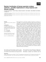
cortical plate collapsed (Figure 1A). As the surrounding neurons migrate along the radial
fibers, the cortical plate rolled inward and became infolded, forming microgyria (Figure 1B).
Thus, the damage to intermediate cortical layers would produce a difference in growth rate
between outer and inner cortical layers, with consequent excessive folding of the cortical
surface (Figure 1C) (Takano et al., 2005).
Fig. 1. A: Cortical lesions 1 day after ibotenate injection shown by vimentin
immunohistochemistry. Note the disrupted neuronal arrangement in the cortical plate and
intermediate zone, lacking the vimentin-positive radial glial fibers (arrow). B: Cortical
infolding mimicking microgyria (arrow) 5 days after ibotenate injection. Hematoxylin-eosin
staining. C: Cerebral cortex illustrating the histogenetic development of the microgyria.
After the radial glial fibers were damaged (small arrows), its corresponding area within the
cortical plate collapsed. As the surrounding neurons migrate along the radial fibers, the
cortical plates roll in and infold. MZ, marginal zone; CP, cortical plate; SP, subplate; IZ,
intermediate zone; VZ, ventricular zone. Scale bar, A = 120 μm, B = 160 μm.
Polymicrogyria: A Clinical and Experimental Approach to Epilepsy
5
3. Congenital bilateral perisylvian syndrome and epilepsy
Several specific syndromes are associated with cerebral polymicrogyria. Congenital bilateral
perisylvian syndrome (CBPS) was first described by Kuzniecky and coworkers (1993), and it
is characterized by pseudobulbar palsy, cognitive deficits, and bilateral perisylvian
abnormalities such as polymicrogyria (Table 1). Pseudobulbar palsy is one of the striking
clinical symptoms of CBPS, however, the oropharyngoglossal dysfunction, such as
abnormal tongue movement and the presence of dysarthric speech, may be difficult to
investigate in young children. Moreover, epilepsy is an additional diagnostic manifestation
of this syndrome, but the mean age at seizure onset has been estimated to be 7.9 years
(Kuzniecky et al., 1994). Therefore, in the pediatric population, CBPS is likely to have
different manifestations than in adults (Gropman et al., 1997).
Table 1. Criteria for the diagnosis of congenital bilateral perisylvian syndrome (CBPS)
(Kuzniecky R, et al. (1993))
Three cases of epilepsy with congenital bilateral or unilateral perisylvian polymicrogyria are
presented as follows.
Case 1: This male child showed complex partial seizures (CPS) at 3 years of age.
Electroencephalogram (EEG) revealed focal spikes on the bilateral frontal areas, and
carbamazepine (CBZ) was started. No feeding difficulties and drooling were observed, but
expressive language development was mildly delayed. Brain computed tomography (CT)
was not able to reveal the cortical abnormalities at 3 years of age. His epileptic seizures were
well-controlled by the administration of CBZ, but CPS reappeared due to withdrawal at 16
years of age. Brain magnetic resonance imaging (MRI) showed narrow and deep sylvian
fissures and their surrounding pachygyric cortex on fluid-attenuated inversion-recovery
Epilepsy in Children – Clinical and Social Aspects
6
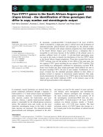
(FLAIR) image (Figure 2A). Although no pseudobulbar disorders have yet been recognized,
his expressive language skills have been still delayed, and he was diagnosed to have
pervasive developmental disorders.
Fig. 2. Brain magnetic resonance imaging (MRI) findings of three patients with perisylvian
polymicrogyria. Fluid-attenuated inversion-recovery (TR/TE/ TI = 8002/133/2000 ms) (A,
C), and T1-weighted MRI (TR/TE = 500/9 ms) (B). A: Case 1. Narrow and deep sylvian
fissures (arrows) and their surrounding pachygyric cortex were found. B: Case 2. The
bilateral perisylvian cortical dysplasia are accompanied with bilateral dysplastic insula
(arrows). C: Case 3. Note the dysplastic right perisylvian cortex with broad and thickened
gyri (arrow).
Case 2: This male child was referred to our hospital because of epileptic seizures which he
suffered at 12 years of age. His school performance was normal, and he has not shown any
developmental abnormalities and pseudobulbar disorders. His seizure type was CPS, which
included behavioral arrest, lateralized tonic posturing with head and eye deviation, and
facial automatisms. Interictal EEG showed focal spikes on the left front-temporal area. These
clinical findings suggested a diagnosis of temporal lobe epilepsy. Brain MRI revealed
bilateral perisylvian cortical dysplasia, accompanying with an abnormality of the insula and
of the parietal cortex on T1-weighted image (Figure 2B). The frequency of his epileptic
seizures was monthly, and partial or transitory improvements have been obtained with
CBZ, zonisamide or phenytoin.
Case 3: This female child manifested generalized tonic-clonic seizures or left partial seizures
during sleep at 4 years of age. Her psychomotor development was mildly delayed,
accompanied with mild left hemiparesis. Initial EEG showed focal slow spikes with frequent
associated diffuse slow spikes and waves. In brain MRI, the right perisylvian cortex was
dysplastic showing the appearance of pachygyria with broad and thickened gyri, suggesting
right perisylvian polymicrogyria (Figure 2C). Her generalized or partial seizures were
refractory to the administration of valproate or CBZ, respectively. Four months later, her
sleep EEG demonstrated continuous bilateral and diffuse slow spike and waves, mainly at
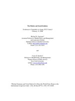
1.5 ~ 2.5 Hz, persisting through all the slow sleep stages (Figure 3). These characteristic
clinical features were considered as the diagnosis of the epilepsy with continuous spikes and
waves during slow sleep.
More immature anomalous brain lesions may be associated with an enhanced capacity for
epilepsy and resultant refractory seizures (Takano et al., 2006). However, the epilepsy
Polymicrogyria: A Clinical and Experimental Approach to Epilepsy
7
related to polymicrogyria may have variable types and severity, including cases with good
outcome and spontaneous remissions, even after a period of intractability. Surgical
treatment of epilepsy may be applicable to a very limited number of patients in whom large
resections are feasible, because the epileptogenic zone in polymicrogyria remains largely
unknown.
Fig. 3. Sleep EEG of Case 3. Note the continuous bilateral and diffuse slow spike and waves.
4. Epileptogenicity in experimental polymicrogyria by freeze lesion model
Polymicrogyria can be modeled in rats with a transcortical prenatal or neonatal freeze
lesion, which mimics the histological characteristics of a human four-layered
polymicrogyria. This experimental model does not have spontaneous epileptiform activity
in vivo, but several investigations have been presented concerning the epileptogenicity of
this malformation.
4.1 Upregulation of glutamate receptor subunits
Glutamate receptors are widespread in the nervous system where they are responsible for
mediating the vast majority of excitatory synaptic transmission in the brain and spinal cord.
The glutamate receptor family is composed of several distinct subtypes, which are
pharmacologically distinguished by four agonists: NMDA, amino-3-hydroxy-5-
methylisoxazoleproprionic acid (AMPA), kainate, and quisqualate. Electrical kindling
stimulation in prenatal freeze lesion rat revealed the significant prolonged after discharges
in both of the cortex and hippocampus, the early development of hippocampal kindling,
Epilepsy in Children – Clinical and Social Aspects
8
and the spontaneous cortico-hippocampal spikes. Immunoreactive expression for NMDA
receptor subunit 1 and 2B was shown to be markedly upregulated not only in the
microgyria, but also in the hippocampus (Takase et al., 2008). These investigations indicate
that dysplastic cortex of microgyria can be highly seizure susceptible lesion by a certain
brain insult such as kindling or excitable cortical stimulation.
4.2 Alterations in ion channels
Na+, K+-ATPase contributes to the asymmetrical distribution of sodium and potassium ions
across the plasma membrane and to maintenance of the membrane potential in many types
of cells (McGrail et al., 1991). A decrease in α3 subunit expression may cause neurons to be
less effective in restoring their normal electrochemical gradient and membrane potential
after repeated membrane depolarization, resulting in hyperexcitability (Li & Stys, 2001;
Vaillend et al., 2002). Alterations in this protein are thought to play a significant role in
many human neurological disorders, including epilepsy. It has been demonstrated that
there was a significant decrease in α3 subunit of Na+, K+-ATPase immunoreactivity in the
neuropil of freeze lesion cortical layer V in paramicrogyral area, where is an area that
typically exhibits evoked epileptiform activity. The significant decrease in Na+, K+-ATPase
in the paramicrogyral cortex is suggested to contribute to epileptogenesis (Chu et al., 2009).
4.3 New excitatory or inhibitory rewiring
The electrophysiological studies by cortical slices demonstrated that the field potentials
evoked by stimulation within a few millimeters of the microgyrus have characteristics
typical of epileptiform activity. These results imply that the epileptiform activity in
polymicrogyria can be generated outside the lesion itself, which is a focal zone adjacent to
the microgyria and called paramicrogyral area (Jacobs et al., 1996; Jacobs et al., 1999). Jacobs
and Prince (2005) recorded isolated whole cell excitatory postsynaptic currents (EPSCs) and
GABA
A
receptor-mediated inhibitory postsynaptic currents (IPSCs) from layer V pyramidal
neurons in the region of paramicrogyral area. They demonstrated that the conductance or
the frequency of IPSCs or EPSCs was significantly larger or greater in paramicrogyral cells
compared with controls. These findings imply that there is an increase in numbers of
functional excitatory synapses on both interneurons and pyramidal cells in the
paramicrogyral cortex, because the cortical afferents unable to find appropriate targets
within the malformed region may instead synapse in the adjacent paramicrogyral area.
4.4 Downregulation of GABA
A
receptor subunits
Synaptic inhibition in the mammalian brain is mediated principally by γ-aminobutyric acid
(GABA) receptors. The most widespread ionotropic receptor activated by GABA is
designated GABA
A
. The majority of GABA
A
receptors contain a variable combination of α, β,
and γ subunits, showing a specific regional and cellular distribution (Fritschy & Mohler,
1995). Functional studies demonstrated that the subunit composition of receptor subtypes
determines their electrophysiological and pharmacological properties (Barnard et al., 1998;
Narahashi, 1999). In adult rats with freeze-lesioned microgyria, widespread regionally
differential reduction of GABA
A
receptor subunits α1, α2, α3, α5, and γ2 was observed within
the microgyral area and the lateral to the dysplastic cortex. It has been also observed that the
downregulation of GABA
A
receptor subunits involved the ipsilateral hippocampal
formation, as well as restricted contralateral neocortical areas, indicating widespread
Polymicrogyria: A Clinical and Experimental Approach to Epilepsy
9
disturbances in the neocortical and hippocampal network (Redecker et al., 2000). The
downregulation of GABA
A
receptor subunits might contribute to the widespread cortical
hyperexcitability in patients with polymicrogyria.
5. Interneurons and epileptogenicity of polymicrogyria
The proper functioning of the cerebral cortex is dependent on two classes of neurons: a)
excitatory , projecting neurons, with pyramidal somatodendritic morphology using
glutamate as a neurotransmitter, which typically send their axons to distant cortical as well
as subcortical targets; b) inhibitory local circuit interneurons, whose axonal arborization is
typically restricted to the neocortex and does not project into the white matter (Druga, 2009).
These neurons primarily use GABA as a neurotransmitter. The majority of cortical neurons
belong to the category of pyramidal cells. Cortical GABAergic interneurons represent about
20-30% of the total number of neocortical neurons (Druga, 2009).
We previously demonstrated the intracerebral injection of ibotenate produces excitotoxic
brain lesions to mimic neuronal migration disorders (Takano et al., 2004). We also reported
that subventricular zone cells play an important role in the formation of cortical dysplasia
(Sawai et al., 2009). Biotinylated dextran amine (BDA) are highly sensitive tools for
anterograde and retrograde pathway tracing studies of the nervous system. The high
molecular-weight BDA yields sensitive and exquisitely detailed labeling of axons and
terminals using preferentially anterograde transport. In the brains injected with BDA to the
ganglionic eminence, BDA-positive fibers were derived from the dorsolateral part of the
subventricular zone (Figure 4A), and BDA-labeled neurons were specifically located within
Fig. 4. Biotinylated dextran amine (BDA) tracer immunohistochemistry with hematoxylin
double staining 5 days after ibotenate injection. A: Numerous BDA-positive radially
oriented fibers extended from the dorsolateral part of the subventricular zone (SVZdl) and
reached the pial surface in the frontparietal cortex. Note the microgyria (arrows). B: Higher
magnification of microgyria in A. Note the BDA-positive neurons in the microgyric cortex
(arrows), which were mobilized out of the ganglionic eminence. Scale bar, A = 120 μm, B =
80 μm.
Epilepsy in Children – Clinical and Social Aspects
10
the polymicrogyric area of the parietal cortex (Figure 4B). This experiment demonstrated
that the interneurons are mobilized to the microgyric area out of the ganglionic eminence,
which thus leads to the construction of a part of the abnormal neuronal arrangement of this
microgyria (Takano et al., 2010). Polymicrogyria is not invariably associated with epilepsy,
and the pathogenetic basis of epileptogenesis in polymicrogyria is also unclear. It is
suggested that one of the factors that might explain why some patients with polymicrogyria
do not develop epilepsy may be due to the fact that a population of aberrantly migrating
inhibitory interneurons are present in the microgyric area.
6. Conclusion
The cortical hyperexcitability in polymicrogyria may be reduced by the inhibitory
neuronal network constructed by a population of aberrantly migrating inhibitory
interneurons, which are mobilized from the ganglionic eminence during the development of
polymicrogyria.
7. Acknowledgment
This work was supported by the Japan Society for the Promotion of Science, a Grant-in-Aid
for Scientific Research (C) (22591125).
8. References
Barnard EA, Skolnick P, Olsen RW, Mohler H, Sieghart W, Biggio G, Braestrup C, Bateson
AN, Langer SZ (1998) International union of pharmacology. XV. Subtypes of
gamma-aminobutyric acid A receptors: classification on the basis of subunit
structure and receptor function. Pharmacol Rev 50: 291-313.
Chassoux F, Landre E, Rodrigo S, Beuvon F, Turak B, Semah F, Devaux B (2008)
Intralesional recordings and epileptogenic zone in focal polymicrogyria. Epilepsia
49: 51-64.
Druga R (2009) Neocortical inhibitory system. Folia Biologica (Praha) 55: 201-217.
Chu Y, Parada I, Prince DA (2009) Temporal and topographic alterations in expression of the
α3 isoform of Na
+
, K
+
-ATPase in the rat freeze lesion model of microgyria and
epileptogenesis. Neuroscience 162: 339-348.
Ferrer I (1984) A Golgi analysis of unlayered polymicrogyria. Acta Neuropathol 65: 69-76.
Ferrer I, Catala I (1991) Unlayered polymicrogyria: structural and developmental aspects.
Anat Embryol 184: 517-528.
French J (1989) Child neurology and developmental disabilities. Brookes, Baltimore.
Fritschy JM, Mohler H (1995) GABAA-receptor heterogeneity in the adult rat brain:
differential regional and cellular distribution of seven major subunits. J Comp
Neurol 359: 154-194.
Graham DI, Lantos PL (2002) Greenfield’s neuropathology. 7th ed. Arnold, London.
Gropman AL, Barkovich AJ, Vezina LG, Conry JA, Dubovsky EC, Packer RJ (1997) Pediatric
congenital bilateral perisylvian syndrome: clinical and MRI features in 12 patients.
Neuropediatrics 28: 198-203.
Polymicrogyria: A Clinical and Experimental Approach to Epilepsy
11
Guerrini R, Filippi T (2005) Neuronal migration disorders, genetics, and epileptogenesis. J
Child Neurol 20: 287-99.
Jacobs KM, Gutnick MJ, Prince DA (1996) Hyperexcitability in a model of cortical
maldevelopment. Cereb Cortex 6: 514-523.
Jacobs KM, Hwang BJ, Prince DA (1999) Focal epileptogenesis in a rat model of
polymicrogyria. J Neurophysiol 81: 159-173.
Jacobs KM, Prince DA (2005) Excitatory and inhibitory postsynaptic currents in a rat model
of epileptogenic microgyria. J Neurophysiol 93: 687-696.
Kuzniecky R, Andermann F, Guerrini R (1993) Congenital bilateral perisylvian syndrome:
study of 31 patients. Lancet 341: 608-612.
Kuzniecky R, Andermann F, Guerrini R (1994) Infantile spasms: an early epileptic
manifestation in some patients with the congenital bilateral perisylvian syndrome. J
Child Neurol 9: 420-423.
Li S, Stys PK (2001) Na
+
, K
+
-ATPase inhibition and depolarization induce glutamate release
via reverse Na
+
-dependent transport in spinal cord white matter. Neuroscience 107:
675-683.
McGrail KM, Phillips JM, Sweadner KJ (1991) Immunofluorescent localization of three Na,
K-ATPase isozymes in the rat central nervous system: both neurons and glia can
express more than one Na, K-ATPase. J Neurosci 11: 381-391.
Narahashi T (1999) Chemical modulation of sodium channels and GABA
A
receptor channel.
Adv Neurol 79: 457-480.
Redecker C, Luhmann HJ, Hagemann G, Fritschy JM, Witte OW (2000) Differential
downregulation of GABAA receptor subunits in widespread brain regions in the
freeze-lesion model of focal cortical malformations. J Neurosci 20: 5045-53.
Sawai C, Takano T, Takeuchi Y (2009) Experimental neuronal migration disorders following
the administration of ibotenate in hamsters: the role of the subventricular zone in
the development of cortical dysplasia. J Child Neurol 24: 275-286.
Sisodiya SM (2004) Malformations of cortical development: burdens and insights from
important causes of human epilepsy. Lancet Neurol 3: 29-38.
Takano T, Sawai C, Takeuchi Y (2004) Radial and tangential neuronal migration disorders in
ibotenate-induced cortical lesions in hamsters: immunohistochemical study of
reelin, vimentin, and calretinin. J Child Neurol 19: 107-115.
Takano T, Sawai C, Sakaue Y, Takikita S, Takeuchi Y (2005) Experimental cortical dysplasia
following ibotenate administration in hamsters: pathogenesis of microgyria and
associated gray matter heterotopia. Congenit Anom 45: 9-13.
Takano T, Sokoda T, Akabori S, Sakaue Y, Sawai C, Takeuchi Y, Ohno M (2006) Enhanced
capacity of epilepsy in brain malformation produced during early development.
Pediatr Neurol 35: 38-41.
Takano T, Sawai C, Akabori S, Takeuchi Y (2010) Polymicrogyria without epilepsy by
aberrantly migrating inhibitory interneurons. Epilepsy Behav 18: 505-506.
Takase K, Shigeto H, Suzuki S, Kikuchi H, Ohyagi Y, Kira J (2008) Prenatal freeze lesioning
produces epileptogenic focal cortical dysplasia. Epilepsia 49: 997-1010.