Báo cáo khoa học " Structure elucidation and antioxidant activity of a novel polysaccharide isolated from Tricholoma matsutake " ppt
Bạn đang xem bản rút gọn của tài liệu. Xem và tải ngay bản đầy đủ của tài liệu tại đây (251.35 KB, 5 trang )
International Journal of Biological Macromolecules 47 (2010) 271–275
Contents lists available at ScienceDirect
International Journal of Biological Macromolecules
journal homepage: www.elsevier.com/locate/ijbiomac
Structure elucidation and antioxidant activity of a novel polysaccharide isolated
from Tricholoma matsutake
Xiang Ding
a,1
, Jie Tang
a,1
, Mei Cao
b,1
, Chun-xiao Guo
a
, Xia Zhang
a
, Jing Zhong
a
, Jie Zhang
a
,
Qun Sun
a
, Su Feng
a
, Zhi-rong Yang
a
, Jian Zhao
a,b,∗
a
Key Laboratory of Biological Resource and Ecological Environment of the Ministry of Education, College of Life Sciences, Sichuan University, Chengdu 610064, PR China
b
Key Laboratory of Sichuan Academy of Medical Sciences, Sichuan Provincial People’s Hospital, Chengdu 610072, PR China
article info
Article history:
Received 20 March 2010
Received in revised form 17 April 2010
Accepted 19 April 2010
Available online 27 April 2010
Keywords:
Polysaccharide structure
Antioxidant assay
Tricholoma matsutake
abstract
In this study, structural features of Tricholoma matsutake polysaccharide (TMP-A) were investigated
by a combination of infrared (IR) spectra, gas chromatography–mass spectrometry (GC–MS), nuclear
magnetic resonance (NMR) spectroscopy. The results indicated that TMP-A had a backbone of 1,4--
d-glucopyranose residue which branches at O-6 based on the experimental results. The branches were
mainly composed of an (1→ 3)-␣-d-galactopyranose residue, and terminated with ␣-d-xylopyranose
residue. The antioxidant activity of TMP-A was evaluated with several biochemical methods, includ-
ing DPPH
−
radical scavenging, hydrogen peroxide scavenging, superoxide anion radical scavenging. The
results indicated that TMP-A showedstrong antioxidant. In the in vitro antioxidant assay by MTT method,
TMP-A could attenuate PC12 cell damage significantly caused by hydrogen peroxide.
© 2010 Elsevier B.V. All rights reserved.
1. Introduction
Oxidation is essential to many organisms for the production of
energy to fuel biological processes. However, the uncontrolled pro-
duction of superoxide anion free radicals is involved in the onset
of many diseases such as cancer, atherosclerosis and degenerative
processes with aging [1]. Thus, it is essential to develop effective
and natural antioxidants so that they can protect the human body
from free radicals and many chronic diseases [2]. Polysaccharides
extracted from mushrooms, such as Lentinus edodes, Ganoderma
tsugae and Cordyceps sinensis, have also exhibited antioxidant prop-
erties by their free radical scavenging ability [3,4].
Tricholoma matsutake is a kind of fungi belonging to Sub-
genus Tricholoma and is widely distributed in Asian countries,
such as China, Japan, and Korea. As a traditional edible fungus
in oriental countries, it has been consumed as a vegetable and
used as a traditional Chinese medicine in single and compound-
ing prescriptions for the prevention and treatment of diseases
for several thousand years [5]. As an extract from T. matsutake,
polysaccharides (TMP) have showed strongly bioactive properties
towards antioxidant and anti-tumor [6]. Thus the determination
∗
Corresponding author at: Key Laboratory of Biological Resource and Ecological
Environment of the Ministry of Education, College of Life Sciences, Sichuan Univer-
sity, Wangjiang Road 29#, Chengdu 610064, PR China. Tel.: +86 28 85460487;
fax: +86 28 85460487.
E-mail address: (J. Zhao).
1
These authors contributed equally to this research.
of the structure of polysaccharides was necessary to establish
the relationship between the biological activities and the struc-
ture.
In this work, one novel water-soluble polysaccharide was
extracted and purified from the fruiting bodies of T. matsutake
using a DEAE-cellulose column chromatography and a Sephadex
G-100 column chromatography. Its chemical structures were char-
acterized for the first time. The antioxidant activity of TMP-A
was evaluated by various antioxidant assay and MTT method.
The result of this study introduced T. matsutake as a possible
valuable source which helped to exhibit unique antioxidant prop-
erties.
2. Materials and methods
2.1. Chemicals
The fruiting bodies of T. matsutake were collected in Xiaojing
country of Sichuan Province, China, and were authenticated by
Prof. Sao-rong Ge (College of Life Sciences, Sichuan University,
Chengdu, China). At the same time, a voucher specimen had been
preserved in Key Laboratory for Biological Resource and Ecolog-
ical Environment of Education Ministry, College of Life Sciences,
Sichuan University. DEAE-cellulose 52 and Sephadex G-100 were
purchased from Sigma–Aldrich (mainland, China). Monosaccharide
standards, Dextran T-500, T-110, T-70, T-40, and T-10, were pur-
chased from Beijing Biodee Biotechnology Co., Ltd. (Beijing, China).
All other reagents used were of analytical grade.
0141-8130/$ – see front matter © 2010 Elsevier B.V. All rights reserved.
doi:10.1016/j.ijbiomac.2010.04.010
272 X. Ding et al. / International Journal of Biological Macromolecules 47 (2010) 271–275
2.2. Extraction, purity and fractionation of polysaccharides from
T. matsutake
After the fruiting bodies (200 g) of T. matsutake were soaked
with 95% EtOH, the residue was dried and then extracted with
boiling water for three times (6 h for each). After the filtrate was
concentrated, dialyzed (MWCO 5000, Sigma), and centrifuged, the
supernatant was added with 3 volumes of 95% EtOH to precipitate
crude polysaccharides (32.8 g, recovery 16.4%). After Sevag method
(Staub [7]) was used for thedeproteination, TMP(8 g) was subjected
to a DEAE-cellulose column (Tris–HCl, pH 7.0, 4.5 cm× 50 cm, Cl
−
)
and eluted stepwise with 0, 0.1, 0.2, 0.3, 0.4, 0.5 and 1.0 M NaCl. The
eluate was monitored by the phenol-sulfuric acid method [8]. The
0 M NaCl eluation was concentrated, lyophilized and purified on
a Sephadex G-100 column (2.6 cm × 60cm). The resulting T. mat-
sutake polysaccharide, named TMP-A, was obtained by the above
processes and the yield rate of TMP-A was 0.22% (0.432 g) for the
starting material.
2.3. Measurement of molecular weight and monosaccharide
composition analysis of TMP-A
High performance gel permeation chromatography (HPGPC)
was carried out to measure molecular weight. Thecolumn was cali-
brated withstandard T-seriesDextran (T-500,T-110, T-70,T-40 and
T-10). The data were processed with Waters GPC (Millennium32
software). Thepolysaccharide TMP-A(5.0 mg) was hydrolyzed with
2 M trifluoroacetic acid (TFA) at 110
◦
C for 6 h on the mecha-
nism of acid-catalyzed hydrolysis [9]. Excess acid was removed
by co-distillation with methyl alcohol (MeOH) after the hydroly-
sis was completed. One part of the hydrolysate (1.0 mg) was used
for thin layer chromatography (TLC) analysis as described previ-
ously. Developing solvent: acetoacetate–pyridine–ethanol–water
solution (8:5:1.5:1); the developer system: diphenylamine–aniline
system (85% phosphoric acid solution 140 mL containing 8 mL
diphenylamine, 8 g aniline) [10], and the other (1.0 mg) was
dissolved in pyridine (0.2 mL). The derivatization reaction was ini-
tiated by addition of hexamethyl-disilazane (0.2 mL) and trimethyl
chloro-silicane (0.2 mL) according to the method described by
Dong [11,12]. The resulting supernatant was examined by GC–MS
at a temperature program of 50–230
◦
C with a rate of 2
◦
C/min
[13].
2.4. Methylation analysis
The polysaccharide, TMP-A (10 mg), was methylated using
methyl iodide (MeI) according to the Hakomori method [14].
After complete methylation, the permethylated polysaccharide
was depolymerized with 90% aqueous formic acid (3 mL) for 10 h
at 100
◦
C in a sealed tube. The methylated sugars were derivatized
using the method described and analyzed by GC–MS.
2.5. UV and infrared (IR) spectra analysis
TMP-A was tested in UV from 200 to 600 nm and infrared
analysis of the samples was obtained by grinding a mixture of
polysaccharide with dry KBr and then pressing in a mold. Spectra
were run in the 4000–400 cm
−1
region.
2.6. Nuclear magnetic resonance (NMR) experiment
1
H NMR spectra and
13
C NMR spectra were recorded on a Var-
ian Unity INOVA 400/45 in D
2
O with tetramethylsilane as internal
standard.
2.7. Determination of 1,1-diphenyl-2-picrylhydrazyl free radical
(DPPH
−
) scavenging activity of TMP-A
The DPPH
−
radical scavenging activity of TMP-A was measured
according to the method described by Braca et al. [15]. The percent-
age scavenging activity was calculated by the following formula:
scavenging effect (%)= (1 − A sample/A control) × 100, where A con-
trol is the absorbance of control (DPPH solution without sample), A
sample is the test sample (DPPH solution plus test sample or pos-
itive control) [16]. Vitamin C (Vc) and butylated hydroxytoluene
(BHT) were used as a positive control in the study.
2.8. Scavenging effect on hydroxyl radicals
The ability of the TMP-A to scavenge hydrogen peroxide was
determined according to the method of Smirnoff and Cumbes [17].
The percentage of scavenging of hydrogen radicals was calcu-
lated as follows: scavenging effect (%) =[1 − (A sample − A sample
blank)/A control] × 100, where A control was the absorbance of the
control group in the hydroxyl radicals generation system, A sample
was the absorbance of the test group and A sample blank was the
absorbance of the samples only. Vc was used as a positive control
in the study.
2.9. Determination of superoxide anion scavenging activity
Superoxide anion scavenging activity was measured accord-
ing to the pyrogallol’s autoxidation method [18]. The inhibition
of superoxide anion production was calculated as follows: scav-
enging effect (%) =(A − B)/A × 100, where A is the change speed of
absorbance of the control group in the superoxide anion generation
system and B is the change speed of absorbance of the test sample.
Vc was used as a positive control in the study.
2.10. Cell lines and culture
PC12 cells (ATCC, American Type Culture Collection, USA) were
maintained in Dulbcco’s Modified Eagle Medium, which contained
10% heat-inactivated horse serum, 5% fetal bovine serum and
antibiotics (100 U/mL penicillin, 100 mg/mL streptomycin) at 37
◦
C
in a humidified atmosphere containing 5% CO
2
.
2.11. Antioxidant activity assay
In this study, PC12 cells were seeded into 96-well plates at the
concentration of 5 × 10
4
cells/mL using Dulbcco’s Modified Eagle
Medium. After 24 h, PC12 cells were pretreated with TMP-A for 2 h
before H
2
O
2
(300 mMsolution) exposurefor 1h. Afterthe H
2
O
2
was
withdrawn, cells were then further incubated in the fresh medium
for another 6 h at 37
◦
C. Then methyl thiazolyl tetrazolium (MTT)
stock solution was added to each well reaching a final concentra-
tion of 0.5 mg/mL. After incubating for 4 h, the supernatants were
aspirated to remove untransformed MTT. Finally, 150 L dimethyl
sulfoxide (DMSO) was added to dissolve the formazan crystals and
the amount of purple formazan was determined by the absorbance
measurement at 570 nm using the Universal Microplate Reader
(Bio-RAD) [19]. The damage inhibitory effect was expressed as:
damage inhibitory effect (%) =[(A
s
− A)]/[(A
0
− A)]× 100% where A
s
is the absorbance in the presence of the sample and H
2
O
2
, A
0
is the
absorbance of the control in the absence of the sample and H
2
O
2
,
and A is the absorbance only in the presence of the H
2
O
2
.
2.12. Statistical analysis
All data were presented as means ± standard deviation (SD) of
three replications. Statistical analyses were performed using Stu-
X. Ding et al. / International Journal of Biological Macromolecules 47 (2010) 271–275 273
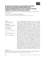
Fig. 1. FTIR spectra of polysaccharide TMP-A.
dent’s t-test and one-way analysis of variance. Values of P < 0.05
were considered to be a statistically significant finding.
3. Results and discussion
3.1. Extraction, purity and composition of polysaccharides
The crude polysaccharide, named TMP, was obtained from
the fruiting bodies of T. matsutake with a yield of 16.4%. After
fractionation on DEAE-cellulose 52 and Sephadex G-100 column
chromatography, 324 mg of TMP-A was obtained from the 0 M NaCl
eluate and detected by the phenol-sulfuric acid assay. The homo-
geneity of the polysaccharide was elucidated by the following tests.
TMP-A waseluted fromgel-filtration chromatography on Sephadex
G-100 column and was detected by the phenol-sulfuric acid assay
as a single peak. No absorption at 280 and 260 nm in UV absorp-
tion spectra of TMP-A demonstrated the absence of protein and
nucleic acid in this polysaccharide and it had the same optical
rotation: [␣]
20
D
−1.648
◦
(c0.5, water) in different low concentra-
tion of ethanol using HK7-SGW-1 automatic optical polarimeter at
room temperature. Weight-average molecular weight was around
8.89 × 10
4
Da. The three monosaccharides, d-glucose, d-galactose,
d-xylose (d-Glc, d-Gal, and d-Xyl) were also identified using the
hydrolysate of TMP-A by GC–MS which was in good agreement
with the TLC with the ratios of 79.37:9.81:10.82.TMP-A was sup-
posed to contain the d-configuration monosaccharide according to
GC–MS analysis.
3.2. Structure elucidation of TMP-A
The intensity of bands around 3408.22 cm
−1
in the IR spectrum
(Fig. 1) was due to the hydroxyl stretching vibration of the polysac-
charide and asexpected they were broad. The bands inthe region of
2923.62 cm
−1
were due to C–H stretching vibration, and the bands
in the region of 1643.29 cm
−1
were due to associated water [20].
Two strong absorption bands at 1075.03cm
−1
, 1041.64 cm
−1
in the
range of 1200–1000 cm
−1
in the IR spectrum suggested that the
monosaccharide in TMP-A had a pyranose-ring [21]. The absorp-
tion at 875.43 cm
−1
indicated that TMP-A had -glucopyranose
linkages, which was indicated by the anomeric proton signals at
ı4.570 in the
1
H NMR (400 MHz) [22]. Moreover, the characteris-
tic absorptions at 799.73 cm
−1
indicated ␣-configurations existing
in the polysaccharide [23], which was in good agreement with the
anomeric proton signals at ı5.182, ı5.107, ı5.060 in the
1
HNMR
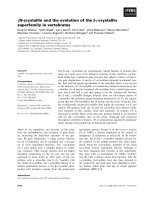
(400 MHz) spectrum. According to the literature [24], the reso-
nances in the region of 98–106 ppm in the
13
C NMR (200 MHz)
spectrum of TMP-A were attributed to the anomeric carbon atoms
of -d-glucopyranose (-d-Glcp), ␣-d-galactopyranose (␣-d-Galp)
and ␣-d-xylopyranose (␣-d-Xylp). In the anomeric carbon region,
signals at ı105.2 could be attributed to C-1 of →4)--d-Glcp-(1→;
Fig. 2. The
13
C NMR spectra of polysaccharide TMP-A.
Table 1
13
C NMR chemical shift data (ı, ppm) for polysaccharide TMP-A.
Sugar residues Chemical shifts, ı (ppm)
C
1
C
2
C
3
C
4
C
5
C
6
→4)--d-Glcp-(1→ 105.239 69.370 78.160 72.941 78.750 63.636
→4,6)--d-Glcp-(1→ 105.423 69.370 78.426 72.121 79.598 63.636
→3)-␣-d-Galp-(1→ 100.762 70.820 77.388 75.439 80.717 63.258
␣-d-Xylp-(1→ 104.194 71.047 75.638 74.305 72.121
Table 2
GC–MS results of methylation analysis of TMP-A.
Methylated sugar Linkage m/z
2,3,6-Me
3
-Glc 1,4- 45,59,73,88,101, 133,146,159,232
2,3-Me
2
-Glc 1,4,6- 45,59,73,88,101,116,133,146,174,232
2,4,6-Me
3
-Gal 1,3- 45,59,73,89,116,146,159,191,204,233
2,3,4-Me
3
-Xyl T- 45,59,73,88,101,116,133,146,174
ı105.4 to C-1 of →4,6)--d-Glcp-(1→; ı100.7 to C-1 of →3)-␣-d-
Galp-(1→; ı104.1 to C-1 of ␣ d-Xylp-(1→, respectively (Fig. 2). All
the assignment of the carbon atoms signals was shown in Table 1.
After methylation according to the Hakomori method for four
times, the methylated polysaccharide was depolymerized and con-
verted into partially methylated ramifications. The analysis of
the methylated monosaccharide was conducted by GC–MS. The
information in MS showed that fragment ion peaks were consis-
tent with the data of d-configuration monosaccharide fragment
ions peaks which can be concluded that the glucose, galactose
and xylose residues were d configuration. Methylation analysis
for TMP-A proved that the -d-glucopyranose residues were 2,3-
bis-substituted and 2,3,6-trisubsituted, the ␣-d-galactopyranose
residues were 2,4,6-trisubsituted, and the ␣-d-xylopyranose
residue was 2,3,4-trisubsituted (Table 2). Results methylated
linkage analysis of TMP-A indicated that (1 → 4)-linked--d-
glucopyranose was one of the largest amounts residue of the
polysaccharide structure, the branched residue was (1 → 4,6)-
linked --d-glucopyranose revealing that (1 → 4)-linked--d-
glucopyranose should be possible to form the backbone structure.
The relative amounts of (1 → 4,6)-linked--d-glucopyranose indi-
cating that approximate branch ratios could theoretically be 12.5%,
namely on average one branching point for each eight residues of
backbone. Residues of branch structure were (1 → 3)-linked-␣-d-
galactopyranose and terminated with
␣-d-xylopyranose residue.
It is concluded that a repeating unit of TMP-A has a backbone of
(1 → 4)--d-glucopyranose residues which branches at O-6 based
274 X. Ding et al. / International Journal of Biological Macromolecules 47 (2010) 271–275
Fig. 3. Predicted chemical structure of polysaccharide TMP-A.
Fig. 4. DPPH
−
radical scavenging effect of TMP-A from Tricholoma matsutake.
on the experimental results. The branch was supposed to be the
composition of an (1 → 3)-␣-d-galactopyranose residue and one
terminated with ␣-d-xylopyranose residue. The predicted struc-
ture of the novel polysaccharide TMP-A was shown in Fig. 3.
3.3. Determination of DPPH radical scavenging activity of TMP-A
DPPH is a useful reagent for investigating the free radical scav-
enging activities of various samples. It is noticeable by eye that
there is a discolouration from purple to yellow induced by antiox-
idants. Fig. 4 illustrates the scavenging activity of the purified
polysaccharide samples on the DPPH radical. These results showed
that the IC
50
value of TMP-A for eliminating DPPH radicals was
about 3.0 mg/mL, which indicated that TMP-A have a noticeable
effect on scavenging DPPH radical,especially at highaddition quan-
tity. However, the inhibiting ability was lower than that of BHT and
Vc.
3.4. Scavenging effect of hydroxyl radical by TMP-A
Hydroxyl radical can easily cause tissue damage or cell death.
Thus, hydroxyl radical removing is important for the protection of
living systems. Fig. 5 shows the percentage hydroxyl radical scav-
enging effects of TMP-A at the dose of 0.5, 1, 2, 3, 4, 5 and 10 mg/mL
and IC
50
value of TMP-A was about 7.1 mg/mL. At the test concen-
trations, TMP-A exhibited scavenging effect on hydroxyl radicals in
a concentration-dependent manner which showed that the purifi-
cation polysaccharides had weaker hydroxyl radical scavenging
effects than Vc of same dose.
3.5. Determination of superoxide anion scavenging effect
Fig. 6 illustrates the superoxide radical scavenging effect of 0.5,
1, 2, 3, 4, and 5 mg/mL of TMP-A in comparison to the same doses of
Vc. At all the concentrations, the polysaccharide samples exhibited
varying degrees of antioxidant effect and IC
50
value of TMP-A was
Fig. 5. Hydroxyl radical scavenging effect of TMP-A from Tricholoma matsutake.
Fig. 6. Superoxide radical scavenging effect of TMP-A from Tricholoma matsutake.
Fig. 7. TMP-A attenuated PC12 cell damage induced by hydrogen peroxide.
about 3.6 mg/mL. Results showed that the purification polysaccha-
rides had weaker hydroxyl radical scavenging effects than Vc of
same dose.
3.6. Antioxidant activity analysis of TMP-A by MTT
In MTT experiments we determined protection effect of TMP-
A on PC12 cell from hydrogen peroxide (H
2
O
2
) induced injury.
After pretreated with 1.25, 2.5, 5, 10 mg/mL of TMP-A, the
PC12 cell could been protected from H
2
O
2
(300 mM) injury in
a dose dependent way, with the cell viability rate of 12.1%,
35.4%, 76.1%, 85.2%, respectively (Fig. 7). Thus, we confirmed
X. Ding et al. / International Journal of Biological Macromolecules 47 (2010) 271–275 275
that TMP-A may attenuate the injury on PC12 cells induced by
H
2
O
2
.
4. Conclusions
According to the results above, it was concluded that the novel
polysaccharide obtained from T. matsutake is a heteropolysac-
charide, namely TMP-A and the purified polysaccharide prepared
(TMP-A) was confirmed of high purity. The present study also
showed that TMP-A consisted of three monosaccharides, namely
d-Glc, d-Xyl and d-Gal and their ratios were 8:1:1 by GC–MS.
Structure study demonstrated that TMP-A has a backbone of
(1 → 4)--d-glucopyranose residues which branches at O-6 based
on the experimental results. The branches were mainly composed
of an (1 → 3)-␣-d-galactopyranose residue, and terminated with
␣-d-xylopyranose residue. Purification polysaccharides prepared
in our work are confirmed of high purity. Anti-oxidation test
in vitro shows that it possesses strong free radical scavenging
activity, which may be comparable to Vc and BHT. In PC12 cell
antioxidant effect assay, we found that TMP-A could significantly
attenuate PC12 cell damage caused by hydrogen peroxide. Overall,
T. matsutake may be one ideal sources of antioxidants develop-
ment.
Acknowledgements
The authors thank Dr. PuSu (Department ofChemistry of Medic-
inal Natural Products and Key Laboratory of Drug Targeting of
Education Ministry PRC, West China College of Pharmacy, Sichuan
University) for helpful assistance in NMR experiments. This project
was supported by Chinese National Programs of High Technology
Research and Development (2007AA021506) and the 11th Five
Years Key Programs for Science and Technology Development of
China (2007BAD81B04).
References
[1] J.L. Mau, S.Y. Tsai, Y.H. Tseng, S.J. Huang, LWT—Food Sci. Technol. 38 (2005)
589–597.
[2] J.E. Kinsella, E.N. Frankel, J.B. German, J. Kanner, Food Technol. 4 (1993) 85–89.
[3] X. Chen, H.Y. Zhong, J.H. Zeng, J. Ge, Carbohydr. Polym. 74 (2008) 445–450.
[4] Y.H. Tseng, J.H. Yang, J.L. Mau, Food Chem. 107 (2008) 732–738.
[5] L. Gurein, L.M. Vaario, N. Matsushita, K. Shindo, K. Suzuki, F. Lapeyrie, Mycol.
Progr. 2 (2003) 37–44.
[6] P. Liu, Med. Biotechnol. 8 (2001) 284–287.
[7] A.M. Staub, Methods Carb. Chem. 5 (1965) 5–6.
[8] M. Dubois, K.A. Gillis, J.K. Hamilton, P.A. Rebers, F. Smith, Anal. Chem. 28 (1956)
350–356.
[9] R.M. Yu, Y. Yin, W. Yang, W.L. Ma, L. Yang, X.J. Chen, Z. Zhang, B. Ye, L.Y. Song,
Carbohydr. Polym. 75 (2009) 166–171.
[10] S.M. Partridge, Nature 164 (1949) 443–446.
[11] Q. Dong, Z.Y. Zhang, Y. Lin, J.N. Fang, Acta Biochim. Biophys. Sin. 27 (1995)
261–265.
[12] L. Guentas, P. Pheulpin, P. Michaud, A. Heyraud, C. Gey, B. Courtois, Carbohydr.
Res. 332 (2001) 167–173.
[13] Y. Chen, M.Y. Xie, S.P. Nie, C. Li, Y.X. Wang, Food Chem. 107 (2008) 231–241.
[14] S. Hakomori, J. Biochem. 55 (1964) 205–208.
[15] A. Braca, N.D. Tommasi, L. Dibari, C. Pizza, M. Politi, I. Morelli, J. Nat. Prod. 64
(2001) 892–895.
[16] H. Ye, K.Q. Wang, C.H. Zhou, J. Liu, X.X. Zeng, Food Chem. 111 (2008) 428–432.
[17] N. Smirnoff, Q.J. Cumbes, Phytochemistry 28 (1989) 1057–1060.
[18] G.L. Zou, X.F. Gui, X.L. Zhong, R.P. Zhu, Prog. Biochem. Biophys. 4 (1986) 71–73.
[19] J.F. Yuan, Z.Q. Zhang, Z.C. Fan, J.X. Yang, Hortic. Carbohydr. Polym. 74 (2008)
822–827.
[20] W. Cao, X.Q. Li, L. Liu, M.C. Wang, H.T. Fan, C. Li, Carbohydr. Res. 341 (2006)
1870–1877.
[21] S.A. Barker, E.J. Bourne, M. Stacey, D.H. Whiffen, J. Chem. Soc. Chem. Commun.
(1954) 171–176.
[22] Y.T. Kim, E.H. Kim, C. Cheong, D.L. Williams, C.W. Kim, S.T. Lim, Carbohydr. Res.
328 (2000) 331–341.
[23] X.M. Wu, P.F. Tu, J. Asian Nat. Prod. Res. 7 (2005) 823–828.
[24] Z.J. Wang, D.H. Luo, Z.Y. Liang, Carbohydr. Polym. 57 (2004) 241–247.