HEALTH AND ENVIRONMENT IN AQUACULTURE Edited by Edmir Daniel Carvalho, Gianmarco Silva David ppt
Bạn đang xem bản rút gọn của tài liệu. Xem và tải ngay bản đầy đủ của tài liệu tại đây (19.85 MB, 427 trang )
Edited by
Edmir Daniel Carvalho,
Gianmarco Silva David
Reinaldo J. Silva
ENVIRONMENT
IN AQUACULTURE
HEALTH
AND
HEALTH AND
ENVIRONMENT
IN AQUACULTURE
Edited by Edmir Daniel Carvalho,
Gianmarco Silva David and Reinaldo J. Silva
Health and Environment in Aquaculture
Edited by Edmir Daniel Carvalho, Gianmarco Silva David and Reinaldo J. Silva
Published by InTech
Janeza Trdine 9, 51000 Rijeka, Croatia
Copyright © 2012 InTech
All chapters are Open Access distributed under the Creative Commons Attribution 3.0
license, which allows users to download, copy and build upon published articles even for
commercial purposes, as long as the author and publisher are properly credited, which
ensures maximum dissemination and a wider impact of our publications. After this work
has been published by InTech, authors have the right to republish it, in whole or part, in
any publication of which they are the author, and to make other personal use of the
work. Any republication, referencing or personal use of the work must explicitly identify
the original source.
As for readers, this license allows users to download, copy and build upon published
chapters even for commercial purposes, as long as the author and publisher are properly
credited, which ensures maximum dissemination and a wider impact of our publications.
Notice
Statements and opinions expressed in the chapters are these of the individual contributors
and not necessarily those of the editors or publisher. No responsibility is accepted for the
accuracy of information contained in the published chapters. The publisher assumes no
responsibility for any damage or injury to persons or property arising out of the use of any
materials, instructions, methods or ideas contained in the book.
Publishing Process Manager Molly Kaliman
Technical Editor Teodora Smiljanic
Cover Designer InTech Design Team
First published April, 2012
Printed in Croatia
A free online edition of this book is available at www.intechopen.com
Additional hard copies can be obtained from
Health and Environment in Aquaculture,
Edited by Edmir Daniel Carvalho, Gianmarco Silva David and Reinaldo J. Silva
p. cm.
ISBN 978-953-51-0497-1
Contents
Preface IX
Part 1 Parasitic Diseases 1
Chapter 1 Transmission Biology of the Myxozoa 3
Hiroshi Yokoyama, Daniel Grabner
and Sho Shirakashi
Chapter 2 Metazoan Parasites of the
European Sea Bass Dicentrarchus labrax
(Linnaeus 1758) (Pisces: Teleostei) from Corsica 43
Laetitia Antonelli and Bernard Marchand
Chapter 3 Parasitic Diseases in Cultured
Marine Fish in Northwest Mexico 63
Emma J. Fajer-Ávila, Oscar B. Del Río-Zaragoza
and Miguel Betancourt-Lozano
Part 2 Bacterial Diseases 95
Chapter 4 Molecular Detection and
Characterization of Furunculosis
and Other Aeromonas Fish Infections 97
Roxana Beaz Hidalgo and María José Figueras
Chapter 5 An Overview of Virulence-Associated Factors
of Gram-Negative Fish Pathogenic Bacteria 133
Jessica Méndez, Pilar Reimundo, David Pérez-Pascual,
Roberto Navais, Esther Gómez, Desirée Cascales
and José A. Guijarro
Part 3 Antibiotics and Probiotics 157
Chapter 6 Antibiotics in Aquaculture –
Use, Abuse and Alternatives 159
Jaime Romero, Carmen Gloria Feijoó
and Paola Navarrete
VI Contents
Chapter 7 The Use of Antibiotics in Shrimp Farming 199
M.C. Bermúdez-Almada and A. Espinosa-Plascencia
Chapter 8 Probiotics in Aquaculture – Benefits to the
Health, Technological Applications and Safety 215
Xuxia Zhou and Yanbo Wang
Chapter 9 Probiotics in Aquaculture of
Kuwait – Current State and Prospect 227
Ahmed Al-marzouk and Azad I. Saheb
Part 4 Applied Topics of Cellular and Molecular Biology 249
Chapter 10 Use of Microarray Technology
to Improve DNA Vaccines in Fish
Aquaculture – The Rhabdoviral Model 251
P. Encinas, E. Gomez-Casado, A. Estepa and J.M. Coll
Chapter 11 Fighting Virus and Parasites
with Fish Cytotoxic Cells 277
M. Ángeles Esteban, José Meseguer
and Alberto Cuesta
Chapter 12 Bacteriocins of Aquatic
Microorganisms and Their Potential
Applications in the Seafood Industry 303
Suphan Bakkal, Sandra M. Robinson
and Margaret A. Riley
Chapter 13 The Atlantic Salmon (Salmo salar) Vertebra
and Cellular Pathways to Vertebral Deformities 329
Elisabeth Ytteborg, Jacob Torgersen,
Grete Baeverfjord and Harald Takle
Part 5 Ecological Impacts of Fish Farming 359
Chapter 14 Ecological Features of Large
Neotropical Reservoirs and Its
Relation to Health of Cage Reared Fish 361
Edmir Daniel Carvalho, Reinaldo José da Silva,
Igor Paiva Ramos, Jaciara Vanessa Krüger Paes,
Augusto Seawright Zanatta, Heleno Brandão,
Érica de Oliveira Penha Zica, André Batista Nobile,
Aline Angelina Acosta and Gianmarco Silva David
Part 6 Work-Related Hazards – Prevention and Mitigation 383
Chapter 15 Aquacultural Safety and Health 385
Melvin L. Myers and Robert M. Durborow
Contents VII
Part 7 Spread of Pathogens from Marine Cage 401
Chapter 16 Spread of Pathogens from Marine Cage
Aquaculture – A Potential Threat for Wild
Fish Assemblages Under Protection Regimes? 403
Antonio Terlizzi, Perla Tedesco and Pierpaolo Patarnello
Preface
Aquaculture is a modality of food production that has been experiencing continuous
expansion in many countries worldwide. This expansion brings the challenge of
developing reliable tools for disease control, to assure high productivity of healthy
seafood. The increase of farmed fish production raises the issue of achieving a
sustainable and environmental friendly aquaculture. The adoption of best
management practices in the whole production chain, based on “state of the art”
scientific knowledge, is the key for sustainable health management. In this book,
experts from several countries bring updated information about some of the main
health issues that currently affects aquaculture. Topics concerning pathogens,
antibiotics, probiotics, cell biology, ecological interactions, and safety are included in
the six sections of this book.
The first section is entitled as “Parasitic diseases”, addressing issues and impacts of
parasites upon aquaculture. The first chapter is the “Transmission biology of the
myxozoa”, which explains about the diseases that some myxozoans cause in marine
and freshwater fish, and how they can be a problem for aquaculture and fishery
industries. It also elucidates the life cycle of myxozoans, that involves invertebrates,
and a vertebrate host that is typically a fish. However, there are no commercially
available chemotherapeutants and vaccines to treat myxozoan infections. This review
summarizes the current knowledge on the transmission biology of myxozoans, which
would be useful for designing management strategies for related diseases. The second
chapter is “Metazoan parasites of cultured European sea bass Dicentrarchus labrax
(Linneaus 1758) from Corsica”. It is a study relating that parasitic infections and
associated diseases have emerged in aquaculture systems in many regions of Europe,
resulting in significant economical losses. This study points out that wild fish are
believed to be the primary reservoirs of parasite infection for fish farmed in cages, and
environmental conditions in culture systems may favor disease transmission,
threatening production activity. In this sense, it is considered that animals reared in
sea-cages are exposed to a large number of parasitic agents. The third chapter is
“Parasitic diseases in cultured marine fish in Northwest Mexico”. This chapter
summarizes the main parasitic diseases that affect marine fish species with
aquaculture potential in the Norwest Pacific coast of Mexico, emphasising proper
strategies for their control. The study shows the need to perform parasite treatment
and control applying prophylactic and therapeutic measures.
X Preface
In the section “Bacterial diseases”, the fourth chapter “Updated information of
Aeromonas infections and furunculosis derived from molecular methods” focuses on
the bacteria Aeromonas salmonicida, the causal agent of furunculosis, considered a
particularly important fish pathogen mainly due to its widespread distribution and
ability to infect a diverse range of hosts, causing massive mortalities and economic
losses. Additionally, climate change has been considered to play a role in the
appearance and impact of furunculosis. The study undertakes molecular techniques in
Aeromonas infections in fish, including significant advances in genomics and taxonomy
of these microorganisms. The fifth chapter is “An overview on virulence-associated
factors of Gram-negative fish pathogenic bacteria”, which addresses the issue of
bacterial outbreaks causing important economic losses for aquaculture. Gram-negative
bacteria have long been recognized as a cause of the most prevalent fish pathologies in
the aquaculture industry. The application of in vivo and in vitro molecular techniques
to fish pathogenic bacteria resulted in the characterization of novel virulence
determinants and allowed to increase the knowledge of bacterial pathogenic
mechanisms. This review deals with representative species of gram-negative fish
pathogenic bacteria in the context of the analysis of well-established virulent factors
produced by these pathogens.
In the section “Antibiotics and probiotics”, the sixth chapter is “Antibiotics in
aquaculture: use, abuse and alternatives”. This study argues that unpredictable
mortalities in aquaculture production may be due to negative interactions between
fish and pathogenic bacteria. To solve this problem, farmers frequently use antibiotic
compounds to treat bacterial diseases. The concerns about the increase in bacterial
resistance and antibiotic residues have aroused great caution in the use of antibiotics
in aquaculture, which has encouraged research to obtain alternatives. The aim of this
chapter is to provide information about the current knowledge in antibiotic use in
aquaculture systems, including information about mechanisms of action and
resistance. The seventh chapter is “The use of antibiotics in shrimp farming”. This is an
important study, considering that shrimp cultivation has been the most expanding
aquaculture activity. Nevertheless, this industry faces major problems with viral and
bacterial diseases, and large quantities of chemical and antibiotic products are
frequently used to counteract this. The study demonstrates the importance of applying
appropriate therapies with antibiotics, seeking greater effectiveness for the control of
bacterial infections. The eighth and ninth chapters, within this section, deal with
probiotics in aquaculture, which has been considered a key factor for fish health
management, due to the increasing demand for environment friendly aquaculture. The
eighth chapter is “Probiotics in aquaculture: benefits to the health, technological
applications and safety”. This study points out that, currently, a number of
preparations of probiotics are commercially available and have been introduced to
fish, shrimp and molluscan farming as feed additives. Thus, there is a commercial and
academic interest of increasing our knowledge in effective preparation, technological
applications, and safety evaluation of probiotics. The ninth chapter is “Probiotics in
aquaculture of Kuwait: current state and prospect”, and mentions the application of
Preface XI
autochthonous probiotics. In this experimental study, a protocol for the isolation,
screening and selection of candidate probiotic bacteria based on several selective
criteria was accomplished. This study showed that the methods were suitable to
certain extent to assess the antagonism ability of probiotic bacteria on pathogenic
bacteria, and these findings can be applied to other cultured fish.
In the section entitled as “Applied topics of cellular and molecular biology”, the tenth
chapter is the “Use of microarray technology to improve DNA vaccines in fish
aquaculture: the rhabdoviral model”. Rhabdovirosis are one of the most important
diseases affecting farmed fish worldwide, and are amongst the few fish diseases for
which there is an efficacious DNA vaccine. Understanding the induced molecular
events occurring after fish immunization with rhabdoviruses and their DNA vaccines
might contribute to improve vaccines to other fish pathogens. This study focus on data
published on the use of microarrays for the identification of rhabdoviral-induced
genes, with properties that make them candidate adjuvants for the improvement of
fish DNA vaccines. The eleventh chapter is “Fighting virus and parasites with fish
cytotoxic cells”, which is a review on the fish cell-mediated cytotoxic activity as the
main cellular immune mechanism against tumors, parasites and viral infections. It also
addresses the modulation of this activity by means of immunostimulants, stress,
pollution, and vaccines. This research contributes to understand fish cytotoxic cells
and their activity from an evolutionary point of view. Furthermore, the lack of
commercial antiviral and anti-parasitic vaccines for fish makes necessary to increase
the knowledge on the cell-mediated cytoxic activity of fish. The twelfth chapter is
“Bacteriocins of aquatic microorganisms and their potential applications in the seafood
industry”. Narrow killing spectrum bacteriocins are recognized as a promising
alternative to broad-spectrum antibiotics, whose efficacy has been compromised by
the evolution of resistant bacteria. This study aims to provide an overview of the
diversity of bacteriocins produced by marine microorganisms, their role in mediating
microbial interactions in the marine environment, and their potential applications in
the seafood industry. The thirteenth chapter is “Molecular characterization of
pathological bone development in Atlantic salmon (Salmo salar)”. This study argues
that spinal disorders are a recurrent problem for aquaculture, and until recently, their
molecular development in fish has received relatively little attention. In this review,
the current knowledge on the cellular and molecular mechanisms for skeletal
homeostasis and aberrant development of bone in the Atlantic salmon vertebrae is
referred.
In the section “Ecological impacts of fish farming”, the fourteenth chapter is
“Ecological features of large Neotropical reservoirs related to health of cage reared
fish”. This study raises the subject of fish cage culture in hydroelectric reservoirs in
Brazil. Wild native fish species and a farmed fish species, Oreochromis niloticus, were
searched for ectoparasites, which showed that the cultured fish presented high rates of
parasitic infection. This research attempted to identify interferences of fish cage
farming upon water quality, wild fish assemblages and parasitic diseases in large
freshwater reservoirs. The fifteenth chapter is “Spread of pathogens from marine cage
XII Preface
aquaculture: a potential threat for wild fish assemblages under protection regimes?”
focusing on the exchange of viruses between farmed and wild populations, and
further, the potential impact on natural ecosystems. The study reviews the effects of a
serious disease, Viral Nervous Necrosis (VNN), which affects more than 40 fish species
worldwide. Likewise, betanodaviruses are the most important viral pathogens
reported in marine aquaculture within the Mediterranean region.
The last section is “Work-related hazards: prevention and mitigation”, with the
sixteenth chapter: “Aquacultural Safety and Health” showing that occupational
hazards in aquaculture are associated with different rearing technologies. Farm
operators are encouraged to adopt or develop inherently safety technologies by first
eliminating, then guarding against, and finally warning about the hazard. A model
safety manual presents contents that can be adapted to aquaculture.
The challenge of editing this book could only be accomplished with the help of some
colleagues. Therefore, we would like to thank Professor Dr. Fernanda Natália
Antoneli, from Federal University of Mossoró (RN, Brazil), who has assisted us with
her background on cell biology; Dra. Fabiana Garcia Scaloppi, from Sao Paulo State
Agency of Agribusiness Technology (APTA at Votuporanga, SP, Brazil) who has
collaborated with her expertise on parasitology; Professor Dra. Mara Renata Dega,
from Marechal Rondon Faculty (at Sao Manuel, SP, Brazil) who has helped with
pharmacology themes. Finally, I would like to give especial thanks to the biologist
Aline Angelina Acosta, a graduate student in Zoology, who has collaborated
throughout the edition process with her English skills.
Dr. Edmir Daniel Carvalho
Dr. Reinaldo J.Silva
Dr. Gianmarco Silva David
Sao Paulo State University
Brazil
Part 1
Parasitic Diseases
1
Transmission Biology of the Myxozoa
Hiroshi Yokoyama
1
, Daniel Grabner
2
and Sho Shirakashi
3
1
The University of Tokyo
2
University of Duisburg-Essen
3
Kinki University
1,3
Japan
2
Germany
1. Introduction
Myxozoans are spore-forming parasites of both freshwater and marine fishes (Lom &
Dyková, 1992, Kent et al., 2001; Feist & Longshaw, 2006). The Myxozoa were previously
classified as protozoans, although the multicellular state and functional specialization of the
cells composing spores were considered to exceed protozoan level (Lom & Dyková, 1992).
Indeed, molecular studies demonstrated that myxozoans are metazoans (Smothers et al.,
1994, Siddal et al., 1995). However, there were two conflicting views concerning the
phylogenetic origin of myxozoans; the Bilateria (Smothers et al., 1994, Schlegel et al., 1996,
Anderson et al., 1998, Okamura et al., 2002) vs. the Cnidaria (Siddal et al., 1995). More
recently, the Cnidaria-hypothesis has been strongly supported by phylogenetic analyses of
protein-coding genes of myxozoans (Jimenez-Guri et al., 2007, Holland et al., 2010). The
phylum Myxozoa, of which more than 2100 species in 58 genera are described to date, is
divided into two classes, Myxosporea and Malacosporea (Lom & Dyková, 2006). Most of
myxozoans are not harmful to host fish, however, some species cause diseases in cultured
and wild fish which are problems for aquaculture and fishery industries worldwide.
Generally, freshwater myxosporeans appear to be specific at the family or the genus level of
the host, while some marine myxosporeans have a low host-specificity. Some examples are
mentioned below.
For freshwater species, myxozoans infecting salmonids have been relatively well studied.
For example Myxobolus cerebralis, the causative agent of whirling disease, Tetracapsuloides
bryosalmonae, the cause of proliferative kidney disease (= PKD), and Ceratomyxa shasta,
causing ceratomyxosis, have fatal effects on farmed salmonid fish (Table 1). Salmonid
ceratomyxosis is a local disease which is restricted only to North America (Bartholomew et
al., 1997), while whirling disease and PKD are widely distributed in the world (Hedrick et
al., 1993, 1998). M. cerebralis infects cartilage tissue and causes a whirling behaviour (tail-
chasing swimming), a black tail, and skeletal deformities of affected fish. Whirling disease
was previously known as a hatchery disease, but recently, it has been recognized as one of
the causes for the decline of natural rainbow trout populations in several western states of
the USA (Hedrick et al, 1998). Symptoms of PKD in salmonid fish are a swollen kidney (Fig.
1A) and anemic gills, evoked by chronic inflammation of the kidney interstitium. The
Health and Environment in Aquaculture
4
Myxozoans Disease names or typical
signs
Fish References
Ceratomyxa shasta
Ceratomyxosis Salmonids
Bartholomew et al.
(1997)
Chloromyxum
truttae
Hypertrophy of gall bllader Salmonids Lom & Dyková (1992)
Henneguya ictaluri
Proliferative gill disease
(PGD)
Ictalurus
punctatus
Pote et al. (2000)
Henneguya
salminicola
Milky condition Salmonids
Awakura & Kimura
(1977)
Hoferellus carassii
Kidney enlargement disease
(KED)
Carassisus
auratus
Yokoyama et al. (1990)
Myxidium giardi
Systemic infection Anguilla spp.
Ventura & Paperna
(1984)
Myxobolus artus
Muscular myxobolosis
Cyprinus carpio
Yokoyama et al. (1996)
Myxobolus cerebralis
Whirling disease Salmonids Hedrick et al. (1998)
Myxobolus cyprini
Malignant anemia
Cyprinus carpio
Molnár & Kovács-
Gayer (1985)
Myxobolus koi
Gill myxobolosis
Cyprinus carpio
Yokoyama et al. (1997a)
Myxobolus
murakamii
Myxosporean sleeping
disease
Oncorhynchus
masou
Urawa et al. (2009)
Myxobolus wulii
Cysts in gill or
hepatopancreas
Carassius auratus
Zhang et al. (2010b)
Parvicapsula
pseudobranchicola
Inflammation and necrosis
of filaments
Salmo salar
Karlsbakk et al. (2002)
Sphaerospora
dykovae
Swimbladder inflammation
(SBI)
Cyprinids Dyková & Lom (1988)
Tetracapsuloides
bryosalmonae
Proliferative kidney disease
(PKD)
Salmonids Hedrick et al. (1993)
Thelohanellus
hovorkai
Hemorrhagic thelohanellosis
Cyprinus carpio
Yokoyama et al. (1998)
Table 1. Economically important freshwater myxosporeans.
causative agent of PKD has not been identified for a long time, and thus the organism was
previously called PKX (Hedrick et al., 1993). It was assigned to the Myxozoa in 1999 and
initially called Tetracapsula bryosalmonae (Canning et al., 1999). Canning et al. (2000) erected
the new class Malacosporea in the Myxozoa, and later, in the course of nomenclature
changes by Canning et al. (2002) Tetracapsula bryosalmonae was renamed to Tetracapsuloides
bryosalmonae (Fig. 1B). Salmonids suffering from ceratomyxosis show abdominal distension
and exophthamia, possibly caused by osmotic imbalance due to C. shasta infection in the
internal organs (Bartholomew et al., 1997). Henneguya salminicola produces cysts in the
musculature of anadromous salmonid fish (Fig. 1C, D). This parasite does not cause a health
Transmission Biology of the Myxozoa
5
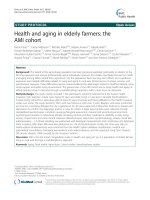
A & B: Proliferative kidney disease of rainbow trout (Oncorhynchus mykiss). Note the swollen kidney
(arrow). Malacospore of Tetracapsuloides bryosalmonae from bryozoans host (B). C & D: Milky condition
of pink salmon (Oncorhynchus gorbuscha). White exudate (arrow) filled with spores of Henneguya
salminicola (D). Photos of courtesy by Dr. T. Awakura. E & F: Hemorrhagic thelohanellosis of common
carp (Cyprinus carpio). Note extensive haemorrhages in mouth and abdomen caused by Thelohanellus
hovorkai (F) in the subcutaneous tissue. G & H: Creamy appearance of enlarged hepatopancreas of
goldfish (Carassius auratus) infected with Myxobolus wulii (H). Scale bars for B, D, F and H are 10μm.
Fig. 1. Myxozoan diseases of freshwater fish and the causative myxozoan parasites.
Health and Environment in Aquaculture
6
problem of the host, but renders the infected fish unmarketable due to the milky condition
of the flesh (Awakura & Kimura, 1977). Myxosporean sleeping disease is caused by
Myxobolus murakamii infecting the peripheral nerve of masu salmon (Oncorhynchus masou).
This disease has been known only in Hiroshima Prefecture, in south-western Japan,
although M. murakamii occurs also in Hokkaido, the northernmost area of Japan. It remains
to be clarified why the sleeping disease does not occur in Hokkaido (Urawa et al., 2009).
Chloromyxum truttae infects the gallbladder of brood stock of rainbow trout (Oncorhynchus
mykiss), while it infects the yearlings of Atlantic salmon (Salmo salar). Affected fish showed
loss of appetite, yellow colouration of body, and hypertrophic gall bladder (Lom & Dyková,
1992). Pseudobranch infection with Parvicapsula pseudobranchicola has been reported in
Atlantic salmon in Norway, showing lethargy, disorganized swimming, exophthalmia and
low-grade to significant mortalities (Karlsbakk et al., 2002). Affected fish exhibited eye
bleeding and cataracts, possibly due to obstruction of the blood supply to the choroid bodies
of the eyes.
Myxobolus koi, Thelohanellus hovorkai, and Sphaerospora dykovae (= S. renicola) are well-known
pathogens in cultured common carp (Cyprinus carpio) in Europe and Asia (Dyková & Lom,
1988, Yokoyama et al., 1997a, 1998). M. koi infects the gills and causes a respiratory
disfunction of carp juveniles. Yokoyama et al. (1997a) reported that there are two types of M.
koi infections; the one forms large-type (pathogenic) cysts in the gill filaments, while the
other forms small-type (non-pathogenic) cysts in the gill lamellae. T. hovorkai infecting the
connective tissue is the causative agent of the hemorrhagic thelohanellosis of common carp
(Yokoyama et al., 1998). Spore dispersion of T. hovorkai in subcutaneous connective tissue
causes extensive hemorrhages and edema, finally resulting in death of affected fish (Fig. 1E,
F). S. dykovae, the cause of swimbladder inflammation (SBI) was previously known as S.
renicola, but has recently been renamed as S. dykovae in association with revised taxonomy of
the genus Leptotheca (Gunter & Adlard, 2010). The target organ (spore forming site) for S.
dykovae is the kidney, but the extrasporogonic stage of S. dykovae proliferates in the
swimmbladder, which causes SBI of carp (Dyková & Lom, 1988). Myxobolus artus produced
rice bean-like cysts in the musculature of common carp. Adult carp (over 1-year old) do not
die of the disease but lose their commercial value. In contrast, juvenile carp (0-year old)
heavily infected with M. artus exhibit hemorrhagic anemia and increased mortality rate.
After degeneration of M. artus cysts in the musculature, spores engulfed by macrophages
are transferred into gills, where numerous spores accumulate and pack within the lamellae.
As a result, the gill epithelia are exfoliated, causing the hemorrhagic anemia (Yokoyama et
al., 1996). Myx
obolus cyprini infecting the skeletal muscle of common carp was also reported
to cause the malignant anemia (Molnár & Kovács-Gayer, 1985), but it is unknown whether
the disease mechanisms are the same as M. artus. Thelohanellus kitauei forms large cysts in the
intestinal mucosa of common carp so that the intestine was occluded to emaciate the
infected fish.
Hoferellus carassii infecting the kidney of goldfish (Carassius auratus) is the causative agent of
kidney enlargement disease (KED). This parasite does not cause a high mortality of affected
fish, but a low marketability as an ornamental fish (Yokoyama et al., 1990). Myxobolus wulii
forms numerous cysts in the gills of goldfish in some cases, whereas large cysts are formed
in the hepatopancreas in other cases (Fig. 1G, H). In both cases, infection of fish results in high
mortality (Zhang et al., 2010b). Gill infections with Henneguya ictaluri and H. exilis are typical
Transmission Biology of the Myxozoa
7
myxosporean diseases in catfish culture. H. ictaluri causes proliferative gill disease of catfish
(Ictalurus punctatus) (Pote et al. 2000). Myxidium giardi infects multiple organs including gills
and kidney of several eel species, Anguilla anguilla, A. rostorata, and A. japonica. Infected elvers
exhibit dropsy, ascites, and swollen kidney (Ventura & Paperna, 1984).
Compared to freshwater myxosporeans, many marine species have a broad host range,
such as Kudoa thyrsites, K. yasunagai and Enteromyxum leei (Table 2). K. thyrsites lowers the
Myxozoans Disease names or typical
signs
Fish References
Enterom
y
xum leei
Enteromyxosis or
myxosporean emaciation
disease
Di
p
lodus
p
untazzo,
Sparus aurata,
Paralichthys olivaceus,
Pagrus major,
Takifugu rubripes
Diamant (1997)
Yasuda et al. (2002)
Enterom
y
xum sco
p
hthalmi
Enteromyxosis Palenzuela et al. (2002)
Henne
g
u
y
a lateolabracis
Cardiac henneguyosis Lateolabrax sp. Yokoyama et al. (2003)
Henne
g
u
y
a
p
a
g
ri
Cardiac henneguyosis
Pa
g
rus ma
j
o
r
Yokoyama et al. (2005a)
Kudoa amamiensis
Kudoosis amami
Seriola
q
uin
q
ueradiata
Yokoyama et al. (2000)
Kudoa iwatai
Cysts in multiple organs
Dicentrarchus labrax,
Lateolabrax japonicus,
Mugil cephalus,
Sparus aurata,
Pagrus major,
Oplegnatus punctatus
Diamant et al. (2005)
Kudoa lateolabracis
Post-mortem
myoliquefaction
Lateolabrax sp.,
Paralichthys olivaceus
Yokoyama et al. (2004)
Kudoa lut
j
anus
Systemic infection
Lut
j
anus er
y
thro
p
terus
Wang et al. (2005)
Kudoa neuro
p
hila
Meningoencephalomyelitis
Latris lineata
Grossel et al. (2003)
Kudoa shiomitsui
Cysts in the heart
Taki
f
u
g
u rubri
p
es,
Thunnus orientalis
Zhang et al. (2010)
Kudoa th
y
rsites
Post-mortem
myoliquefaction
Salmo salar,
Paralichtys olivaceus,
Coryphaena hyppurus
Moran et al. (1999a)
Kudoa
y
asuna
g
ai
Abnormal swimming
Lateolabrax
j
a
p
onicus,
Oplegnathus fasciatus,
Seriola quinqueradiata,
Takifugu rubripes,
Thunnus orientalis,
Plotosus lineatus
Zhang et al. (2010a)
My
xobolus acantho
g
obii
Myxosporean scoliosis or
skeletal deformity
Seriola
q
uin
q
ueradiata
,
Scomber japonicus
Yokoyama et al. (2005b)
S
p
haeros
p
ora e
p
ine
p
heli
Disorientation, hemorrhage
E
p
ine
p
helus
malabaricus
Supamattaya et al. (1991)
S
p
haeros
p
ora
f
u
g
u
(= Leptotheca fugu)
Myxosporean emaciation
disease
Taki
f
u
g
u rubri
p
es
Tin Tun et al. (2000)
Table 2. Economically important marine myxosporeans (see also Fig. 2).
Health and Environment in Aquaculture
8
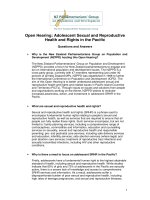
A & B: Skeletal deformity (A) of Japanese mackerel (Scomber japonicus) infected with Myxobolus
acanthogobii (B) in the brain. C & D: Enlarged bulbus arteriosus (C) of Chinease seabass (Lateolabrax sp.)
infected with Henneguya lateolabracis (D) in the heart. E & F: Myxosporean emaciation disease (E) of tiger
puffer (Takifugu rubripes) infected with developmental stages (arrows) of Enteromyxum leei (F) in the
intestine. Diff-Quik stain (F). G & H: Cysts (arrows) in the skeletal muscle (G) of red sea bream (Pagrus
major). Cysts are packed with spores of Kudoa iwatai (H). Scale bars for B, D, F and H are 10 μm.
Fig. 2. Myxosporean diseases of marine fish and the causative myxozoan parasites.
Transmission Biology of the Myxozoa
9
commercial value of various cultured marine fish species, particularly Atlantic salmon
(Salmo salar) in North America, by causing post-mortem myoliquefaction (Moran et al.,
1999a). K. yasunagai forms numerous cysts in the brain, probably causing disorder of
swimming performance of many fish species (Zhang et al., 2010a). Recently, enteromyxosis
or myxosporean emaciation disease, caused by E. leei, has emerged as a new threat in
various cultured marine fish, e.g. gilthead sea bream (Sparus aurata) in Mediterranean
countries and tiger puffer (Takifugu rubripes) in Japan (Diamant, 1997, Yasuda et al., 2002). In
contrast, Enteromyxum scophthalmi and Sphaerospora fugu (= Leptotheca fugu) have been found
only in the intestine of turbot (Psetta maxima) and tiger puffer (Takifugu rubripes),
respectively, although the signs of the disease appear to be similar to E. leei infection (Tin
Tun et al., 2000, Palenzuela et al., 2002). Heart infections have been documented such as
Henneguya lateolabracis, H. pagri, and Kudoa shiomitsui. The former two species are highly
pathogenic to Chinese sea bass (Lateolabrax sp.) and red sea bream (Pagrus major),
respectively (Yokoyama et al., 2003, 2005a), whereas the pathogenic effects of K. shiomitsui
are not clear (Zhang et al., 2010a). Many Kudoa infections in skeletal muscle may render the
infected fish unmarketable by producing cysts (e.g., K. amamiensis and K. iwatai) or causing
myoliquefaction (e.g., K. lateolabracis and K. neothunni). K. neurophila has become an
impediment to the juvenile production of striped trumpeter (Latris lineata) in Tasmania, due
to meningoencephalomyelitis of hatched larvae (Grossel et al., 2003). Myxobolus acanthogobii
infects the brain and causes the myxosporean scoliosis in yellowtail (Seriola quinqueradiata),
while infected Japanese mackerel (Scomber japonicus) exhibits the lordosis (dorso-ventral
deformity) and infected goby (Acanthogobius flavimanus) is subclinical (Yokoyama et al.,
2005b). Sphaerospora epinepheli infects the kidney of Epinephelus malabaricus, which shows
disorientation of the body and hemorrhages (Supamattaya et al., 1991).
2. Myxosporeans
The class Myxosporea is comprised of the two orders, Bivalvulida and Multivalvulida.
Bivalvulids include 52 genera with more than 2100 species described from freshwater and
marine fishes, while multivalvulids contain 5 genera with more than 60 species
predominantly from marine fish (Lom & Dyková, 2006). Morphology, life cycle, phylogeny,
and biology of myxosporeans are summarized below.
2.1 Morphology of myxosporean
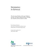
Myxosporean spores are co
mposed of shell valves, sporoplasms, and polar capsules
containing coiled polar filaments (Fig. 3). Number of valves and polar capsules,
arrangement of the polar capsules, and ornamentation of spores allow the genus-level
diagnosis of myxosporeans. Identification at the species-level is based on spore dimensions.
Species description of myxospores should follow the guidelines of Lom & Arthur (1989). For
bivalvulids, spore length and spore width in frontal view, spore thickness in side view,
length and width of polar capsules are measured (Fig. 3). If ornamentations such as the
caudal appendages for Henneguya are present, the length is also measured. For
multivalvulids, spore length (including the apical projections, if present) in side view, spore
width and spore thickness in top view, length and width of polar capsules are determined.
Care must be taken to avoid confusion of thickness and width of spores, because
multivalvulids are radially symmetrical.
Health and Environment in Aquaculture
10
PC: polar capsule, SP: sporoplasm, SV: shell valve, SL: sutural line, L: spore length, W: spore width, T:
spore thickness, PCL: polar capsule length, PCW: polar capsule width.
Fig. 3. Diagrams of bivalvulid (A: frontal view, B: side view) and multivalvulid (C & E, top
view, D: side view) myxosporean spores.
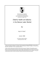
2.2 Life cycle of myxosporeans
The first myxozoan life cycle was discovered for M. cerebralis by Wolf & Markiw in 1984 and
was later confirmed by many other researchers, who reported similar life cycles for more than
30 myxosporean species. These life cycles involve an annelid invertebrate (mainly oligochaetes
for freshwater species and polychaetes for marine species) and a vertebrate host which is
typically a fish (Fig. 4). In the latter, myxosporean spore stages (= myxospores) develop.
Myxospores are ingested by annelids, in which the polar filaments extrude to anchor the spore
to the gut epithelium. Opening of the shell valves allows the sporoplasms to penetrate into the
epithelium. Subsequently, the parasite undergoes reproduction and development in the gut
tissue, and finally produces usually eight actinosporean spore stages (= actinospores) within a
pansporocyst. After mature actinospores are released from their hosts they float in the water
column (El-Matbouli & Hoffmann, 1998). Upon contact with skin or gills of fish, sporoplasms
penetrate through the epithelium, followed by development of the myxosporean stage.
Myxosporean trophozoites are characterized by cell-in-cell state, where the daughter
(secondary) cells develop in the mother (primary) cells. The presporogonic stages multiply,
migrate via nervous or circulatory systems, and develop into sporogonic stages. At the final
site of infection, they produce mature spores within mono- or disporic pseudoplasmodia, or
polysporic plasmodia (El-Matobouli & Hoffmann, 1995).
Transmission Biology of the Myxozoa
11
A: The polar filaments are extruded to anchor the spore to the gut epithelium, followed by opening of
shell valves of myxospore. B: Gametogony. C: Sporogony of actinosporean phase. D: Mature
actinospore stages develop in a pansporocyst, and actinospores are released into the water. E: Upon
contact of actinospores with the skin or gills of the fish host, polar filaments extrude to anchor the spore
to the skin or gills, facilitating invasion of the sporoplasms into the fish. F: Presporogonic multiplication
in a cell-in-cell state. G: Sporogony of myxosporean phase.
Fig. 4. Diagram of the life cycle of myxosporean alternating fish and annelid hosts.
2.3 Morphology of actinospores
Actinospores that are formed in the invertebrate hosts have a triradiate form with
exclusively 3 polar capsules and mostly 3 caudal processes (Figs. 5 & 6). To characterize
actinosporean stages, researchers should follow the guidelines of Lom et al. (1997); shape of
the caudal processes (straight, curved or branched), presence of the style (small stalk below
the spore body) and formation of spore nets (pattern of connection between several spores),
number of daughter cells in the spore body, and measurements of the spore body, style,
polar capsules and processes (Fig. 6).
Health and Environment in Aquaculture
12
A: Raabeia-type actinospores of Myxobolus cultus from oligochaete Branchiura sowerbyi, B:
Neoactinomyxum-type actinospore from B. sowerbyi. C: Triactinomyxon-type actinospore of M. arcticus
from oligochaete Lumbriculus variegatus, D: Echinactinomyxon-type actinospore from B. sowerbyi, E:
Aurantiactinomyxon-type actinospore of Thelohanellus hovorkai from B. sowerbyi, F: Sphaeractinomyxon-
type actinospores from unidentified marine oligochaete, which was collected in May 1990, on the coast
of Mie Prefecture, the middle part of Japan. Arrow shows an actinospore released from a pansporocyst
which develops 8 actinospores. Scale bars for A, C and D are 100 μm, and those for B, E and F are 50
μm.
Fig. 5. Several morphotypes of actinosporean spores.