ADVANCES IN MICROFLUIDICS pdf
Bạn đang xem bản rút gọn của tài liệu. Xem và tải ngay bản đầy đủ của tài liệu tại đây (26.03 MB, 250 trang )
ADVANCES IN
MICROFLUIDICS
Edited by Ryan T. Kelly
Advances in Microfluidics
Edited by Ryan T. Kelly
Published by InTech
Janeza Trdine 9, 51000 Rijeka, Croatia
Copyright © 2012 InTech
All chapters are Open Access distributed under the Creative Commons Attribution 3.0
license, which allows users to download, copy and build upon published articles even for
commercial purposes, as long as the author and publisher are properly credited, which
ensures maximum dissemination and a wider impact of our publications. After this work
has been published by InTech, authors have the right to republish it, in whole or part, in
any publication of which they are the author, and to make other personal use of the
work. Any republication, referencing or personal use of the work must explicitly identify
the original source.
As for readers, this license allows users to download, copy and build upon published
chapters even for commercial purposes, as long as the author and publisher are properly
credited, which ensures maximum dissemination and a wider impact of our publications.
Notice
Statements and opinions expressed in the chapters are these of the individual contributors
and not necessarily those of the editors or publisher. No responsibility is accepted for the
accuracy of information contained in the published chapters. The publisher assumes no
responsibility for any damage or injury to persons or property arising out of the use of any
materials, instructions, methods or ideas contained in the book.
Publishing Process Manager Martina Durovic
Technical Editor Teodora Smiljanic
Cover Designer InTech Design Team
First published February, 2012
Printed in Croatia
A free online edition of this book is available at www.intechopen.com
Additional hard copies can be obtained from
Advances in Microfluidics, Edited by Ryan T. Kelly
p. cm.
978-953-51-0106-2
Contents
Preface IX
Part 1 Fluid Dynamics 1
Chapter 1 Microfluidic Transport Driven by Opto-Thermal Effects 3
Matthieu Robert de Saint Vincent and Jean-Pierre Delville
Chapter 2 Hydrodynamic
Focusing in Microfluidic Devices 29
Marek Dziubinski
Chapter 3 Analysis of a
Coupled-Mass Microrheometer 55
David Cheneler
Part 2 Technology 75
Chapter 4 Droplet-Based Microfluidic Scheme
for Complex Chemical Reactions 77
Venkatachalam Chokkalingam, Ralf Seemann,
Boris Weidenhof and Wilhelm F. Maier
Chapter 5 Mesoscopic Simulation Methods for Studying
Flow and Transport in Electric Fields
in Micro- and Nanochannels 97
Jens Smiatek and Friederike Schmid
Chapter 6 Smart Microfluidics: The Role of
Stimuli-Responsive Polymers in Microfluidic Devices 127
Simona Argentiere,
Giuseppe Gigli, Mariangela Mortato,
Irini Gerges and Laura Blasi
Chapter 7 Robust Extraction Interface
for Coupling Droplet-Based and
Continuous Flow Microfluidics 155
Xuefei Sun, Keqi Tang, Richard D. Smith and Ryan T. Kelly
VI Contents
Part 3 Applications 171
Chapter 8 Microfluidics in
Single Cell Analysis 173
Caroline Beck and Mattias Goksör
Chapter 9 A Tunable Microfluidic
Device for Drug Delivery 193
Tayloria Adams, Chungja Yang, John Gress,
Nick Wimmer and Adrienne R. Minerick
Chapter 10 Microfluidizer Technique for Improving Microfiber Properties
Incorporated Into Edible and Biodegradable Films 219
Márcia Regina de Moura, Fauze Ahmad Aouada,
Henriette Monteiro Cordeiro de Azeredo and
Luiz Henrique Capparelli Mattoso
Preface
When the field of microfluidics emerged in the early 1990s, it was primarily focused
on the development of analytical microdevices. Since then, microfluidics has expanded
its influence into virtually every branch of science and engineering. There are many
driving forces behind this explosive growth. To name a few:
• Scaling properties afforded by miniaturization are desirable for many
applications. For example, enhanced mass transfer and heat dissipation
enable faster chemical separations without sacrificing separation
performance.
• Sample and reagent requirements can be greatly reduced.
• The unique properties of fluids when confined to small channels (e.g.,
laminar flow) make novel applications possible.
• Photolithographic patterning provides tremendous design flexibility. Rather
than manually coupling different components and capillaries to create a
microsystem, microfluidic design relies on the creation of photomasks that
are drawn using computer aided design software.
These favorable conditions have led to a positive feedback loop in which new
applications drive additional technology development and vice versa. Of course, such
developments are ongoing, and we will undoubtedly continue to see brisk growth in
both the research environment and in commercial settings for many years to come.
This book provides a current snapshot of the field of microfluidics as it relates to a
variety of sub-disciplines. The chapters have been divided into three sections: Fluid
Dynamics, Technology, and Applications, although a number of the chapters contain
aspects that make them applicable to more than one section. It is hoped that this book
will serve as a useful resource for recent entrants to the field as well as for established
practitioners.
Ryan T. Kelly, Ph.D.
Senior Research Scientist
Pacific Northwest National Laboratory,
Richland, Washington,
USA
X Preface
Part 1
Fluid Dynamics
0
Microfluidic Transport Driven
b y Opto-Thermal Effects
Matthieu Robert de Saint Vincent and Jean-Pierre Delville
Univ. Bordeaux, LOMA, UMR 5798, F-33400 Talence,
CNRS, LOMA, UMR 5798, F-33400 Talence
France
1. Introduction
Microfluidic applications to biology and chemistry rely on precise control over the transport
of (bio-)molecules dissolved in tiny volumes of fluid. However, while the rigid environment
of a microfluidic chip represents a convenient way to impose flows at the micrometer scale,
an active control of transport properties usually requires the action of an external field
(Squires & Quake, 2005).
Can light provide such control? Light indeed has several specific assets. First, as optical
methods are contact-free, they are intrinsically sterile. Second, light fields can be tightly
focused, providing by the way a very local and selective action. Third, light excitation can be
totally disconnected from the chip (even though integration is possible (Monat et al., 2007)),
therefore no microfabrication or specific treatment of the chip are required. This also provides
a high degree of reconfigurability and versatility. The interest of applying optical fields to
lab-on-a-chip devices is therefore evident.
Optical forces, which rely on the exchange of momentum between a light beam and a material
object at a refractive index discontinuity (Ashkin, 1970), have led to the development of
optical tweezers (Ashkin et al., 1986), themselves having opened a huge field of applications
(Jonáš & Zemánek, 2008). However, the use of optical forces in the scope of microfluidic
transport is limited by their very weak amplitude—typically, in the picoNewton range.
To circumvent this limitation, several alternatives have been proposed. The basic idea is to
use light to induce hydrodynamic forces. A convenient means of doing this is to use a light
source as a localized heater. The light beam thus provides a direct transfer of energy, rather
than a transfert of momentum. Indeed, as the photon momentum equals its energy divided
by the velocity of light, the total impulsion which can be communicated to an object is weak
at given energy per photon. A direct transfer of energy therefore appears more favorable than
a transfer of momentum to provide mechanical effects.
Besides the assets mentioned above, the use of focused light as a heating source has two extra
advantages. On the one hand, it allows for producing very strong temperature gradients
with a moderate heating. On the other hand, the possible disconnection from the chip, and
the ability to duplicate or displace at will a laser beam (through galvanometric mirrors or
holographic methods) provide two complementary ways of using the heating source: (i) a
1
2 Will-be-set-by-IN-TECH
‘remote controlled’ mode, in which the source is static, and (ii) a ‘writing’ mode, involving a
continuously moving source. This complementarity opens the way to various opportunities.
How can the heating affect the transport properties of a fluid, or of a solute carried by this
fluid? A first method consists in directly tuning the concentration of the solute, providing
that the thermally-induced transport is strong enough to overcome the natural Brownian
diffusion. Alternatively, the manipulation of the carrier fluid provides another way to
control the transport of reagents. Such a manipulation can be achieved by tuning the fluid
properties, density and viscosity, which are both temperature-dependent. On the other
hand, diphasic flows are particularly relevant in lab-on-a-chip applications since they allow
for the manipulation of calibrated volumes of reagents, while preventing from potential
cross-contamination according to the immiscible character of the fluids involved (Song et al.,
2006; Theberge et al., 2010). From the viewpoint of fluid manipulation, diphasic flows add
another degree of freedom, namely, the interfacial tension, toward the control of fluid
transport. Finally, a last possibility consists in performing phase changes, involving liquid-gas
or gas-liquid transitions.
These two families of flows—mono- or diphasic flows—build the structure of the
present chapter, basically constituting its two main parts, inside which we overview the
main approaches developed in the literature. The scope of this review includes the
transport of fluids and macromolecules of biological interest in the view of—proven or
potential—lab-on-a-chip applications. Our purpose is not to give an exhaustive overview
of the literature (especially, the manipulation techniques of small molecules, colloids, and
nanoparticles, are not included in the present chapter), but rather to give a comprehensive
survey, centered on the main physical mechanisms, and then to bridge the gap between the
highly diverse opto-thermal approaches.
2. One-fluid flows
This section reviews the main transport phenomena involved in monophasic flows. We will
first remind the main principles involved, then we will show two major research directions
combining these methods, namely, the generation of channel-free microfluidic flows, and the
manipulation of biological molecules.
2.1 Basic principles and methods
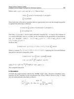
Three basic mechanisms, as summarized in Fig. 1, are involved in monophasic solutions:
thermophoresis, thermal convection, and thermoviscous expansion. A more anecdotic
alternative, involving a thermally-induced sol-gel transition, will also be briefly presented.
2.1.1 Thermophoresis
Thermophoresis, also called thermodiffusion, or Ludwig-Soret effect, takes place in solutions
submitted to a temperature gradient (Piazza & Parola, 2008; Würger, 2010). The macroscopic
effect is the creation, at steady state, of a concentration gradient overtaking the natural
smoothing due to the Brownian diffusion (Fig. 1(a)). While this effect has been
discovered in the mid-nineteenth century (independently by Ludwig and Soret), its theoretical
4
Advances in Microfluidics
Microfluidic Transport Driven by Opto-Thermal Effects 3
Hot
Cold
Thermophoretic drift
(a)
g
Diverging laser beam
Convection rolls
(b)
Scanning laser beamTemperature
Fluid
contraction
Fluid
expansion
Net flow
Temperature Scanning laser beam
Fluid
contraction
Fluid
expansion
Temperature-independant viscosity
Viscosity decreasing with temperature
(c)
Fig. 1. Schematic illustration of the three main mechanisms involved in monophasic
opto-thermal transport. (a) Thermophoresis of (here thermophobic) molecules, (b)
laser-induced convection, and (c) thermoviscous expansion (adapted after Weinert & Braun
(2008b)).
understanding is still controversial. The recent review by Würger (2010) provides significant
insight on the different mechanisms which can be involved.
From a phenomenological point of view, the motion of particles submitted to a temperature
gradient can be described as a thermophoretic drift of velocity
u
Soret
= −D
T
∇T,(1)
with D
T
the thermophoretic mobility. Note that the word ‘particle’ should be understood
here in a generic meaning, including both molecules, nanoparticles, microbeads, etc. Indeed,
while biomolecules will mainly be considered in the following, thermodiffusion applies to a
broad range of systems. Even though fundamental differences exist in the involved physical
mechanisms (more details can be found in (Würger, 2010)), the phenomenological description
we provide here keeps its generality.
Comparing thermophoresis to the Brownian diffusion leads to the definition of the Soret
coefficient,
S
T
=
D
T
D
,(2)
with D the Brownian diffusivity. This coefficient has the dimension of the inverse of a
temperature. It can be either positive or negative and then determines both the direction and
amplitude of the overall particle drift. To date, no unified theory is able to predict either
the sign or the order of magnitude of the Soret coefficient, which have been observed to
5
Microfluidic Transport Driven by Opto-Thermal Effects
4 Will-be-set-by-IN-TECH
usually depend on both solute and solvent parameters, as well as external conditions such
as temperature (Piazza & Parola, 2008; Würger, 2010). The theoretical background aiming
at describing the fluidic thermophoresis is built upon two main approaches. On the one
hand, hydrodynamic descriptions rely on the hypothesis of quasi-slip flow at the particles
boundary (Weinert & Braun, 2008a; Würger, 2007). On the other hand, at the microscopic
scale, thermodynamic approaches assume the local thermodynamic equilibrium to account
for solvent diffusivity and fluctuations (Duhr & Braun, 2006b; Würger, 2009).
A positive value of the Soret coefficient thus corresponds to a migration toward the colder
regions (‘thermophobic’ behavior, as shown on Fig. 1(a)). Conversely, a solute with a
negative Soret coefficient will be said ‘thermophilic’. For DNA in aqueous buffer solution
Braun & Libchaber (2002) measured S
T
= 0.14 K
−1
at room temperature, but this coefficient
has been observed to change of sign with temperature (Duhr & Braun, 2006b).
From an experimental point of view, the study of thermophoresis requires (i) to apply a
temperature gradient to the test cell, and (ii) to detect and measure the resulting concentration
distribution. Optical methods are indeed well suited to fulfill these two requirements. First,
as already pointed out, a much higher temperature gradient can be produced by direct laser
heating of the fluid than by externally heating the cell boundary. Second, the same laser
beam can also be used to characterize the concentration gradient. One possible method relies
on the thermal lensing effect: as the concentration gradient created by thermal diffusion
modifies locally the refractive index of the solution, the transmitted beam is either focused
or spread (effect called ‘Soret lens’), depending on the direction of the solute migration
(Giglio & Vendramini, 1974). An alternative method makes use of a fluorescent marker
grafted to the particles of interest, or of the particles fluorescence themselves if applicable,
to reconstruct the concentration profile in real time by microscope imaging (Duhr et al., 2004).
Moreover, the temperature profile can also be monitored by using a temperature-dependant
fluorescent marker.
2.1.2 Thermoconvection
Thermoconvection relies on the difference of density of an homogeneous fluid heated
inhomogeneously. As density usually decreases with temperature, the local heating of a fluid
leads to its dilatation. Considering the heating induced by a collimated laser beam with radial
symmetry, the thermal expansion would also be axisymmetric, and no net flow would appear
even in the case where the laser beam moves. Inducing a net flow in this case would require to
break the heating symmetry. This can be done if the laser beam is divergent, as shown in Fig.
1(b): the fluid more heated at the bottom side raises up by buoyancy, then loses its heat and
falls down, creating convective rolls (Boyd & Vest, 1975). This mechanism, generally known as
Rayleigh-Bénard convection, is involved in many processes at the macroscopic scale, ranging
from the cooking of pasta to atmospheric currents. However, in the micrometer-scale, gravity
is not the predominant force, and the heating symmetry breakup induced by gravity is rather
limited because the Rayleigh number, which controls the convection onset, behaves as the
cube of the heated layer width. In that sense, thermal convection is usually not relevant at this
scale. Microfluidic applications of thermoconvection can nevertheless be developed provided
that the sample is thick enough, or that the other forces (essentially, viscous or capillary) can
be efficiently reduced.
6
Advances in Microfluidics
Microfluidic Transport Driven by Opto-Thermal Effects 5
2.1.3 Thermoviscous expansion
Another elegant means of breaking the heating symmetry to induce a net flow has
recently been proposed by Weinert & Braun (2008b). This method relies on the temperature
dependance of viscosity of the fluid submitted to a scanning heating beam (Fig. 1(c)).
Let us consider a confined fluid, in which the influence of gravity is negligible. We first assume
that the fluid viscosity does not depend on temperature, as shown on the top part of Fig. 1(c).
As the laser beam moves, the fluid at the front of the spot scanning expands due to its decrease
in density while, on the other hand, the fluid at the rear of the spot scanning contracts as well.
As this thermal expansion is a linear process, expansion and contraction balance, and no net
fluid flow is produced.
Let us now add the temperature dependence of fluid viscosity. As viscosity usually decreases
with temperature, the expansion and contraction processes will be favored in the heated
regions, as shown on the bottom part of Fig. 1(c). This dissymmetry results in a net flow,
directed in the direction opposite to the scanning.
As thermal diffusion is faster, by several orders of magnitude, than the fluid flow, the fluid
warms and cools down in milliseconds, so scanning can be operated at rates in the kiloHertz
range. The resulting pump velocity can be expressed in a simple manner, dropping a
numerical prefactor of order unity, as (Weinert & Braun, 2008b)
u
thermoviscous
= scanning velocity × thermal expansion × thermal decrease of viscosity
= − f
th
αβT
2
.(3)
In this expression f is the scanning rate,
th
the heating spot length scale, T the temperature
rise, α
=(1/ρ)(∂ρ/∂T) and β =(1/η)(∂η /∂T) the thermal expansion coefficient and
temperature dependance with temperature, respectively. For water, α
= −3.3 × 10
−4
K
−1
and β = −2.1 × 10
−2
K
−1
, then considering a heating spot size of 30 μm, a scanning rate of
5 kHz and a temperature rise of 10 K lead to a pump velocity of 104 μms
−1
.
2.1.4 Thermally-induced sol-gel transition
An alternative way, inducing a local phase transition in the fluid, should also be mentioned.
As the thermoviscous expansion presented above, this approach relies on a thermally-induced
change in the fluid viscosity, but in the framework of a phase change. Krishnan et al. (2009)
used a thermorheological fluid (water containing 15 % w/w of Pluronic F127, a tribloc
copolymer) flowing in a channel including an absorbing substrate. The laser heating induced
a reversible gelation of the fluid, resulting in the interruption of the flow. A flow switch
without any moving part was then achieved. A similar approach was also used to perform
fluorescence-activated cell sorting (Shirasaki et al., 2006).
2.2 Channel-free microfluidic flows
The direct manipulation of volumes of fluid allows for the controlled creation of arbitrary
flows without the need of a rigid microfluidic channel. In particular, Weinert & Braun (2008b)
have shown that flows can be driven along complex patterns by thermoviscous pumping
(Fig. 2). As illustrated in Fig. 2(a), an infrared laser beam writing the words ‘
LASER PUMP’can
7
Microfluidic Transport Driven by Opto-Thermal Effects
6 Will-be-set-by-IN-TECH
(a)
(b) (c)
Fig. 2. Examples of channel-free flows performed by thermoviscous expansion. (a) A water
flow is induced by a laser beam scanning in the opposite direction, and visualized by
fluorescent tracer particles. From Weinert & Braun (2008b). (b) Thermoviscous motion in an
ice sheet: the ice melts in front of the heating spot, and moves in the same direction due to
the thermal expansion. The image is a superposition of frames of the molten spot along a
curved path, shown in dashed line. From Weinert et al. (2009). (c) Optical creation of a
dilution series of biomolecules. The system is composed of two neighboring gels, one of
them (at the bottom) containing fluorescein-marked biomolecules. The laser beam first
creates a liquid channel including three chambers of distinct volumes (upper panel), then
mixes the content of these chambers with the ambiant fluid by pumping along a pattern
alternating horizontal and vertical stripes. From Weinert & Braun (2008b).
produce a flow, in a 10-μm-thick water layer, in the direction opposite to the laser scanning.
Due to the very small thickness of fluid involved, the thermoconvection cannot be invoked as
a driving mechanism in this case.
The thermoviscous paradigm has also been extended to the case of melting ice (Weinert et al.,
2009). In that particular case, the scanning laser first melts the ice, the liquid motion is then
driven by thermal expansion, and finally the liquid refreezes (Fig. 2(b)). The motion can be
described as a thermoviscous pumping in the case where the water does not freeze in the
channel, when the chamber is cooled above 0
◦
C. However, as the water density increases
with temperature below 4
◦
C the fluid flow takes same direction as the scanning. Pumping
velocities of several cm s
−1
can be reached.
The creation of fluid flows along arbitrarily complex patterns can, in principle, provide an
alternative to the design of rigid dedicated channels. To highlight the potentialities of the
method for (bio)chemical applications, Weinert & Braun (2008b) created a dilution series by
8
Advances in Microfluidics
Microfluidic Transport Driven by Opto-Thermal Effects 7
thermoviscous expansion. To this aim, they used a drop of agarose gel, gelated at room
temperature, and molten by moderate heating (Fig. 2(c)). Biomolecules (30 kDa dextran
marked with fluorescein) were added at the bottom part of the drop only, with a large amount
of saccharose in order to avoid diffusion across the interface between the two halves of the
gelated drop. The laser first draws a liquid channel along the two parts of the gel, creating in
particular three liquid chambers of 65, 40, and 20 pL, respectively, in the upper part (initially
without biomolecules). This step is represented in the upper row of Fig. 2(c). In a second step
(lower row of Fig. 2(c)), the laser scans the gelated zones surrounding these chambers, along
successive crossing lines. This scan enlarges the actual chambers, and dilute the biomolecules
by mixing them with the molten agarose gel. As a result, a dilution series is created, with
volume ratios of 4:1, 1:1, and 1:4 in equal volumes.
2.3 Manipulation of biological molecules: Diluting, trapping, replicating, and analyzing
Besides setting in motion a fluid, manipulating directly molecules of biological interest which
might be dissolved in it is also of particular relevance. Such direct manipulation should indeed
allow for precise tuning of the molecule concentration, and, further, for inducing particular
reactions (especially, DNA replication).
2.3.1 DNA dilution or accumulation
As stated above, DNA exhibit a thermophobic behavior at room temperature
(Braun & Libchaber, 2002). Therefore, the laser heating of a buffer solution of DNA
deplete the zone at the vicinity of the spot due to the DNA thermophoretic drift, as illustrated
in the right image of Fig. 3(a). Thermophoresis is therefore a convenient way of locally
diluting a DNA solution. However, the most relevant issue is rather to concentrate molecules
at a given point. Duhr & Braun (2006b) observed that the thermophoretic behavior of
DNA could be reversed by simply cooling the sample: at 3
◦
C, the DNA molecules become
thermophilic and can therefore be trapped at the hot spot (left image of Fig. 3(a)). Besides this
very simple method, several alternatives exist to perform effective DNA trapping.
One elegant way consists in opposing a liquid flow to the thermophoretic drift (Duhr & Braun,
2006a). This method seems particularly relevant in the lab-on-a-chip context due to its
easy integrability into microfluidic channels. A 16-fold increase in DNA concentration
was reached, at about 10 μm upstream from the beam axis, with a peak flow velocity of
0.55 μms
−1
. However, the time required to reach the equilibrium concentration profile is
about 15 min, which limits the potentiality of the method for high-throughput applications.
By increasing the vertical temperature gradient effects, thermal convection can become
significant. Figure 3(b) represents the effective DNA trapping by the interplay between these
two mechanisms, as observed by Braun & Libchaber (2002). They considered a 50-μm-thick
chamber, in the center of which a heating beam was focused. The top and bottom walls of the
chamber were cooled to enhance the axial thermal gradient. The trapping mechanism is made
of four main steps. First, the lateral thermophoresis drives the DNA molecules away from
the heating spot (step 1). Then, the convection rolls carry the molecules downward, as the
upward part of the rolls occur in the depleted zone close to the beam axis (step 2). The axial
thermophoresis holds the DNA molecules at the chamber floor (step 3), where they finally
accumulate at a radial position which result from the balance between lateral thermophoresis
9
Microfluidic Transport Driven by Opto-Thermal Effects
8 Will-be-set-by-IN-TECH
(a) (b)
Fig. 3. Optothermal dilution and trapping of DNA. (a) Use of the temperature dependance of
the Soret coefficient to write complex DNA-enhanced or DNA-depleted patterns. When the
microfluidic chamber is cooled down to 3
◦
C the DNA molecules are thermophilic (left
picture), while they are thermophobic at room temperature (right picture). From
Duhr & Braun (2006b). (b) DNA trapping by a combination of thermophoresis and
thermoconvection. From Braun & Libchaber (2002).
and convection (step 4). The DNA molecules are therefore trapped in a ring-shaped pattern
around the laser beam axis. Braun & Libchaber (2002) observed a 60-fold local increase in
concentration at steady state, which is reached within 60 s, for a mean temperature of about
80
◦
C in the chamber. They even increased significantly the trapping efficiency, by using
a thicker chamber (500 μm) and a divergent laser beam, in a scheme comparable to that
represented in Fig. 1(b). The enhancement of the convection effect compared to the lateral
thermophoresis leads to a point-like trapping pattern along the beam axis. After 180 s, a
concentration increase by a factor of 2,450 was measured (Braun & Libchaber, 2002).
Another interesting trapping mechanism involves the combination of thermophoresis and a
bidirectional flow induced by thermoviscous expansion in a very thin (2 μm) liquid layer
(Weinert & Braun, 2009). Let us consider a vertical slice of this liquid layer, along the scanning
path of the laser beam. As the laser scans, say, from the right to the left, then the lower part of
the fluid will flow from the left to the right. However, the mass conservation applied together
with lateral boundary conditions impose a symmetric counterflow in the upper part of the
slice. Let us now reproduce at high rate the same scanning pattern, in such a way that each
slice draws a radius of a circular fluid pancake, as illustrated in Fig. 4(a). The resulting flow
pattern is therefore a toroidal roll, with a centrifugal flow at the bottom of the cell, and its
centripetal counterpart at the top. In the meantime, the vertical temperature gradient drives
the DNA molecules upward by thermophoresis. It means that the centrifugal flow concerns
rather DNA-depleted fluid, while the centripetal flow advects more DNA molecules toward
the center, where they accumulate.
As the scanning pattern is radial, the average trapping position is stationary. However, it can
be still moved by displacing the average position of the scanning laser, allowing to collect
particles over longer ranges. The so-called optothermal conveyor has been demonstrated
efficient with small beads as well as DNA molecules (Fig. 4(b)).
10
Advances in Microfluidics
Microfluidic Transport Driven by Opto-Thermal Effects 9
(a)
(b)
Fig. 4. Optothermal molecule conveyor (Weinert & Braun, 2009). (a) A radial centripetal laser
scanning leads to the efficient trapping of molecules by a combination of thermophoresis and
thermoviscous flow. The heating is provided at the bottom surface of the chamber by the
laser absorption by a thin chromium coating. Typical warm spot radius is 35 μm. (b) Optical
conveyor: the optothermal trap is moved along arbitrary patterns in order to collect particles
(40 nm polystyrene beads, top row) or DNA molecules (bottom row, DNA concentration is
given in color scale). After the laser has been switched off, the trapped objects are released
and diffuse freely (right frames). The scale bars are 100 μm.
2.3.2 DNA replication
The investigations reported above highlight the possibility of DNA accumulation by purely
thermal mechanisms. On the other hand, thermal convection has been shown to be able
to drive the replication of DNA. The most popular DNA replication mechanism used by
molecular biologists is polymerase chain reaction (PCR). Basically, each DNA molecule first
dissociates into two single strands when heated at 95
◦
C. In a second step (in the 55-65
◦
C
temperature range), pairs of short DNA fragments called primers, exhibiting sequences
complementary to that to be amplified, anneal at the end of each single strand. Each of
the two so-generated double helices finally elongate, by enzyme-activated replication of
the complementary part of the initial strand (72
◦
C). The process is then repeated cyclicly,
each reaction cycle doubling the concentration of DNA exhibiting the targeted sequence.
As pointed out by Braun et al. (2003), DNA molecules carried along a circular convection
streamline experience a cycling change in temperature which mimic the temperature pattern
11
Microfluidic Transport Driven by Opto-Thermal Effects
10 Will-be-set-by-IN-TECH
of a PCR. Mast & Braun (2010) then combined the trapping (by thermoviscous expansion
and thermophoresis) and convective replication mechanisms in a capillary. Beyond the
biotechnological interest of such a combination, it opens new perspectives for fundamental
studies on the molecular evolution of life. Indeed, the two pillars on which the Darwinian
evolutional theory relies, namely, the duplication of genetic material, and its storage against
molecular diffusion, are retrieved. Moreover, inhomogeneously heated microfluidic chambers
can be viewed as model systems reproducing the pores of hydrothermal rocks in the deep
oceanic floor, in which life could have originated (Braun & Libchaber, 2004).
2.3.3 Analysis of biomolecular binding
Understanding the interactions between biomolecules, or between a particular biomolecule
and its environment, is of crucial relevance for medical applications. Very recently, Braun
and co-workers have proposed the use of thermophoresis as a probe of these interactions.
As the thermophoretic properties of a solute depend on its interactions with the solvent (or,
more generally, with its environment), they indeed proposed to quantify the biomolecular
binding by accurately measuring the corresponding changes in thermophoretic depletion.
This method has thus been used to quantify the aptamer-target interactions in a buffer solution
(Baaske et al., 2010), and then generalized to various protein-protein and protein-ion binding
in buffer solutions as well as in more complex biological liquids (Wienken et al., 2010).
2.3.4 Single molecule s tretching
Besides the transport and analysis of DNA samples, several studies investigated the
stretching of individual DNA molecules under the action of a laser-induced thermal gradient.
Ichikawa et al. (2007) characterized the elongation of long DNA chains by the hydrodynamic
stresses arising from thermal convection. Jiang & Sano (2007) anchored a DNA molecule
by one or two ends and observed its deformation when located at a given distance from a
heating laser. One-end-anchored molecules appear elongated along the direction opposite
to the temperature gradient, while the two-end-anchored exhibit an arc-like shape when the
laser is approached between the two bonded points. As the authors ensured the convection
to be negligible, they interpreted the deformations to result from the thermophoretic drift of
the movable parts of the molecule. From their observations, they calculated a tension force of
about70fNfora3Kμm
−1
temperature gradient. These methods for studying the physical
properties of biopolymers compete with others, such as AFM, optical or magnetic tweezers,
by their contact- and probe-free characters.
3. Two-fluid flows
Let us now turn to the case of diphasic flows. We consider here, more generally, the case
of a liquid droplet (a possible microreactor) immersed in, or floating on, another fluid, both
of them being not miscible with each other. Controlling the microfluidic transport should
here mean, essentially, controlling the motion of these microreactors. Then, hereafter is an
overview of techniques to push, pull, divert, sort, or broadly speaking, manipulate droplets.
This section is divided into two parts. The first part deals with the direct manipulation of
droplets through interfacial tension effects. The second one gathers different approaches
involving one (or several) change(s) of state, in which at least two fluid phases are involved.
12
Advances in Microfluidics
Microfluidic Transport Driven by Opto-Thermal Effects 11
Liquid drop
Solid
HotCold
(a)
Solid block
Liquid pool
(b)
Liquid 2 Liquid (or gas) 1
Cold Hot
(c)
Fig. 5. Principle of thermocapillary migration, considering a surface (or interfacial) tension
decreasing with temperature. (a) A liquid partly wetting a heated solid substrate is driven
toward the colder regions by ‘rolling’ on the surface (as for a tracked vehicle). (b) A solid
block floating on a liquid pool and heated at its end is propelled in the opposite direction (the
schematic velocity field is in the laboratory frame). (c) An immersed droplet (or bubble)
migrates to the warm by a ‘swimming’ motion.
3.1 Optocapillary effect
A spatial unbalance of surface tension along a liquid surface creates surface stresses. This
effect, responsible for example for the phenomenon of ‘wine tears’, which can be observed
above the free surface of wine in a clean glass, has been evidenced by Marangoni in 1871 and
named after him. Over the past decade, this effect has known a renewed interest as it opens
important vistas on the fluid manipulation at small scales. These potentialities are justified by
the increased surface-to-volume ratio at small scales, which tends to favor interfacial effects
compared to volume effects.
Basically, Marangoni effect appears as soon as a gradient of interfacial tension
1
is created,
which can be achieved essentially through gradients of chemical composition or temperature.
The resulting effect is then related as ‘solutocapillary’ or ‘thermocapillary’, respectively.
For example, wine tears result from a gradient in ethanol concentration due to the excess
evaporation in the rising wetting film, which increases the surface tension in these zones.
Inhomogeneities in surfactant interfacial concentration can play the same role. Even though
solutocapillary effect can be particulary relevant in microfluidic systems, this section will
focus on thermocapillary effect—more precisely, ‘optocapillary’, i.e., thermocapillary effect
in the special case where the temperature gradient is optically induced. Nevertheless, the role
of surfactants can be significant, as we will see hereafter, to understand experimental features.
3.1.1 Thermocapillary migration: Basic principles
Creating an interfacial stress allows for setting in motion a fluid element (bubble, drop, liquid
film), which we call thermocapillary migration, providing that a condensed surrounding
medium can support this motion. For example, what follows would never happen if
1
The term ‘interfacial tension’ is related to a liquid-liquid interface, when ‘surface tension’ is used for
liquid-gas free surfaces. However, the phenomena presented in the following under the denomination
‘interfacial tension’ can be generalized to the free surface case.
13
Microfluidic Transport Driven by Opto-Thermal Effects
12 Will-be-set-by-IN-TECH
considering a free levitating droplet in vacuum. Thermocapillary migration can therefore
occur in the following cases, as reported in Fig. 5: a liquid element on a solid substrate
(Fig. 5(a)), or conversely a solid element floating on a liquid surface (Fig. 5(b)), or a fluid
element (either liquid or gas) in a liquid (Fig. 5(c)). In all cases considered here, the interfacial
tension is supposed to decrease with temperature, so the driving temperature gradient leads
to an inverted interfacial tension gradient—and therefore, interfacial stresses opposed to the
temperature gradient.
In the first case (Fig. 5(a)), a partly wetting liquid drop is on an inhomogeneously heated
substrate—note that the drop could as well be heated instead of the substrate. The surface
stresses drive a surface flow at the droplet free surface, an opposite counterflow then results
from mass conservation. Due to the no-slip condition at the solid-liquid boundary, this
flow sets the drop in motion toward the high surface tension regions (Cazabat et al., 1990;
Darhuber et al., 2003). Note that the particulary case of liquids on solid surfaces offers several
additional degrees of freedom to spatially modulate the surface tension, including surface
texturation, chemical patterning, or electrocapillarity. Further improvements can be found in
the review by Darhuber & Troian (2005).
The symmetric case, less documented in the literature, involves an inhomogeneously heated
solid, floating on a liquid surface (Fig. 5(b)). The surface stresses symmetrically drive the fluid
on either sides of the hot point (creating a deeper counterflow as well). However, as the solid
is heated from one side, the surface flow carries it toward the less heated direction.
The case of immersed bubbles (Fig. 5(c)) has been first treated half of a century ago by
Young et al. (1959), then extended to droplets (Barton & Subramanian, 1989; Hähnel et al.,
1989). It seems to be the most relevant to droplet microfluidic systems as no solid part is
involved, avoiding any difficulty related to the liquid wetting (Chen et al., 2005). Here, the
interfacial stresses drive an interfacial flow, in both sides of the interface, toward the colder
part of the droplet. In the droplet, this flow creates internal rolls.
2
In the surrounding
medium, this flow drags the bulk fluid, which in turn propels the droplet in the opposite
direction according to the action-reaction principle. A simple analogy can be made with a
swimmer, who drags fluid backward and then moves forward. Young et al. (1959) quantified
the thermocapillary migration velocity of a bubble, and extended their calculation to droplets,
in an infinite medium:
u
thermocapillary
= −
2
3η
1
+ 2η
2
R
2 + Λ
1
/Λ
2
∂σ
∂T
∇T.(4)
Here, R is the drop radius, Λ the thermal conductivity, and σ the interfacial tension. The
subscripts 1 and 2 denote the droplet and surrounding phases, respectively. Considering a
water droplet (η
1
= 1mPasandΛ
1
= 0.6 W K
−1
m
−1
), of radius 100 μm, in silicone oil
(η
2
10 mPa s and Λ
2
0.1 W K
−1
m
−1
), with ∂σ/∂T ∼−0.1 mN m
−1
K
−1
, a temperature
gradient of 1 K mm
−1
leads to a migration velocity of 110 μms
−1
.Thisvaluerisesupto
5mms
−1
in the case of a gas bubble in water.
Beyond this ideal background, two main disturbing effects must be taken into account in
microfluidic environments. First, the confining effect of channel walls would quantitatively
2
The bubble case significantly differs: as gas is inviscid, the interfacial flow does not diffuse in the bulk
gas phase, and then no flow is produced.
14
Advances in Microfluidics
Microfluidic Transport Driven by Opto-Thermal Effects 13
or qualitatively modify the physics of thermocapillary migration. Considering a squeezed
bubble in a Hele-Shaw cell, Bratukhin & Zuev (1984) showed that the friction to walls
reduces the migration efficiency, but the scaling u
thermocapillary
∼−R∇T remains valid.
The case of elongated bubbles in capillary tubes has then been treated both theoretically
(Mazouchi & Homsy, 2000; 2001) and experimentally (Lajeunesse & Homsy, 2003). In the case
of a polygonal cross-sectioned channel, they evidenced the strong influence of liquid flow
through both corners and wetting films on the migration velocity. Especially, for a rectangular
cross section the migration velocity then varies non-trivially with the channel aspect ratio,
and no longer depends on the bubble size, due to the fluid recirculation through the corners.
Furthermore, the recirculating flows through the wetting films add nonlinear corrections.
On the other hand, diphasic microfluidics often involves surfactants. Several
investigations, both experimental (Barton & Subramanian, 1989; Chen et al., 1997) and
theoretical (Kim & Subramanian, 1989a;b), have shown that insoluble surfactants tend to
reduce the thermocapillary migration efficiency. A recent theoretical study by Khattari et al.
(2002) has proposed to account for the surfactant effect by writing an ‘effective’ variation of
interfacial tension with temperature, which for insoluble surfactants reads as
∂σ
∂T
eff
=
∂σ
∂T
Γ
+
∂σ
∂Γ
T
∂
2
σ
∂Γ
2
T
1
T
∂σ
∂Γ
T
−
∂
2
σ
∂T∂Γ
,(5)
with Γ the interfacial concentration. Although difficult to link with measurable quantities,
the corrective term accounting for surfactant effect is expected to be positive as it reduces the
effective variation of interfacial tension.
What was described above is related to thermocapillary effect in general. Let us now focus
more specifically on optocapillary effect.
3.1.2 Optocapillary propelling
The three configurations depicted in Fig. 5 have been experimentally explored in an
optocapillary scheme, as illustrated in Figs. 6 and 7.
The optocapillary migration of a liquid object (a film) on a heated solid surface is represented
in Fig. 6(a). Garnier et al. (2003) considered an horizontal film, inhomogeneously heated by
a light pattern which superimposes a gradient of intensity perpendicular to the contact line,
and a sinusoidal intensity fluctuation along it. As a result, the contact line, mostly the less
enlighten part, moves toward the darker edge, drawing the wavy-like pattern shown on Fig.
6(a). By varying the spatial periodicity of the illumination, the authors studied quantitatively
the contact line instability.
Besides this study, relatively few publications are related to the optocapillary migration in
the ‘rolling droplet’ configuration. However, this configuration is well adapted to the optical
heating through surface plasmon decay (Farahi et al., 2005; Passian et al., 2006).
Thermocapillary migration of a solid floating object, within a scheme as represented in Fig.
5(b), is practically difficult to achieve since this requires the heating of a small movable
object—heating the pool itself is however possible. Laser-induced heating is therefore a
15
Microfluidic Transport Driven by Opto-Thermal Effects