Autonomic Nervous System in Old Age pot
Bạn đang xem bản rút gọn của tài liệu. Xem và tải ngay bản đầy đủ của tài liệu tại đây (3.65 MB, 148 trang )
Autonomic Nervous System in Old Age
Interdisciplinary Topics in
Gerontology
Vol. 33
Series Editors
Patrick R. Hof, New York, N.Y.
Charles V. Mobbs, New York, N.Y.
Editorial Board
Constantin Bouras, Geneva
Christine K. Cassel, New York, N.Y.
Anthony Cerami, Manhasset, N.Y.
H. Walter Ettinger, Winston-Salem, N.C.
Caleb E. Finch, Los Angeles, Calif.
Kevin Flurkey, Bar Harbor, Me.
Laura Fratiglioni, Stockholm
Terry Fulmer, New York, N.Y.
Jack Guralnik, Bethesda, Md.
Jeffrey H. Kordower, Chicago, Ill.
Bruce S. McEwen, New York, N.Y.
Diane Meier, New York, N.Y.
Jean-Pierre Michel, Geneva
John H. Morrison, New York, N.Y.
Mark Moss, Boston, Mass.
Nancy Nichols, Melbourne
S. Jay Olshansky, Chicago, Ill.
James L. Roberts, San Antonio, Tex.
Jesse Roth, Baltimore, Md.
Albert Siu, New York, N.Y.
John Q. Trojanowski, Philadelphia, Pa.
Bengt Winblad, Huddinge
Autonomic Nervous
System in Old Age
Volume Editors
George A. Kuchel, Farmington, Conn.
Patrick R. Hof, New York, N.Y.
11 figures and 9 tables, 2004
Basel · Freiburg · Paris · London · New York ·
Bangalore · Bangkok · Singapore · Tokyo · Sydney
George A. Kuchel, MD FRCP
UConn Center on Aging
University of Connecticut Health Center
Farmington, Conn., USA
Patrick R. Hof, MD
Associate Professor, Kastor Neurobiology of Aging Laboratories
Dr. Arthur Fishberg Research Centre for Neurobiology
Mount Sinai School of Medicine
New York, N.Y., USA
Library of Congress Cataloging-in-Publication Data
Autonomic nervous system in old age / volume editors, George A. Kuchel, Patrick R. Hof.
p. ; cm. – (Interdisciplinary topics in gerontology, ISSN 0074–1132 ; v. 33)
Includes bibliographical references and index.
ISBN 3–8055–7685–4 (hard cover : alk. paper)
1. Autonomic nervous system–Pathophysiology–Age factors. 2. Autonomic nervous
system–Aging. I. Kuchel, George A. II. Hof, Patrick R. III. Series.
[DNLM: 1. Autonomic Nervous System–physiology–Aged. 2. Aging–physiology. WL
600 A939545 2004]
HQ1060.I53 vol. 33
[RC347]
362.6 s–dc22
[612.8Ј9]
2003064038
Bibliographic Indices. This publication is listed in bibliographic services, including Current Contents® and
Index Medicus.
Drug Dosage. The authors and the publisher have exerted every effort to ensure that drug selection and
dosage set forth in this text are in accord with current recommendations and practice at the time of publication.
However, in view of ongoing research, changes in government regulations, and the constant flow of information
relating to drug therapy and drug reactions, the reader is urged to check the package insert for each drug for
any change in indications and dosage and for added warnings and precautions. This is particularly important
when the recommended agent is a new and/or infrequently employed drug.
All rights reserved. No part of this publication may be translated into other languages, reproduced or
utilized in any form or by any means electronic or mechanical, including photocopying, recording, microcopying,
or by any information storage and retrieval system, without permission in writing from the publisher.
© Copyright 2004 by S. Karger AG, P.O. Box, CH–4009 Basel (Switzerland)
www.karger.com
Printed in Switzerland on acid-free paper by Reinhardt Druck, Basel
ISSN 0074–1132
ISBN 3–8055–7685–4
Contents
VII Preface
1 Age-Related Sympathetic Autonomic Neuropathology.
Human Studies and Experimental Animal Models
Schmidt, R.E. (St. Louis, Mo.)
24 Clinical and Therapeutic Implications of Aging Changes in
Autonomic Function
Ford, G.A. (Newcastle upon Tyne)
32 Normal and Pathological Changes in Cardiovascular
Autonomic Function with Age
Attavar, P.; Silverman, D.I. (Farmington, Conn.)
45 The Autonomic Nervous System and Blood Pressure
Regulation in the Elderly
Bourke, E. (Brooklyn, N.Y.); Sowers, J.R. (Columbia, Mo.)
53 Aging, Carbohydrate Metabolism and the Autonomic
Nervous System
Madden, K.M.; Meneilly, G.S. (Vancouver)
67 Aging and the Gastrointestinal Tract
Pilotto, A. (San Giovanni Rotondo); Franceschi, M. (Schio);
Orsitto, G.; Cascavilla, L. (San Giovanni Rotondo)
78 Structure and Function of the Aged Bladder
Tannenbaum, C. (Montréal); Zhu, Q.; Ritchie, J.; Kuchel, G.A. (Farmington, Conn.)
V
94 Impact of Aging on Reproduction and Sexual Function
Beshay, E.; Rehman, K.-u.; Carrier, S. (Montreal)
107 Aging of the Autonomic Nervous System. Pain Perception
Lussier, D. (Montreal); Cruciani, R.A. (New York, N.Y.)
120 Aging and Thermoregulation
McDonald, R.B.; Gabaldón, A.M.; Horwitz, B.A. (Davis, Calif.)
134 Author Index
135 Subject Index
Contents
VI
Preface
In recent years, all western industrialized countries, and to a growing extent
even many developed and developing Asian nations, have witnessed a remarkable
growth in numbers of older people [1]. Future projections anticipate continued
increases, particularly in numbers of individuals who are 85 years and older [1].
Although US statistics have indicated recent declines in disability trends [2], overall numbers of older individuals living with disability and functional dependence
are likely to increase given projected increases in life expectancy [3]. For example,
average life expectancy for women born today in the United States is nearly 80; for
men, it is nearly 75 [1]. With these considerations in mind, many investigators have
begun to pay increasing attention to identifying factors which may predict the
transition from health and independence to disability and dependence in older
individuals, eventually providing useful targets for interventions [3, 4].
Neurodegenerative disorders such as Alzheimer’s and Parkinson’s diseases
are both common and important causes of cognitive and motor deficits in later
life. Moreover, the presence of cognitive and motor deficits resulting from these
disorders represents a major risk for the development of disability, dependence
and need for institutionalization among older individuals [1]. Thus, it is not at all
surprising that the central nervous system has received far more research attention than has the peripheral nervous system. Nevertheless, age-related changes
and diseases involving the peripheral nervous system, particularly its autonomic
elements, do frequently play determining roles in late life health and functional
independence.
Homeostasis, the need for the body to maintain a constant internal milieu,
was first defined by Claude Bernard in the mid 19th century [5]. In a 1932 book,
VII
Walter B. Cannon clearly recognized that as the body ages its ability to maintain
normal homeostasis in response to common challenges is altered [6]. In fact,
many of the physiologic parameters discussed by Cannon – temperature, blood
sugar and blood pressure – are all closely regulated by autonomic function and
are discussed in some detail in this book. However, our understanding of autonomic system aging and its role in human health and disability has increased a
great deal since the time of Bernard and Cannon.
Above all, modern clinical investigators typically study autonomic aging in
healthy older individuals and are thus able to dissect the contribution being made
by aging from that caused by disease. Such studies clearly indicate that while
basal sympathetic activity increases with normative aging, there is evidence of
considerable dysregulation in terms of the ability of the aging sympathetic
nervous system to respond to a variety of challenges. Moreover, markers of
elevated sympathetic activity appear to predict increased mortality among ill
[7, 8], as well as community dwelling independent older individuals [9, 10].
Although many questions remain unanswered, recent conceptual and technological advances have provided both the clinician and investigator with much
new information drawn from clinical, as well as basic research. In the following pages, investigators from several different disciplines discuss aging of the
autonomic nervous system from a variety of perspectives. Given the fact that
aging of the parasympathetic elements of the autonomic nervous system is not
nearly as well understood as that of its sympathetic portions, greater emphasis
has been placed on the latter. Some authors are basic scientists, while others are
clinical investigators, yet efforts have been made by all to begin bridging the
barriers between the two perspectives in a fashion that is meaningful to both.
In the first chapter, Dr. Schmidt discusses the major neuropathological and
cellular changes that have been described during autonomic aging in both
animal and human studies. Dr. Ford addresses the impact of physiologic
changes involving the autonomic nervous system, but does so from the point of
view of a clinical pharmacologist and clinician in describing the impact of agerelated changes in autonomic function on responses to common medications. In
Chapter 3, Drs. Attavar and Silverman discuss the impact of autonomic aging
on cardiac performance and the management of common cardiac conditions.
Drs. Bourke and Sowers focus their discussion on autonomic mechanisms
involved in the regulation of blood pressure and the impact of age-related
changes on the management of both hypertension and hypotension in older
individuals. Aging is associated with specific deficits in the body’s capacity to
handle glucose and the role of autonomic aging in these changes is addressed
by Drs. Madden and Meneilly. Many aspects of gastrointestinal function,
particularly motility, are closely influenced by autonomic function. Drs. Pilotto,
Franceschi, Orsitto and Cascavilla discuss the role of autonomic changes on
Preface
VIII
gastrointestinal performance in late life. Urinary incontinence is a major cause
of morbidity and disability in older individuals. Drs. Tannenbaum, Zhu, Ritchie
and Kuchel provide an overview of age-related changes in the autonomic
elements that closely regulate bladder performance and discuss their potential
roles in maintaining continence in older women and men. As discussed by
Drs. Beshay, Rehman and Carrier, both reproductive function and sexual
performance decline in advanced age, with autonomic changes providing a
contribution to both. The management of pain is a crucial element in improving the quality of life older patients and, as discussed by Drs. Lussier and
Cruciani, autonomic changes are among the many important considerations
needed to be brought into the assessment of an older individual in pain. Finally,
the inability of many older individuals to appropriately regulate their body temperatures in response to both high and low extremes of environmental temperature is a major risk factor for death. Drs. McDonald, Gabaldón and Horwitz
provide an excellent overview addressing a number of clinically important
questions by highlighting key clinical and basic research studies.
Clearly, the years since Claude Bernard’s first presentation of the concept
of homeostasis and Cannon’s comments regarding the influence of aging on
these mechanisms have witnessed a tremendous growth in our knowledge. At
the same time, the coming decade should lead to an even better understanding
of this area. This will take place as more ambitious and well-defined clinical
studies are undertaken and as the power of basic research is harnessed, particularly in terms of using genetically modified animals, with real efforts made to
move or translate knowledge between the two fields.
George A. Kuchel, Farmington, Conn.
Patrick R. Hof, New York, N.Y
References
1
2
3
4
5
6
Guralnik JM, Ferrucci L: Demography and epidemiology; in Hazzard WR, Blass JP, Halter JB,
Ouslander JG, Tinetti ME (eds): Principles of Geriatric Medicine and Gerontology. New York,
McGraw-Hill, 2003, pp 53–76.
Fries JF: Measuring and monitoring success in compressing morbidity. Ann Intern Med 2003;139:
455–459.
Guralnik JM, Fried LP, Salive ME: Disability as a public health outcome in the aging population.
Annu Rev Public Health 1996;17:25–46.
Fried LP, Tangen CM, Walston J, Newman AB, Hirsch C, Gottdiener J, et al: Frailty in older adults:
Evidence for a phenotype. J Gerontol A Biol Sci Med Sci 2001;56:M146–M156.
Grande F, Visscher MB: Claude Bernard and Experimental Medicine. Cambridge, Mass.,
Schenkman, 1967.
Cannon WB: The aging of homeostatic mechanisms; in Cannon WB (ed): The Wisdom of the
Body. New York, Norton, 1932, pp 202–215.
Preface
IX
7
8
9
10
Semeraro C, Marchini F, Ferlenga P, Masotto C, Morazzoni G, Pradella L, et al: The role of
dopaminergic agonists in congestive heart failure. Clin Exp Hypertens 1997;19:201–215.
Goldstein DS: Plasma catecholamines in clinical studies of cardiovascular diseases. Acta Physiol
Scand Suppl 1984;527:39–41.
Seeman TE, McEwen BS, Singer BH, Albert MS, Rowe JW: Increase in urinary cortisol excretion
and memory declines: MacArthur Studies of Successful Aging. J Clin Endocrinol Metab 1997;82:
2458–2465.
Reuben DB, Talvi SL, Rowe JW, Seeman TE: High urinary catecholamine excretion predicts mortality and functional decline in high-functioning, community-dwelling older persons: MacArthur
Studies of Successful Aging. J Gerontol A Biol Sci Med Sci 2000;55:M618–M624.
Preface
X
Kuchel GA, Hof PR (eds): Autonomic Nervous System in Old Age.
Interdiscipl Top Gerontol. Basel, Karger, 2004, vol 33, pp 1–23
Age-Related Sympathetic Autonomic
Neuropathology
Human Studies and Experimental Animal Models
Robert E. Schmidt
Division of Neuropathology, Department of Pathology and Immunology,
Washington University School of Medicine, Saint Louis, Mo., USA
Autonomic dysfunction is an increasingly recognized complication of
human aging and, as a result of the rising mean age of the human population,
has widespread ramifications for health care. Age-related autonomic neuropathy may produce clinical symptoms directly or result in subclinical disease,
complicate therapeutic intervention in a variety of diseases (e.g., sympatholytic drugs in hypertension, aggressive insulin therapy in diabetes) or
decrease the safety margin upon which superimposition of additional insults
(e.g., diabetes) produce symptomatic disease. Early studies of the function and
neuropathology of the autonomic nervous system in aged human subjects were
largely anecdotal and often contradictory. However, recent systematic studies
by a number of investigators have contributed substantively to the understanding of age-related autonomic dysfunction and its neuropathologic substrate.
The development and use of animal models of human aging have begun to
address pathogenetic mechanisms and intervention strategies in age-related
autonomic dysfunction.
The Aging Human Autonomic Nervous System
Clinical Studies
Clinical studies [reviewed in ref. 1–5] support a role for age-related autonomic dysfunction in: (1) temperature regulation and sudomotor responses [6]
which may lead to life threatening hypo- or hyperthermia; (2) bowel motility
[7, 8], presenting as ‘major gastrointestinal dysfunction’ in 27% of one series of
hospitalized elderly [8, 9]; (3) visual abnormalities [10, 11]; (4) bladder function; (5) fat metabolism; (6) water and electrolyte regulation; (7) maintenance
of blood pressure [12], and (8) cardiovascular reflexes [4, 10]. Cardiovascular
dysfunction in aging is particularly complex and multifactorial, involving
sympathetic [13] and parasympathetic [14] components as well as superimposed endorgan impairment [15–19]. Exposure of the aged sympathetic
nervous system to a variety of controlled experimental stresses may result in
diminished [20] or unchanged [21] responses, or, surprisingly, produce an
abnormally exaggerated [22, 23] (hyperadrenergic) response, observations hard
to reconcile with the simple loss of sympathetic or parasympathetic ganglionic
neurons. Alternatively, age-related autonomic dysfunction may involve interference with the complex integration of autonomic functions within autonomic
reflex pathways, which may take place in peripheral sympathetic ganglia [24]
or at a number of other sites in the autonomic nervous system.
Neuropathology
The neuronal populations of aged human paravertebral superior cervical
ganglion (SCG, fig. 1) and the prevertebral celiac (CG) and superior mesenteric
(SMG) ganglia, are well preserved in aged human subjects, a result supported
by a large autopsy series [25–27] and previous reports [28–30], although most
human studies to date have not used unbiased stereologic counting techniques.
Neuronal alterations described in aged human ganglia include decreased
catecholamine fluorescence, accumulation of lipopigment and, in some studies
[31], neurofibrillary tangles. The demonstration of perivascular and parenchymal lymphocytic infiltrates in postmortem sympathetic ganglia, widely interpreted as evidence of an autoimmune process (e.g., diabetic autonomic
neuropathy, idiopathic orthostatic hypotension), failed to correlate statistically
with age, sex, diabetes or any other disease parameter and may largely reflect
normal lymphocyte trafficking or a common aspect of the perimortem
course [27].
In contrast to the apparent preservation of sympathetic ganglionic neurons,
structural alterations in dendrites, axons and synapses have been consistently
identified in aged human sympathetic ganglia [25–27, 32–36]. The hallmark
pathologic alteration in aged sympathetic ganglia is neuroaxonal dystrophy
(NAD), a distinctive axonopathy characterized by dramatic 5–30 m axonal
swellings (fig. 2). Dystrophic axons arise from delicate preterminal axons as a
distal axonopathy or ‘synaptic dysplasia’ and displace the perikarya of principal
sympathetic neurons or their primary dendrites [25]. Two types of dystrophic
axons have been identified in aged human SMG [37]: most commonly, dystrophic axons contain neurofilamentous aggregates with a specific immunophenotype; and, less frequently, tubulovesicular elements. Quantitative studies
Schmidt
2
Brainstem
Eye
Salivary
glands
Cx
Superior cervical
ganglion
Heart
Greater splanchnic nerve
Stellate
ganglion
Th
Celiac ganglion
Stomach,
small intestine,
liver, spleen
L
Small intestine,
colon
Inferior
mesenteric
ganglion
Colon
bladder
Superior mesenteric
ganglion
Prevertebral ganglia
Sympathetic paravertebral
(chain) ganglia
(All chain ganglia provide vasculomotor,
pilomotor, and sudomotor innervation)
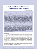
Fig. 1. The sympathetic nervous system. Only one of two paravertebral chains of
ganglia are depicted. Figure modified from M.B. Carpenter: Human Neuroanatomy, ed 7,
Baltimore, Williams & Wilkins, 1976, p 192.
Age-Related Sympathetic Autonomic Neuropathology
3
a
b
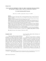
Fig. 2. Neuroaxonal dystrophy in aged human SMG. A markedly swollen dystrophic
axon (a, arrow) is intimately applied to the perikaryon of a principal sympathetic neuron.
Higher magnification demonstrates skeins of misoriented neurofilaments and a peripheral
rim of dense core granules (b, arrow). a 2,740ϫ; b 8,300ϫ.
[27] have demonstrated a progressive increase in the frequency of NAD as a
function of age (increasing particularly after the age of 60), gender (males had
3-fold more dystrophic axons than females) and diabetes (suggesting a shared
pathogenetic mechanism between diabetes and aging).
Nerve terminals in the prevertebral ganglia represent the contribution of
neurons originating in the spinal cord, dorsal root ganglia, parasympathetic nervous system, other sympathetic ganglia or as intraganglionic sprouts, and from
retrogradely projecting intramural alimentary tract myenteric neurons, many of
which have a distinctive neurotransmitter or neuropeptide signature. Dystrophic
axons in aged human SMG are immunoreactive for tyrosine hydroxylase (TH),
dopamine--hydroxylase (DH) and neuropeptide Y (NPY) as well as trkA and
p75NTR (high-affinity NGF and low-affinity neurotrophin receptors, respectively) but not substance P, GRP/bombesin, CGRP or enkephalins [25, 26, 38,
39]. This immunophenotype is most compatible with an origin of dystrophic
axons from sympathetic neurons, intrinsic or extrinsic to the SMG. The total
number of NPY-containing delicate nondystrophic axons and nerve terminals
and perisomal DBH-containing processes of all sizes actually increased in the
aged SMG, a result which may reflect intraganglionic collateral axonal sprouting as well as axonal regeneration. NPY released by sympathetic nerve
terminals has been shown to inhibit presynaptic release of acetylcholine from
intracardiac parasympathetic nerve terminals [40], a process which, if operative
in sympathetic ganglia, could interfere with integration of nerve impulses
derived from a variety of sources. Age-related loss of preganglionic neurons in
Schmidt
4
the intermediolateral nucleus [41] may also contribute to the loss of subpopulations of axon terminals surrounding principal sympathetic neurons [29].
The neurofilaments (NF) which accumulate in dystrophic sympathetic
nerve terminals of aged human SMG consist almost exclusively of extensively
phosphorylated 200-kD NF-H epitopes [42]. Antisera directed against NF-L,
NF-M and nonphosphorylated epitopes of 200-kD NF-H preferentially label
sympathetic neuronal perikarya and principal dendrites and do not label
dystrophic axons, evidence against the origin of NAD from principal dendrites
or proximal perisomal portions of axons. Simultaneous immunolabeling of
phosphorylated NF-H proteins (dystrophic axons) and MAP-2 protein (a marker
for dendrites and cell bodies) also failed to demonstrate colocalization.
Peripherin, a 58-kD cytoskeletal element distinct from any NF subunit, colocalized with phosphorylated NF-H immunoreactivity in many dystrophic elements
in aged sympathetic prevertebral ganglia, a result which suggests a shared defect
in a degradative mechanism or the accumulation of a possible hybrid filament.
Recent work on cytoskeletal changes in diabetic somatic sensory neuropathy
have identified a similar hyperphosphorylation of NF protein, thought to reflect
increased activity of several MAP kinases [43].
The Autonomic Nervous System of Aged Experimental Animals
A variety of animal models have been developed in an attempt to determine
the pathogenetic mechanisms underlying age-related autonomic neuropathy.
Pathophysiological and Biochemical Studies
Heart rate and arterial blood pressure are abnormal in aged rats [44], a
finding thought to reflect age-related degeneration of cardiac noradrenergic
innervation [45], altered norepinephrine turnover [46], or loss of functioning
Ca2ϩ channels [47]. Thermoregulative abnormalities are a function of increased
sympathetic nerve traffic to brown fat in the presence of defective postreceptor
signal transduction [48]. Increased colonic transit time [48] in aged rats may
reflect dysfunction of local reflexes underlying effective peristaltic activity,
which are dependent on connections integrated in sympathetic prevertebral
ganglia. Abnormal bladder function in aged rats may reflect reduced afferent
input [49]. The sympathetic response of aged rats to a variety of experimental
stressors (e.g., reserpine, fasting, heating, immobilization stress) may reveal
pathology not present at their unstressed baseline [50–56].
Norepinephrine content (a coarse measure of sympathetic ganglionic
health) has been reported to be decreased in aged rat CG, SMG and hypogastric ganglia [57, 58], although the activities of catecholamine synthetic enzymes
Age-Related Sympathetic Autonomic Neuropathology
5
a
b
Fig. 3. Neuroaxonal dystrophy in aged rat SMG. A dystrophic axon (arrow, a)
containing a variety of subcellular organelles (seen at higher magnification in 2b) is adjacent to a principal sympathetic neuron and enveloped in a satellite cell process. a 4,310ϫ;
b 18,210ϫ.
TH and DH are not decreased [54, 59]. Choline acetyltransferase, an enzyme
marker predominantly located in presynaptic cholinergic elements, is variously
reported as unchanged or increased in aged rat SCG [54, 59]. Decreased activity of succinate dehydrogenase [60], an important enzyme involved in oxidative
phosphorylation, has been reported in aged rat SCG and CG/SMG and may
represent increased glycolytic pathway activity intended to compensate for
decreased oxidative metabolism; however, more recent studies have not found
an expected change in baseline cytochrome oxidase activity [61].
The sympathetic nervous system does not operate in a vacuum and its
alteration may interplay with the age-related changes in the parasympathetic
nervous system (e.g., cardiac-vagal chemoreflex hyperresponse and baroreflex
hypofunction [62]) which is understudied in aged experimental animals.
Neuropathology
The pathologic alterations of aged rat neurons of the sympathetic intermediolateral column prominently involved their dendritic structure [63, 64] rather
than neuron loss. The neuronal complement of the sympathetic ganglia and
hypogastric ganglia (a mixed sympathetic and parasympathetic ganglion) of
aged rodents is well preserved [65–69] as is the preganglionic trunk to the SCG
[70], evidence of preservation of the preganglionic sympathetic neurons.
As in humans, NAD represents a consistent hallmark of the aged sympathetic nervous system in rats [71] (fig. 3a, b), Chinese hamsters [72], and mice
[73, 74]. Sympathetic ganglia of aged rodents are valid models of aging in
human sympathetic ganglia. Both aged rodents and man: (1) develop NAD, but
Schmidt
6
not substantive neuron loss, involving preterminal axons and synapses in aged
sympathetic ganglia; (2) demonstrate a selectivity of NAD for prevertebral SMG
and CG relative to paravertebral SCG and stellate ganglia; (3) develop neuropathologic changes ultrastructurally, immunohistochemically and anatomically
identical to those in diabetics, and (4) demonstrate a predilection for NAD to
target some subpopulations of nerve terminals while completely sparing others.
In addition to NAD, there also may be concomitant alterations in the numbers of
normal intraganglionic nerve terminals [75], either increased or decreased
numbers, admixed with NAD. The dendritic arborization of intracellularly
labeled CG/SMG neurons of young adult mice was significantly more complex
and extensive than that of the SCG, and aged animals showed a relatively wellpreserved CG/SMG dendritic apparatus [73]. Aged mouse SCG neurons, however, appeared significantly smaller with regard to total dendritic length and
branching, in comparison to those of young animals, and exhibited short, stunted
dendritic processes, results which have also been reported in aged rat SCG [76].
Studies of the aged rat hypogastric ganglion, which is composed of an unusual
admixture of sympathetic and parasympathetic neuronal cell bodies, showed
decreased numbers of synapsin-immunoreactive nerve terminals in relation to
individual sympathetic neurons but normal numbers of nerve endings on parasympathetic neurons [75]. A detailed study of the sympathetic/parasympathetic
composite major pelvic ganglion and preganglionic elements in aged male rats
similarly identified reduction in the number of sympathetic preganglionic
neurons, alterations in their dendritic structure and complexity, and reduced
glutaminergic (but not glycinergic or GABA-immunoreactive) synaptic contact
nerve endings on sympathetic preganglionic neurons but not on parasympathetic
preganglionic neurons [77]. Serotonin- and TRH-immunoreactive nerve terminals were decreased on sympathetic preganglionic neurons innervating aged rat
major pelvic ganglion but not on parasympathetic spinal nuclei [77].
Recent studies in aged mice [73, 74] have demonstrated a novel,
pathologically distinct, marked dilatation of neurites (involving mostly axons but
including dendrites as well) by numerous vacuoles which has been designated
‘vacuolar neuritic dystrophy’ (VND) and is essentially confined to the aged
mouse SCG. Although the cervical sympathetic trunk (the preganglionic projection to the SCG) distant from the SCG never contained VND lesions, the majority of VND lesions in the aged SCG were lost following surgical interruption of
the cervical sympathetic trunk, a result which is consistent with a distal process
directed selectively against terminal axons and synapses. Intraneuronal injection
experiments also demonstrated loss of dendritic arborization and focal dendritic
swellings in the aged mouse SCG [73]. Sequential sectioning of ganglia and
ultrastructural demonstration of dendritic characteristics of some dystrophic
elements, suggested that VND in aged mouse SCG was not confined to axons
Age-Related Sympathetic Autonomic Neuropathology
7
and presynaptic elements. Rarely, VND arose from principal dendrites or from
aberrant spine-like processes directly from the neuronal perikarya. VND was
30- to 100-fold more frequent in the aged mouse paravertebral SCG than in the
prevertebral CG/SMG sympathetic ganglia of the same animals, again suggesting that the response of the sympathetic nervous system to age-related insults is
heterogeneous. Sequential sections of aged ganglia heavily involved by VND
demonstrated that most principal sympathetic neurons were contacted at some
point by NAD, that the majority of dystrophic lesions arose from preterminal
axons of essentially normal caliber and that multiple dystrophic elements often
arose from a single axon and surrounded individual neurons as a basket. The
ultrastructural appearance of individual VND lesions was identical in young and
aged mice, differing only in frequency. Surprisingly, the frequency of VND in
22- to 27-month-old NIA-supplied mice was strain dependent, varying as much
as 30-fold between DBA and C57BL6 strains, which represent the most and
least VND-involved strains, respectively. VND exhibited a prominent gender
effect (males had 3-fold more severe VND than females of a comparable age).
Caloric restriction in mice, which significantly extends lifespan, presumably as
a function of decreased oxidative stress, resulted in 70% fewer VND lesions than
in age- and sex-matched controls fed ad libitum [74].
In addition to dystrophic alterations involving axon terminals contacting
prevertebral principal sympathetic neurons, investigators have also reported an
apparent decrease in distal postganglionic sympathetic noradrenergic axons and
nerve terminals in a variety of target tissues including the rat heart, middle
cerebral artery, ileum, kidney, bladder, pineal gland, spleen, mystacial pad and
the cholinergic sympathetic innervation of sweat glands but not the iris or
submandibular gland [45, 65, 78–86]. Interestingly, the loss of norepinephrine
and serotonin innervation of aged guinea pig vasculature was accompanied by
an increase in the vasodilator neurotransmitters VIP and CGRP [87], suggesting
that attempting to correlate functional consequences of the loss of populations
of sympathetic axons in isolation may be problematic. A recent study of agerelated alterations in the innervation of gastrointestinal sphincters shows an
increase in the density of excitatory neurotransmitters norepinephrine and substance P as well as a decreased density of inhibitory substances VIP and CGRP
[88]. Other studies of aged rats demonstrated dendritic atrophy of the SCG
neurons innervating the middle cerebral artery – which was reversed by local
application of NGF [89] –, but not of those neurons innervating the iris [90].
A similar pattern of decreased NF gene expression has also been demonstrated
for SCG neurons projecting to the middle cerebral artery but not those distributed to the iris [90]. There is, therefore, no compelling evidence that sympathetic autonomic aging in rats is uniform, resulting in a global loss of peripheral
sympathetic endorgan innervation.
Schmidt
8
Alimentary dysfunction in aged rats may also reflect loss of enteric
neurons [91], which may vary in degree from one level of the gut to another
[92]. In addition, multiple subpopulations of enteric neurons may be differentially targeted by the aging process. In aged rats, significant loss in calbindinimmunoreactive neurons, which may represent intrinsic neurons with a sensory
function, contrasts with the relative preservation of serotonin-immunoreactive
myenteric neurons [93].
Postulated Mechanisms of Autonomic Nervous System
Damage with Age
There is little evidence for the wholesale loss of significant numbers of
neurons in aged autonomic ganglia. Instead, reproducible significant ganglionic
pathology involves dendritic alterations, changes in synapse number or structure
and NAD. Ganglionic pathology may be further complicated by the superimposition of significant losses of postganglionic sympathetic axonal projections or
synapse-selective processes, which may vary from one endorgan to another.
Although NAD is characteristic of age-related changes in sympathetic
ganglia, its distinctive pathology is not confined to aged sympathetic ganglia,
and may be found in a variety of other age-related (gracile nucleus), toxic
(bromophenylacetylurea, zinc pyridinethione intoxications), degenerative
(Alzheimer’s disease), genetic (infantile neuroaxonal dystrophy, HallervordenSpatz disease), metabolic (vitamin E deficiency) and neurotraumatic disorders
involving the central and peripheral nervous system of man and experimental
animals [94]. Mechanisms relevant to the pathogenesis of NAD in the relatively
simple aging peripheral nervous system may be extrapolated to a variety of
more complex disease processes in the central nervous system.
The mechanisms underlying age-related damage to the peripheral nervous
system remain largely unknown; however, several hypotheses have been
advanced [32].
Oxidative Injury
Oxidative stress results from a variety of physiologic and pathophysiologic
pathways (e.g., mitochondrial function, catecholamine metabolism, ischemia,
formation of glycated proteins) that may generate increased amounts of reactive
oxygen species in aged animals, particularly in nerve terminals. Coupled with a
reduction in antioxidant defenses (e.g., decreased levels of reduced glutathione,
glutathione peroxidase and superoxide dismutase activities) increased amounts
of reactive oxygen species are thought to contribute to a variety of age-related
insults to the nervous system. Experimental lipid peroxidation of rat brain
Age-Related Sympathetic Autonomic Neuropathology
9
synaptosomes results in alterations in membrane fluidity, lipid composition and
Naϩ-KϩATPase activity, similar to changes produced by aging itself, which
result in greater susceptibility of aged synaptic membranes to additional in vitro
lipid peroxidation [95]. Oxidative stress may directly damage the mitochondrial
genome resulting in dysfunctional mitochondria that produce increased amounts
of free radicals which leak into the surrounding cytoplasm or produce further
mitochondrial damage [4, 96, 97]. In support of this, reactive oxygen species
have been reported to produce oxidized proteins which accumulate in synaptic
mitochondria in old mice [98]. Increased indices of oxidative stress (tissue levels
of malondialdehyde, 4-hydroxynonenal (4-HNE), protein carbonyls and
decreased levels of GSH) have also been reported in the diabetic rat peripheral
nervous system [99] which develops ganglionic pathology similar to that in aged
ganglia. In a normal state, superoxide is degraded by superoxide dismutase;
however, if the amount of superoxide produced overwhelms this capacity, superoxide is converted to hydroxyl radical, a potent oxidant which targets a variety
of intracellular macromolecules, chief among them polyunsaturated fatty acids
resulting in the generation of 4-hydroxynonenal (4-HNE) [100, 101]. 4-HNE
binds to several amino acids in a variety of intracellular proteins, interfering with
their function. In addition, treatment of cultures with 4-HNE has been reported
to interfere with the function of proteosomes, nonlysosomal cytosomes that
function in the degradation of abnormal proteins [102], which may represent a
link between oxidative damage and accumulation of intra-axonal organelles that
represents a conspicuous characteristic of NAD.
Oxidative stress is closely associated with the development of NAD in
several clinical and experimental conditions. Deficiency of the antioxidant
vitamin E results in the premature and exaggerated development of NAD in
aged human and rat primary sensory axon medullary terminals [103], which is
sensitive to antioxidants and free radical scavengers. Studies in diabetic rats,
which develop NAD identical in ganglionic distribution and ultrastructural
appearance to that in aged rats, have provided additional support for oxidative
stress in the pathogenesis and treatment of diabetic neuropathy. Recent studies
[104] of diabetic autonomic neuropathy in rats have demonstrated that inhibitors
of selected portions of the polyol pathway result in substantially decreased
NAD (aldose reductase inhibitors) or significant worsening of NAD (sorbitol
dehydrogenase inhibitors), a result which parallels the known effect of these
agents to diminish or increase, respectively, markers of oxidative stress [105,
106]. Restriction of caloric intake (known to decrease oxidative damage in
rodents) [107], significantly decreases dystrophic synaptic pathology in aged
mouse SCG [74]. We have shown that increased sympathetic NAD in diabetic
rats is nearly eliminated by IGF-I treatment in doses too small to significantly
affect blood glucose levels [108], a result consistent with, although not limited
Schmidt
10
to, the antioxidant effect of IGF-I. IGF-I has also been reported to protect dorsal
root ganglion neurons from glucose-induced injury, a mechanism also known
to involve oxidative stress [109].
Deficiency of Neurotrophic Substances and Aging in the
Peripheral Nervous System
It has been proposed that the trophic support of endorgans on their innervating neurons may decline in old age due to decreased availability of targetderived neurotrophic substances [110–114] or alterations in receptor expression.
Transplantation of aged or young endorgan targets into the anterior eye chamber
of aged or young rats has demonstrated both target [109, 115]- and neuronderived defects [116]. Other studies have reported deficient sympathetic sprouting into aged hippocampus [117] or sweat glands [115]. Exogenous treatment
with NGF increased the sympathetic innervation density on both young and old
targets, although not to the same degree [116, 118]. SCG neurons giving rise to
the noradrenergic innervation of the middle cerebral artery, which decreases its
total innervation by half with age, are reported to show NGF-reversible dendritic
atrophy [119] in the absence of a decrease in NGF protein levels in the circle of
Willis [120]. NGF content of blood vessels, pineal gland, submandibular glands
and iris is not generally reduced in aged animals and age-related changes in
endorgan nerve density do not correlate accurately with endorgan NGF content
[114, 115, 120, 121]. Reinnervation of transplanted blood vessels by aged
neurons is increased by exogenously administered NGF, but to a lesser extent
than with young host neurons [116], which may reflect age-related decreased
neuronal plasticity. The aged sympathetic nervous system may show an impaired
response to low doses of NGF [114], although other studies suggest little decline
in the capability of aged neurons to respond to intraventricular NGF [122].
Exposure of sympathetic neurons to anti-NGF is reported to produce atrophy of
aged but not mature neurons, suggesting a decreased ability to scavenge NGF
with age [123]. Decreased levels of p75NTR (the low-affinity neurotrophin receptor) as well as mRNA for p75NTR and trkA, the high-affinity receptor responding primarily to NGF [112, 124] have been reported in aged sympathetic
ganglia. Other studies of aged rats have demonstrated dendritic atrophy and
decreased NF gene expression of the SCG neurons innervating the middle cerebral artery (reversed by local application of NGF) [89], but not of those neurons
innervating the iris [89, 90]. Neurons which innervate blood vessels are smaller
and exhibit lower levels of NGF uptake (which declines with age) in contrast to
iris-projecting neurons which are larger and take up greater amounts of NGF
(a process which does not decline with age) [125].
Some of the apparent discrepancies between experiments identifying a
target- or endorgan-derived defect in aged animals may reflect the differences
Age-Related Sympathetic Autonomic Neuropathology
11
between impaired collateral reinnervation in old animals [116], a process which
is neurotrophin sensitive [126], and the retained capacity for axonal regeneration in aged rats [127], a neurotrophin-insensitive process [111, 128]. Animals
with deficiency of sensory collateral sprouting (but not axonal regeneration),
result from the administration of a course of anti-NGF into neonatal rats or by
targeted disruption of p75NTR in mice [129]. Septal lesion-induced collateral
sprouting of sympathetic axons into the aged rat hippocampus is also reduced
in the presence of diminished hippocampal NGF upregulation [113, 130].
A physiologic defect in sprouting of uninjured noradrenergic fibers within the
pineal gland following extirpation of one SCG has been reported in aged in
comparison to young rats [131]. Cycles of synaptic degeneration and regeneration may have more in common with collateral sprouting than long distance
regeneration in terms of neurotrophin sensitivity, particularly if turnover involves
replacement of degenerated terminals with adjacent axonal sprouts. Synaptic
maintenance, plasticity, turnover, and collateral sprouting of axons may make
use of shared basic processes which are differentially sensitive to a variety of
neurotrophic substances.
Insulin and the insulin-like growth factors support the development and
growth of sympathetic neurons in culture [132]. Insulin-like growth factor I
(IGF-I) is thought to contribute to synaptic development, axonal sprouting and
regeneration [133–136]. Administration of exogenous IGF-I to diabetic rats
with established NAD in the SMG resulted in nearly complete reversal of NAD
after 2 months [108] in the absence of a salutary effect on the severity of
diabetes. The injury-induced increase in IGF-I content in the distal stump of
axotomized sciatic nerve is reportedly blunted in aging [137]. IGF-I deficiencies identified in both aging and diabetes [138, 139] could contribute to
abnormal synaptic turnover and the development of ganglionic NAD in both
conditions. Significantly, IGF-I is also known to protect DRG neurons against
oxidative insult by reactive oxygen species in vitro [109]. However, recent work
[reviewed in ref. 140] has suggested that the relationship of aging insults to
decreased signaling by IGFs may be more complex since reduced signaling by
insulin-like peptides has been shown to increase the life span of a number of
experimental species.
Neurotrophic Substances in Excess as a Pathogenetic
Mechanism for NAD
Alternatively, excessive amounts of neurotrophic substances may induce
uncontrolled neuritic growth. This mechanism has been suggested to explain the
neuritic swellings and apparent axonal sprouts in senile plaques of Alzheimer’s
disease which are rich in fibroblast growth factor (FGF) [141]. Neonatal sympathetic ganglia treated with 6-hydroxydopamine and high doses of NGF in vivo
Schmidt
12
a
b
Fig. 4. Association of NAD and regenerative axonal sprouts in aged rat SMG. A massively swollen dystrophic axon (arrow, a) is associated with regenerative axonal sprouts
(arrowheads, a), seen better at higher magnification in 3b (arrowheads). These delicate
(0.1–0.2 m) structures, similar those which originate from an axotomized parent axon in
peripheral nerve regeneration, presumably subserve a similar function within sympathetic
ganglia, although perhaps without the orientation supplied by Schwann cell tubes of regenerating peripheral nerve axons. a 4,950ϫ; b 32,420ϫ.
develop large intraganglionic swellings containing a variety of subcellular
organelles which are reminiscent of NAD and suggest a pathogenetic role for
coupled peripheral injury and increased ganglionic NGF [142]. Studies of autonomic neuropathy in diabetic rats have demonstrated that NAD identical to that
found in aged rat ganglia develops prematurely and with increased severity in the
diabetic prevertebral SMG and CG but not SCG [143]. Measurement of endogenous ganglionic NGF by ELISA [144] showed a doubling of NGF content in the
diabetic CG and SMG but no consistent effect in the SCG, a distribution which
parallels the development of ganglionic NAD. Systemic administration of
exogenous NGF to adult control rats for 3 months has been shown to produce a
doubling of NAD in the SMG [145]. Axonopathy may interfere with the retrograde transport of neurotrophic substances further contributing to a local excess
in endorgans and the development of a self-perpetuating cycle. Increased NGF
and other neurotrophins have also been shown to potentiate free radicalmediated neuronal death in some experimental paradigms [146–148].
Regenerative Mechanisms (Axonal Regeneration, Collateral Axonal
Sprouting, Synaptic Plasticity)
The ultrastructural resemblance of some dystrophic axons to growth cones
[94], the terminal motile tips of developing and regenerating axons, the frequent
association of NAD with regenerative axonal sprouts [149, 150] (fig. 4) and its
induction by frustration of peripheral axonal regeneration [151] suggest a relationship of NAD to abnormal axonal regeneration/collateral sprouting.
Age-Related Sympathetic Autonomic Neuropathology
13
Synaptic turnover, a continuous normal process which may represent the structural equivalent of synaptic remodeling or ‘plasticity’ [152, 153], may share
mechanisms with collateral sprouting (i.e., neurotrophin-sensitive sprouting of
uninjured axons into denervated targets) and axonal regeneration (neurotrophininsensitive regrowth of previously injured axons) [128]. Axonal regeneration
and, particularly, collateral sprouting are deficient in various organs of aged
animals [117, 126, 131, 154, 155]. Synaptic turnover in autonomic ganglia may
be further complicated in pathologic states by superimposed postganglionic
axotomy, which itself results in the detachment, swelling and retraction of
presynaptic elements, a process which may represent an exaggerated form of
normal synaptic turnover and may represent the substrate from which NAD
develops. Finally, regeneration of nerve terminals must eventually cease (i.e.,
initiate a ‘stop’ program) to reform a stable nerve terminal. The inhibition of the
stop program has been reported to result in swollen nerve terminals, reminiscent of NAD [156].
Synaptic Degradation of Organelles
NF undergo orthograde transport to the nerve terminal but are not returned
intact and, instead, undergo degradation by calcium-activated neutral proteases
(calpains). Postsynthetic modification of NF by glycosylation resulting in the
formation of advanced glycosylation endproducts [157, 158], a process which
is thought to operate in both aging and diabetes, or by excessive phosphorylation may change the sensitivity of NF to calpains and other proteases, which
could result in their excessive accumulation in axonal terminals.
Extracellular Matrix
Detailed studies [159] of the normal process of removal of supernumerary
neuromuscular junctions suggest a seminal role for alterations in the matrix and
postsynaptic elements in the loss of presynaptic nerve terminals. Neural cell
adhesion molecule (NCAM) may promote or inhibit synaptic plasticity or
stability as the result of alternative splicing or postranslational polysialation
[160]. Cultured aged SCG neurons exhibit diminished responsiveness to laminin
in the presence of NGF [161, 162] and reduced laminin immunoreactivity is
reported to correlate with decreased innervation (possibly due to a defect in
collateral sprouting) of middle cerebral artery walls of aging rats in vivo [163,
164]. Age-related alterations in the extracellular matrix are, thus, also capable of
affecting nerve terminal structure, function and plasticity. Conversely, sympathetic neurons cultured on an aged or young central nervous system frozen
section substrate (an environment with extracellular matrix and possible bound
neurotrophic substances) show region-specific but not age-related differences [165].
Schmidt
14