OPTICAL BIOSENSORS PRESENT AND FUTURE - PART 2 (end) pot
Bạn đang xem bản rút gọn của tài liệu. Xem và tải ngay bản đầy đủ của tài liệu tại đây (26.73 MB, 325 trang )
Optical Biosensors: Present and Future
F.S. Ligler and C.A. Rowe Taitt (editors)
9 2002 Elsevier Science B.V. All fights reserved
CHAPTER 10
GENETIC ENGINEERING OF SIGNALING
MOLECULES
AGATHA FELTUS, PH.D., AND SYLVIA DAUNERT, PHARM.D., PH.D.
Departments of Chemistry and Pharmaceutical Sciences
University of Kentucky
Lexington, KY, USA
In order to expand the capabilities of biosensors, there is a need to develop new
signaling molecules. This chapter focuses on molecules, produced through
genetic engineering, that combine the recognition element with a signaling
element (such as a fluorophore) in an effort to optimize the signal caused by the
binding of the analyte to the recognition element. These systems, while not
necessarily originally developed for an optical fiber, can be immobilized at the
tip of the fiber either through chemical attachment or entrapment behind a
membrane. Three different systems will be examined: fluorophore-labeled
binding proteins, FRET-based systems, and bacteria-based sensors. These
systems use optical signaling methods to reveal the binding event, taking
advantage of molecular biological techniques to optimize the signal. The
advantages and disadvantages of each system will be discussed, as well as the
current state of the art of these biosensors.
1. Technical Concept
In its simplest terms, a biosensor is a sensing system composed of a biological
recognition element and a transducer. Under the strictest definition of the term,
the transducer is responsible for converting the binding event into an electrical
signal. Bacteria that fluoresce upon analyte binding and fluorophore-labeled
binding proteins have been refered to as "reagentless biosensors" or "reagentless
biosensing systems" even though they are used as assays, rather than
307
Feltus and Daunert
immobilized in a sensor. There is no reason these sensing systems cannot be
used as the recognition/signaling element of a fiber optic biosensor, however.
These systems will be discussed in both contexts, i.e., both free in solution as
assays and after adapation to a fiber optic probe. In order to avoid confusion, we
will refer to the source of the optical signal as the signaling molecule.
I.I. Fluorophore-labeled binding proteins
As the name implies, a fluorophore-labeled binding protein consists of two
distinct moieties: the fluorophore and the binding protein. The fluorophore is
covalently attached to the binding protein through the protein's amino acid side
chains. Binding of the analyte to the binding protein causes a conformational
change in the protein structure which may result in altered optical characteristics
of the fluorophore (Figure 1).
Depending upon the actual environmental change experienced by the
fluorophore, this could result in an increase or decrease in the fluorescence
intensity, a change in the emission wavelength, or a change in the lifetime of the
fluorophore, depending upon the characteristic to be measured. These changes
are generally caused by two separate phenomena: an increased or decreased
polarity in the environment surrounding the fluorophore or rotational constraint
of the fluorophore. For example, if the fluorophore moves from a position of
high polarity (exposure to the buffer or the presence of local side chains from
polar amino acids) to one of low polarity (a position inside the protein), the
fluorescence will increase due to decreased quenching by the solvent molecules
or dissolved oxygen in the solvent. Likewise, rotationally constraining the
fluorophore's motion by trapping it inside the protein will increase fluorescence
by removing frictional energy loss caused by fluorophore movement. These
changes are difficult to predict beforehand and are often only revealed once the
protein has actually been labeled.
Having said this, even if the direction of fluorescence change cannot be
predicted, knowledge of the protein structure serves as a good starting point for
choosing the placement of the fluorophore. From the point of view of signal-to-
noise ratio, it is most advantageous to have a single fluorophore placed in a
location where a large environmental change can occur. Often, the most likely
location for such a change is near the binding site. Therefore, most initial studies
are conducted by labeling at a site near the binding site as determined from
observations of the crystal structure or from mutational studies.
Selective labeling of the binding protein is usually accomplished via labeling
through cysteine residues using sulfhydryl-selective fluorophores. (For examples
of commonly employed fluorophores, see Figure 2.) In order to create one-to-
one conjugates of fluorophore to protein, molecular biology is often necessary to
create recombinant proteins with unique cysteine residues. Using recombinant
308
Genetic Engineering of Signalling Molecules
Figure 1. Schematic of a fluorescently labeled protein sensing system. The protein is
labeled with an environmentally-sensitive fluorophore such that the binding of the
analyte changes the conformation of the protein, altering the solvation of the fluorophore.
a) In this example, amino acid 197 of phosphate binding protein (PBP) is located near
the binding pocket and will undergo a change in environment as PBP closes around its
ligand, phosphate. This can result in either b) an increase or c) a decrease in fluorescence
upon ligand binding. In some cases, the emission wavelength of the fluorophore can also
change.
DNA techniques, such as site-directed mutagenesis, all other cysteines in the
protein are removed and other residues that will be the site of attachment are
individually changed to cysteines. In doing so, care must be taken not to alter
any amino acids necessary for the proper functioning of the protein, such as those
residues involved in analyte binding or in oligomerization of the protein. This
entire process will be examined in greater detail in Section 3.1.
309
Feltus and Daunert
H2C-' C~ ~C~
"
II
0
N(CH 3)2
(Q-t 3(:1-t
2)2 N ~O H O_
O~ N~
II
0
IVD(X3
(OH 3OH 2)2 N
~
) O
O
HN ~ N C CH21
8Doll
1 ,,54 ~/g,~
Figure 2. Structures of some cysteine-reactive environmentally sensitive fluorophores.
The reaction with the protein takes place through the maleimide or iodoacetimide groups.
1.2. FRET-based systems
Another method which has been used extensively to develop sensing systems,
particularly in small volumes and inside living cells, is the use of FRET-based
sensing systems (Giuliano and Post, 1995; Giuliano and Taylor, 1998) (Figure 3).
Fluorescence resonance energy transfer (FRET) occurs when one fluorophore, a
donor, nonradiatively transfers its energy to a second fluorophore, the acceptor.
The acceptor then relaxes normally, producing light at its emission wavelength.
In order for this to occur, there must be a significant overlap between the
emission spectrum of the donor and the excitation spectrum of the acceptor. An
important property of FRET is that the rate of energy transfer between the donor
and the acceptor is proportional to the inverse sixth power of the distance
between the two fluorophores (FRET ~ l/r6). For most pairs the F~3rster radius,
310
Genetic Engineering of Signalling Molecules
Figure 3. FRET-based sensing system for cAMP based on protein kinase A (PKA). This
system consists of a cell line transfected with a vector coding for two fusion proteins:
PKA regulatory subunit-BFP (blue fluorescent protein) and PKA catalytic subunit-GFP
(green fluorescent protein). In the absence of cAMP, the regulatory and catalytic
subunits associate, bringing the BFP and GFP moieties in close proximity and allowing
FRET. The presence of cAMP dissociates the complex of regulatory and catalytic
subunits, disrupting FRET. Adapted from Zaccolo et al., 2000.
the distance at which the efficiency of energy transfer is 50%, is between 20A
and 50/~ (Lakowicz, 1983). This distance is comparable to the size of most
proteins, which allows FRET to be used when the distance between the two
fluorophores will be significantly changed by the binding event. This can occur
if either both fluorophores are attached to the same protein molecule and binding
of a ligand to the protein causes a conformational change that either shortens or
lengthens the distance between them, or if the donor is attached to one of the
binding molecules and the acceptor to the other. In the latter case, a donor
fluorophore attached to one of the components can transfer its energy to an
acceptor fluorophore attached to the other only while the two are closely
associated. An example of this is given in Figure 3.
FRET as a detection methodology has a number of advantages for biosenor
applications. Because the system employs the excitation wavelength of the
acceptor and the emission wavelength of the donor, the Stokes shift is more
pronounced than for fluorescence, leading to a lower background. Another
advantage of FRET is that the ratio of fluorescence intensities of the two
311
Feltus and Daunert
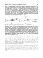
Figure 4. Schematic of a bacteria-based sensing system. The bacteria are transformed
with a plasmid containing the reporter gene under the control of an analyte-sensitive
promoter. In the presence of the analyte, the regulatory protein is released from the
promoter region, allowing transcription of the reporter gene. The mRNA is then
translated into protein, which can be assayed. The amount of protein produced is
proportional to the amount of analyte present, although there is amplification at each step
so that there are many more proteins present than reporter genes. Sometimes it is
necessary to also place the gene for the regulatory protein on the plasmid as well as the
reporter gene, as the native levels of reporter protein within the bacteria are insufficient
for proper regulation of transcription.
fluorophores can be used; this ratiometric technique is more accurate than
measuring just one fluorescence signal.
1.3. Bacteria-based sensing systems
Amplification-based methods take advantage of the high turnover of substrates to
produce a large number of product molecules. This is the basis of such
techniques as PCR and RT-PCR. In these cases, DNA or RNA molecules are
selectively amplified to quantify the numbers of their parent strands. Whole-cell
sensing systems take this one step further by first producing DNA, which is then
amplified again during the transcription to RNA, and finally amplified a third
time by translation to protein.
312
Genetic Engineering of Signalling Molecules
Table 1. Reporter proteins used in whole cell sensing systems.
Detection
Protein Gene Catalyzed reaction method*
chloramphenicol
acetyltransferase Cat acetylation of chloramphenicol RI, FL
CR, FL, EC,
[3-galactosidase LacZ hydrolysis of 13-galactosides CL
firefly luciferase Luc
bacterial luciferase LuxAB
aequorin AQ440
green fluorescent
protein GFP
luciferin + O2 + ATP
BL
oxyluciferin + AMP + PPi + h v
FMNI-I2 + R-CHO+ 02 -~ FMN +
BL
HE0 + RCOOH + h v
coelenterazine + 02 + Ca 2+ ~
BL
coelenteramide + CO2 + h t,
posttranslational formation of an
internal chromophore FL
* RI, radioisotope; FL, fluorescence; CR, colorimetric; EC, electrochemical; CL,
chemiluminescence; BL, bioluminescence.
A typical whole-cell sensing system consists of an organism, generally a
bacterium, that is transformed with a plasmid containing a reporter gene under
the control of a promoter responsive to the analyte of interest (Daunert et al.,
2000; Lewis et al., 1998; Ramanathan et al., 1997a). This plasmid may also
contain genes that will produce the necessary accessory proteins for the
promoter, such as the regulatory proteins. These additional genes are sometimes
necessary, as the number of promoters on the plasmid may greatly outnumber the
usual number of promoters; a larger number of regulatory proteins is necessary to
regulate these plasmid-borne promoters. Once the cells are exposed to analyte,
transcription of the reporter gene will begin (Figure 4). After transcription, the
RNA molecules are translated into protein. Amplification occurs at each of these
steps to produce many more protein molecules than there are reporter genes. If
desired, an extra level of amplification can be achieved if the reporter protein is
an enzyme that will turn over large numbers of substrate molecules. However, if
this further amplification is not required, then a protein such as the green
fluorescent protein (GFP) can be used. GFP does not require addition of an
external substrate, as the protein itself emits green fluorescence upon excitation
at 490 nm. Another way to obviate adding a substrate is to use the entire lux
cassette, instead of just
luxAB,
to produce bacterial luciferase. In this way all the
accessory proteins to produce the substrates necessary for bacterial luciferase
activity are also transcribed (Manen et al., 1997).
313
Feltus and Daunert
The sensitivity of these systems is determined by a number of factors. The
response of the promoter must be taken into account, but the largest effect of the
promoter/repressor protein is upon the selectivity of the system. The more
controllable factor is the choice of reporter protein, since there is often only a
limited choice of promoters for a given analyte. The ideal reporter protein will
be easy to use, have an easily discernable signal over the background, and have a
wide dynamic range and high sensitivity (Daunert et al., 2000). Examples of
reporter proteins that have been used to develop whole-cell sensing systems are
given in Table 1. Sensitivity can be a function of several factors, including the
detection method, efficiency of expression, reporter protein turnover number (if
the protein is an enzyme), and, if applicable, the endogeneous levels of the
reporter protein. For this reason, bioluminescent reporter proteins are a popular
choice because bioluminescence is not found in most organisms, and is a very
sensitive method of detection.
2. History
These three types of fluorescent signaling systems emerged from the need of
researchers in the biological sciences to study protein response to the binding of
various ligands. For example, bacteria-based sensing systems are the result of
experiments on regulation of transcription at various promoters.
2.1. Fluorophore-labeled proteins and FRET-based systems
These two systems share a common ancestry in studies of protein function. One
way of examining the structural changes in proteins upon ligand binding,
dimerization, or denaturation is by measuring in the native fluorescence of
tryptophan residues. This approach since they might not be close can be used to
measure binding only when the tryptophan is proximal to the active site. This
limitation led to the use of fluorescent cofactors and substrates, such as flavin
mononucleotide, to study changes occurring within the binding pocket. Later,
proteins were labeled with extrinsic fluorophores. Such labeled proteins have
been used for a number of applications, including microinjection into cells to
study protein localization and solution studies of protein structural changes.
Initially, biochemists used these fluorophore-labeled proteins to gain information
about the alterations in size, shape, and binding properties of proteins. However,
with the development of environmentally sensitive fluorophores and the ability to
produce mutated recombinant proteins, the fluorophore-labeled sensing system as
it stands today was born. Table 2 gives several examples of analytes that have
been measured using these systems. Most of the currently-developed sensing
systems of this type depend upon molecular biology to either create a unique site
for fluorophore attachment, to translate the protein such that it incorporates non-
native fluorescent amino acids, or to fuse a GFP to the protein.
314
Genetic Engineering of Signalling Molecules
Table 2. Examples of fluorophore-labeled protein sensing systems. In vitro/in vivo
refers to whether the protein is used in situ after being produced by the cells or whether
the proteins are expressed, isolated, and purified prior to use, and used as a sensing
system.
In vitro~in
Analyte Protein* vivo Reference
P~ PBP In vitro Brune et al., 1994, 1998
fatty acids I-FABP In vitro
Richieri et al., 1992
maltose MBP In vitro Gilardi et al., 1994
biotin Streptavidin In vitro
Murakami et al., 2000
Ca2+ CaM-YFP In vitro~in Baird et al., 1999
fusion vivo
Ca 2+ CaM-EGFP- in vitro~in Nakai et al., 2001
vivo
M 13 fusion
Ca z+ CaM in vitro
Co 2+, Zn 2+,
Carbonic
in vitro
Cu 1+ anhydrase
Salins et
al.,
1998;
Schauer-Vukasinovic et
al., 1997
Thompson et al., 1998
Salins et al., 2001;
glucose GGBP in vitro
Tolosa et al., 1999
Dattelbaum and
glutamine GlnBP in vitro
Lakowicz, 2001
*Abbreviations: PBP, phosphate binding protein; I-FABP, intestinal fatty acid binding
protein; MBP, maltose binding protein; CaM, calmodulin; YFP, yellow fluorescent
protein; EGFP, enhanced green fluorescent protein; GGBP, galactose/glucose-binding
protein; GlnBP, glutamine binding protein
FRET-based systems can be considered as a subclass of the fluorophore-labeled
proteins, different only because they depend upon the proteins being labeled with
two fluorophores rather than one. Because FRET is a distance-dependent
phenomenon, it was originally used to study assembly of multi-subunit protein
complexes, such as ribosomes, or interaction between a protein and cellular
membranes. In the 1990s, however, FRET-based systems started to be used for
analytical purposes (Table 3). The most recent trend is to use GFP and its
wavelength-shifted mutants as the donor and acceptor molecules.
315
Feltus and Daunert
Table 3. FRET-based assays using labeled proteins.*
Analyte Protein I)'onor Acceptor Reference
factor Xa factor Xa site " BFP rsGfp Mitra et ai.',' 1996
caspase-3 Caspase-3 site BFP GFP Xu et., 1998
caspase-3 Caspase-3 site CFP YFP Jones et al., 2000
Zn 2+
Ca 2+
zinc finger Lissamine rhodamin Godwin and Ber,
peptide e 1996
aequorin Aequorin GFP Baubet et al., 2000
Ca 2+ CaM BFP GFP
Romoser et al., 1997
Ca 2+
PKA
Cam/M13 BFP/CFP GFP/YFP Miyawaki et al.,
1997
KID B FP GFP Nagai et al., 2000
CAMP PKA fluorescei rhodamin Adams et al., 1991
n e
CAMP PKA BFP/CFP GFP/YFP Zaccolo et al., 2000
*Abbreviations: BFP, blue fluorescent protein; GFP, green fluorescent protein; CFP,
cyan fluorescent protein; YFP, yellow fluorescent protein; CaM, calmodulin; PKA,
protein kinase A; KID, kinase inducible domain.
Because these fluorophores are proteins themselves, plasmid constructs can be
made that fuse the GFP to the sensing protein, allowing these proteins to be
produced within a cell and used
in situ
as sensors. This is advantageous, since
the analytes of interest are generally intracellular second messengers. Moreover,
the need to microinject purified chemically-labeled proteins is avoided.
2.2. Bacteria-based sensing systems
Sensing systems of this type trace their origin back to bioassays in which
nutrient-deficient or antibiotic-resistant strains were plated on media containing
various concentrations of the analyte and the surviving number of cells counted.
From this methodology evolved the non-specific bacteria-based sensing systems.
These bacteria constitutively express a reporter protein while alive, but as toxins
begin tokill the bacteria, the protein is no longer produced, giving a lower signal.
At the same time, the bacterial operons were discovered, and reporter genes were
316
Genetic Engineering of Signalling Molecules
Table 4. Promoters used to develop whole-cell sensing systems
Analyte/target
response
i ii
Promoter References
antimonite/arsenite Ars
copper cupl
cadmium Cad
lead Pbr
mercury Mer
linear alkanes
PatkB
toluene Pu
isopropylbenzene ipbo/p
naphthalene/salicylate
Pnah
chlorocatechols Pc~c
L-arabinose
PBAD
[3-lactose
Plac
DNA damage recA, uvrA,
umuC
Corbisier et al., 1993; Ramanathan et al.i
1997b, 1998; Scott et al., 1997;
Tauriainen et al., 1997
Corbisier et al., 1999; Shetty et al., 2000
Tauriainen et al., 1998
Corbisier et al., 1999
Virta et al., 1995
Sticher et al., 1997
de Lorenzo et al., 1993; Ikariyama et al.,
1997; Willardson et al., 1998
Selifonova and Eaton, 1996
Heitzer et al., 1994; King et al., 1990
Guan et al., 2000
Shetty et al., 1999 ~
Daunert et al., 2000; Shrestha et al., 2000
Billinton et al., 1998; Ptitsyn et al., 1997;
Rettberg et al., 1997, 1999; van der Lelie
et al., 1997
placed under their control in order to study gene expression. The first of the
bacteria-based sensing systems for specific analytes used metal- or toxin-
resistance promoters, presumably because these operons are generally carried on
plasmids rather than within the bacterial genome and because expression is
tightly regulated and induction occurs in response to the presence of the toxin or
metal. Since then, other analytes have been targeted, including sugars (Table 4).
Because fluorescent report proteins have been shown to work well, the newest
trend is to use fluorescent reporter proteins, rather than bioluminescent ones,
317
Feltus and Daunert
because fluorescent proteins require no addition of substrates. There is also a
need to develop sensing strains that can respond to more than one analyte. This
has been accomplished by using two separate promoters and reporter proteins.
3. State of the Art
In the previous section we discussed the history of how the fluorescent signaling
systems emerged. In this section, we Will focus on the more advanced forms of
these systems, and describe particular sensing systems in detail to give the reader
an idea of the full scope of the system, some considerations of how the system is
designed, and the part that molecular biology plays in it.
3.1. Fluorophore-labeled binding proteins
This particular mechanism has been exploited to develop a number of
fluorescence assays for a variety of analytes (Table 2). Molecular biology is
often employed in order to control the site of fluorophore attachment to give the
greatest change upon ligand binding. In several cases, genetic engineering was
employed to introduce a unique cysteine in the binding protein. This residue was
then specifically labeled with a thiol-reactive environmentally-sensitive
fluorescent probe (Brune et al., 1998; Gilardi et al., 1994; Salins et al., 1998;
Schauer-Vukasinovic et al., 1997; Thompson et al., 1998). In the case of I-
FABP, this was unnecessary as acrylodan reacted only at lysine 27 and produced
large changes in fluorescence upon addition of free fatty acids such as oleate,
palmitate, and arachidonate (Richieri et al., 1992). Murakami et al. (2000) used a
different approach, introducing a unique L-2-anthrylalanine into the amino acid
sequence of streptavidin by an
in vitro
transcription method, thus creating a
sensing system for biotin.
The optimal site of fluorophore location can be determined through examination
of the crystal structure of the protein or through NMR studies of the free and
bound forms of the protein. If these have not been determined, then an educated
guess can be made through other studies, such as mutagenesis to determine the
location of the analyte binding site or through examination of a closely related
protein. This protein engineering strategy allows for the specific attachment of a
fluorophore to the protein at a site which undergoes a large change in its
environment upon ligand binding. In some cases, it may be necessary to try
several sites and fluorophores before the optimal response of the system can be
established.
The development of the sensing system for
Ca 2§
based on calmodulin (CAM) by
Schauer-Vukashinovic et al. (1997) is a good example. The protein calmodulin
binds four calcium ions located in pairs in each of two domains: an N-terminal
domain and a C-terminal domain that are linked by a long helix (Babu et al.,
318
Genetic Engineering of Signalling Molecules
1988; Kuboniwa et al., 1995). In the absence of calcium, the structure is more
disordered. When calcium binds, two hydrophobic pockets open, one in each
domain, for binding to proteins such as myosin light chain kinase (MLCK) or to
drugs such as trifluoropiperazine and phenothiazine. Figure 5 shows a close-up
view of the C-terminal binding pocket in both the presence and absence of
calcium (Finn et al., 1993). Several mutants of CaM were produced with unique
cysteine residues at positions 38, 81,109, and 113 (Schauer-Vukashinovic et al.,
1997). The last three are seen in Figure 5 near the pocket; residue 38 is in the N-
terminal domain. Several combinations of thiol-reactive fluorophores and
labeling sites were examined. The best results were obtained with a CaM109-
MDCC conjugate (96% increase in fluorescence upon Ca 2+ binding). When the
other sites were labeled with MDCC, the amount of increase was only 15%, 16%,
and 28%, respectively. As seen in Figure 5, residues 81, 109, and 113 are located
quite close to each other in the structure of CaM, but the three residues
apparently have very different environmental changes upon Ca 2+ binding. There
are also differences when the fluorophore at position 109 is exchanged for
another. If, instead of MDCC, the related fluorophore CPM (Figure 2) is used,
the amount of change is only 25 %. If fluorescein is used, then there is no change
in signal upon Ca 2+ binding.
The limit for detection of calcium using CaM109-MDCC is 2
x 10 -9
M
Ca 2+.
A
random labeling at lysine residues (of which CaM has 9) with fluorescein
isothiocyanate shows a lower amount of change in fluorescence (23% increase
upon Ca 2+ binding) and a higher detection limit (5 x 10 .8 M) (Blair et al., 1994).
The reason is that the change in fluorescence is highly dependent upon the
location of the fluorophore within the protein. Nonspecific labeling with
multiple fluorophores increases the background signal, giving a smaller relative
increase in fluorescence upon calcium binding. This reduces the ability to
detect lower levels of Ca 2§ Similar effects have been seen in the systems for
maltose (Gilardi et al., 1994) and phosphate (Brune et al., 1994), indicating that
the best detection limits are obtained when a unique fluorophore is properly
positioned.
An alternate strategy has recently been employed using GFP instead of a small
organic fluorophores. These studies use circular permutations of GFP (cpGFP);
the C-terminus is fused to the N-terminus. Baird et al. (1999) inserted CaM into
a circular YFP at amino acid 145. The fluorescence of the YFP was retained
while giving rise to a Ca2§ fusion protein with a detection limit of
approximately 2
x 10 "6
M
Ca 2+.
Nakai et al. (2001) used a similar construct in
which GFP was circularized to produce a new N-terminus at residue 149 and C-
terminus at residue 144. The calmodulin-binding peptide M13 was attached to
the new N-terminus and CaM to the new C-terminus. The resulting protein
showed an increase in fluorescence of up to 4.5-fold upon addition of Ca 2+
because the CaM moiety bound the M13 moiety, altering the conformation of
GFP. The detection limit for this system was one order of magnitude better
319
Feltus and Daunert
Figure 5. Alterations in the C-terminal hydrophobic pocket of calmodulin upon calcium
binding. Residue 109 is closer to the pocket than either residue 81 or 113. Labeling
mutant calmodulins gives the most change with an MDCC-CaM109 conjugate. Labeling
at 81 or 113 does not give as much fluorescence change upon calcium binding,
presumably because the two residues are further from the hydrophobic pocket than amino
acid 109. Adapted from Schauer-Vukasinovic et al. (1997).
(1 x 10 "7 M) than the system developed by Baird et al., although the Nakai system
has a narrower dynamic range. Both systems were also shown to be useful in
detecting calcium fluxes in vivo (Baird et al., 1999; Nakai et al., 2001).
3.2. FRET-based systems
FRET systems have been used to detect analytes and biological functions as
varied as protease activity, ions, cyclic AMP, myosin II phosphorylation, and
insulin-receptor signaling. As seen in Table 3, these assays can either be
performed in vitro or in vivo by microinjection or transfection with genes to
transcribe the sensing systems in situ. Molecular biology is used to create the
GFP or other fusion proteins necessary for each sensor. For biosensing purposes,
these labeled proteins can first be produced in vivo, then purified and
immobilized at the tip of a fiber optic probe. It is also possible that the cells
themselves could be immobilized, obviating the purification step.
One of the most important contributions of molecular biology to these systems
has been the creation of GFP mutants that can act as FRET pairs. Native
Aequorea GFP absorbs blue light and emits green light. Its usual FRET donor is
the blue fluorescent protein (BFP), a variant of GFP mutated in several residues
320
Genetic Engineering of Signalling Molecules
Figure 6. FRET-based sensing system for Ca 2§ based on a BFP/GFP pair bridged with a
MLCK CaM binding site. The FRET donor BFP is separated from the acceptor GFP by a
CaM recognition sequence from myosin light chain kinase (MLCK). CaM can only bind
this sequence in the presence of Ca 2§ increasing the distance between the fluorophores
and decreasing the amount of FRET. The system, therefore, responds to the amount of
Ca 2§ present. Adapted from Miyawaki et al. (1997).
in and around the chromophore of the protein; these changes shift the excitation
to UV wavelengths and the emission to blue. This provides a spectral overlap
with GFP, allowing FRET. The other FRET pair used, cyan fluorescent protein
(CFP, donor) and yellow fluorescent protein (YFP, acceptor), is composed of two
mutant GFPs created in the same way. In this pair, CFP absorbs in the blue
region and emits in the blue-green region, overlapping with YFP's absorption
spectrum. YFP then emits in the yellow. A discussion of the many different
mutants of GFP can be found in a review paper by Tsien (1998).
The assays of protease activity do not depend upon a binding event, but rather the
physical separation of tethered fluorophores. In these systems, EGFP and BFP
are connected by a short peptide sequence containing a cleavage site for the
protease of interest. Before the protease acts at its site, EGFP and BFP are kept
at a fixed distance from each other; cleaving the bond within the cut site causes
the fluorophores to drift apart, thus disrupting the FRET. For trypsin, the amount
of change in the fluorescence emission ratio was 4.6-fold and for factor Xa it was
3-fold (Mitra et al., 1996). Since EGFP, the linker, and BFP are all genetically
encoded, an
in vivo
assay can be developed by transfecting cells with DNA to
produce the sensor inside the cells. This was demonstrated by Xu et al. (1998) in
their system for caspase-3. Activation of caspase-3 destroyed FRET between the
GFPs. Such assays are of particular value in the high-throughput screening of
apoptosis-inducing drugs, since caspase-3 is activated during apoptosis. Indeed,
recently Jones et al. (2000) reported that this system could reliably identify
apoptosis-inducing drugs, such as staurosporine, camptothecin, and etoposide.
Cell signaling events, such as Zn 2§ or Ca 2§ release and cAMP accumulation, have
also been found to be good targets for FRET-based systems. An
in vitro
system
321
Feltus and Daunert
for Zn 2§ developed by Godwin and Berg (1996) uses a zinc finger peptide as the
sensing element. Zinc fingers bind zinc tightly and have a great selectivity for
Zn(II) over Co(II), Fe(II), and Ni(II). Godwin and Berg (1996) engineered a zinc
finger with a lissamine donor at the N-terminus and a fluorescein acceptor at the
C-terminus. Binding of Zn 2§ to the peptide brings together the two fluorophores,
resulting in FRET. This system has the ability to detect Zn 2+ at levels of 5 x 10 -7
M (Godwin and Berg, 1996). Ramoser et al. (1997) developed a sensing system
for Ca ~+ by connecting two GFP variants, BFP and RGFP, with a peptide linker
containing the calmodulin binding sequence from myosin light chain kinase.
Binding of (Ca2+)4-CaM to the sensor increases the inter-fluorophore distance
from ~25/~ to ~65A, effectively eliminating FRET (Figure 6). The change in the
fluorescence emission ratio is dose-dependent for both Ca 2§ and (Ca2+)4-CaM and
is shown to work well when microinjected into cells, as well as
in vitro.
A similar system was developed by Miyawaki et al. (1997) using BFP or CFP,
CaM, CaM-binding peptide M13, and GFP. Binding of Ca 2+ causes the CaM
moiety to wrap around the M13 peptide, decreasing the distance between the
pairs and aligning them properly for FRET. This results in a 70% increase in the
fluorescence emission ratio and a wide detection range of three orders of
magnitude from
10 -7 tO 10 -4 M,
with a detection limit of 2.5 x 10 -8 M Ca 2§ By
transfecting the DNA for this sensor into mammalian cells, it was found that a
different pair of GFPs (CFP and YFP) worked better by improving the brightness
(CFP fluoresces more intensely than BFP) and signal-to-noise ratio. However,
the overall amount of change was only 1.5-fold for the CFP/BFP pair versus 1.8-
fold for the BFP/GFP pair, due to bleedthrough of CFP emission into the YFP
spectrum.
A bridged GFP chimera has also been developed for sensing of cAMP-related
effects in cells. Nagai et al. (2000) created a sensor based on a bridge of kinase-
inducible domain of CREB (cAMP response element binding protein). This
domain is phosphorylated by cAMP-dependent protein kinase A (PKA), which
results in a conformational change. BFP and RGFP were again used as the
donor and acceptor, located at the two ends of the bridge.
In vitro
experiments
with this system showed that the emission ratio increased from 0.68 to 0.83 when
incubated with PKA and ATP. Transfection of the chimera into COS-7 cells
showed an increase in fluorescence upon PKA activation while administration of
PKA inhibitor H-89 significantly inhibited FRET within the cells.
Cyclic AMP itself has been monitored
in vivo
using fluorescently-tagged PKA.
PKA consists of regulatory and catalytic subunits that dissociate upon cAMP
binding. This disrupts FRET between the donor on the regulatory subunit and
the acceptor on the catalytic subunit (Figure 2). The original sensor developed
by Adams et al. (1991) used a fluorescein/rhodamine pair. This sensor worked
very well, but required expression and purification of the protein subunits,
in
vitro
labeling, purification, and microinjection. Zaccolo et al. (2000) developed
322
Genetic Engineering of Signalling Molecules
a completely
in vivo
system using BFP and GFP (Figure 2). In a population of
transfected COS-7 cells treated with 10 #M isoproterenol, the emission ratio
increased from 1.7 to 2.0, and was completely reversed by incubation with 10
#M propranolol. This reaction is almost instantaneous upon introduction of
isoproterenol. These systems thus offer an extremely fast response to their
analytes.
3.3. Bacteria-based sensing systems
Bacteria-based sensing systems have been developed for a variety of analytes.
As shown in Table 4, there are a number of promoters that have been used for
either the specific sensing of a particular analyte, a family of compounds, or a
stress response, such as starvation. Many are used to detect toxic substances,
such as heavy metals, carcinogens, or organic pollutants. For example, sensing
systems have been developed for arsenic/antimony (Corbisier et al., 1993;
Ramanathan et al., 1997b, 1998; Scott et al., 1997; Tauriainen et al., 1997),
copper (Corbisier et al., 1999; Shetty et al., 2000), cadmium/lead (Corbisier et
al., 1999; Tauriainen et al., 1998), chromium (Peitzsch et al., 1998), aluminum
(Guzzo et al., 1992), and mercury (Virta et al., 1995). The original purpose of
these promoters is to produce proteins that either sequester the metal ions,
transport them outside the cell, or enzymatically detoxify them (Brown et al.,
1998; Nies, 1999). Experience has shown that these systems are extremely
sensitive to very small amounts of the metal being present. In fact, in one case,
by coupling the
ars
promoter with the gene coding for bacterial luciferase,
Ramanathan et al. (1997b) found that a detection limit of 10 ~5 M arsenite could
be obtained with high selectivity for antimonite and arsenite over other metals
such as bismuth, cadmium, and cobalt.
High selectivity was also seen in a sensing system for L-arabinose developed
using the PBAD promoter and the gene for GFP developed by Shetty et al. (1999).
In cases of low glucose levels,
E. coli
can use other sugars as an energy source.
This system could detect 1
x 10 "7
M L-arabinose while it did not respond to other
pentose sugars or their corresponding D-isomers. These bacteria were also
immobilized at the tip of a fiber optic. A small sleeve was placed over the tip of
the fiber optic, creating a small space in which the bacterial suspension was kept.
The opening was covered with a dialysis membrane to prevent the bacteria from
diffusing out of the sensing range of the fiber optic, but still allow the analyte to
pass through and interact with the bacteria. The sensor had a detection limit one
order of magnitude less sensitive than the solution-based system; this decrease in
sensitivity was attributed to changes in the instrumental setup (i.e., a lower-
powered light source, decreased coupling efficiency, a less sensitive PMT, and an
increased diffusion time). Another study using this system showed the probable
direction that these systems will take in the future. A dual-detection system for
L-arabinose and 13-lactose was developed by combining the arabinose system
described above with a similar system to detect lactose (Daunert et al., 2000;
323
Feltus and Daunert
Shrestha et al., 2000). The lactose system employed the gene for BFP, which
emits in the blue region. Thus, two analytes could be measured at the same time
by simultaneously monitoring the fluorescence emission at two different
wavelengths.
In order to survive in heavily polluted environments, certain organisms have also
developed the capability of using organic pollutants as carbon sources.
Promoters from operons metabolizing these environmental pollutants have been
used to develop biosensing systems for the monitoring of the bioavailable
amounts of chemicals such as alkanes (Sticher et al., 1997), benzene derivatives
(de Lorenzo et al., 1993; Ikariyama et al., 1997; Selifonova and Eaton, 1996;
Willardson et al., 1998), chlorocatechols (Guan et al., 2000), and PCBs (Layton
et al., 1998). Likewise, sensing systems for carcinogens, such as the SOS-lux
system, are capable of monitoring genotoxins by responding to actual DNA
damage of the
cda
promoter by the environmental toxin by producing bacterial
luciferase in a dose-dependent manner (Ptitsyn et al., 1997; Rettberg et al., 1997).
It is important to note that this system responds not to the concentration of the
carcinogen within the cell, but its activity. The SOS-/ux system also has
advantages over the Ames test in that results are available within 1-2 h and
kinetic effects of the toxin can be studied.
4. Advantages and Limitations
Of the systems described in this chapter, the two with the fastest response times
are the binding protein-based systems. Compared to FRET-based probes,
fluorophore-labeled binding proteins usually have greater fluorescence changes,
presumably because they depend upon a direct action upon the fluorophore rather
than on the more indirect method of energy transfer. Also, because these systems
usually are based on using single-chain proteins, they are capable of being
covalently immobilized on a solid surface. This is more difficult in FRET-based
systems because the two component proteins must be free to interact with or
dissociate from one another.
Despite these advantages, fluorophore-labeled binding proteins are more difficult
to optimize, as several different immobilization sites and fluorophores must be
tested. This represents a substantial amount of molecular biology, and can take
quite a long time. The main reason for this is the difficulty in predicting the
environmental change at a specific location on the protein. Although clues can
be obtained from crystallographic and NMR structures, even small changes in
location make a very large difference to the sensitivity of the system. It is much
easier to predict whether the distance between two fluorophores will change upon
the binding of the analyte.
324
Genetic Engineering of Signalling Molecules
One of the main advantages of FRET-based sensing systems is that they employ
a very small number of reagents. In fact, in some cases they use no additional
reagents, as both the fluorophores (GFP, YFP, etc.) and the binding proteins can
be genetically encoded and produced within the cells. Like the whole-cell based
systems, they may be extremely cost-effective, since the transformed cells can be
frozen and a new batch of sensitive cells regrown at any time.
FRET as a detection methodology has additional advantages that make it
attractive for use in biological systems. Because the system uses the excitation
wavelength of the acceptor and the emission wavelength of the donor, the Stokes
shift is more pronounced than for fluorescence, resulting in a lower background.
Another advantage of FRET is that the ratio of fluorescence intensities can be
used; this technique is more accurate than measuring a single fluorescence
intensity. Ratiometric methods are also independent of path-length, accessible
volume, and local concentration, points that become more important as we
consider decreasing the assay volume (Giuliano and Taylor, 1998). Having said
this, it is not always possible to develop a FPdET-based system due to the
necessity of having some sort of change in distance occur between the
fluorophores. Also, because of the association/dissociation of the protein
components, FRET may, in some cases, be more susceptible to matrix effects if
some component of the sample causes premature dissociation.
The least susceptible system to matrix effects is probably the whole-cell based
sensing system. In order for transcription activation to occur, the analyte must be
taken up by the bacteria and then interact at the promoter to induce expression of
the reporter gene. Not only is it unlikely that an interferent will mimic these
steps, but the bacterial cell wall gives the bacteria a high tolerance to pH changes
and to other environmental extremes. A high level of selectivity is also found in
these systems for the same reasons; the interfering species must not only be able
to enter the cell, but it must cause the proper conformational changes in the
promoter's regulatory protein to initiate transcription. Another advantage is the
improved sensitivity of the system due to the number of amplification steps.
The amount of molecular biology required to develop a whole-cell sensing
system is less than for either of the protein-based systems described above, as
there is no need to mutate the reporter protein. In addition, the system is
continuously renewable. If a new batch is needed, it is simply regrown; no
purification is necessary. The main disadvantage of these systems is the long
response times. Because the bacteria must be alive and growing, it may be
necessary to incubate the ceils at 37~ for several hours in order to take up the
analyte and produce a properly-folded reporter protein. Protein-based systems,
on the other hand, bypass this step and require only minutes of incubation.
Notwithstanding the more extended time requirement, the bacteria-based systems
are limited only by the ability to find a promoter that responds to an analyte of
interest. With proper choice of reporter gene and detection method, highly
sensitive and selective systems can be developed to study not only the
325
Feltus and Daunert
concentrations of various analytes, but, more importantly, to study their actual
activities.
5. Potential for Expanding Current Capabilities
The range of analytes that can be measured using the systems described in this
chapter is limited only by the availability of recognition elements. For example, '
the selectivity of bacteria-based systems is controlled by the selectivity of the
binding proteins regulating the promoter's activity. Not only is it possible that
new promoters will be discovered, but that by mutation and selection of bacterial
strains, new promoters can be created, as they have been naturally over the
course of time by new stresses placed on microorganisms living in harsh or
nutrient-depleted environments. New binding proteins as the basis of FRET- or
fluorophore-labeled systems can be created through random synthesis or DNA
shuffling. These can serve to either create binding proteins for new analytes or to
increase the selectivity or binding affinity of known binding proteins.
FRET-based systems depending upon two GFPs as the donor and acceptor
molecules may also be improved through the creation of new GFP variants. In
the past, there has been a focus on creating GFPs that are brighter, have higher
quantum yields, are more stable, and have different absorption and emission
wavelengths than the wild-type protein (Tsien, 1998). This approach also has the
potential to expand whole-cell sensing systems into the multi-analyte area.
Another area in which these systems can find use is in small-volume analyses.
Many high-throughout screening (HTS) applications are beginning to take
advantage of advances in microfluidics and microfabrication to shrink the size of
assays. Since FRET-based systems are already performed
in vivo
and observed
in single cells, they are already proven to be applicable to small volumes.
Fluorophore-labeled binding proteins could also be of use, not only in small
volumes and microfluidic platforms, where decreasing the number of aliquots
will decrease the error, but also in single cells, where injection of a very limited
number of assay components is necessary to prevent the cell from bursting. In
time, ways will be found that obviate these microinjections, so that the sensing
protein will be transcribed
in situ
within the cells, as FRET-based systems are
today. This will further expand their use in biological systems.
6. References
Adams, S.R., A.T. Harootunian, Y.J. Buechler, S.S. Taylor and R.Y. Tsien, 1991,
Nature 349, 694.
Babu, Y.S., C.E. Bugg and W.J. Cook, 1988, J. Mol. Biol. 204, 191.
326
Genetic Engineering of Signalling Molecules
Baird, G.S., D.A. Zacharias and R.Y. Tsien, 1999, Proc. Natl. Acad. Sci. U. S. A.
96, 11241.
Baubet, V., H. Le Mouellic, A.K. Campbell, E. Lucas-Meunier, P. Fossier and P.
Brulet, 2000, Proc. Natl. Acad. Sci. U.S.A. 97, 7260.
Billinton, N., M.G. Barker, C.E. Michel, A.W. Knight, W.D. Heyer, N.J.
Goddard, P.R. Fielden and R.M. Walmsley, 1998, Biosens. Bioelectron.
13, 831.
Blair, T.L., S T. Yang, T. Smith-Palmer and L.G. Bachas, 1994, Anal. Chem.
66, 300.
Brown, N.L., J.R. Lloyd, K. Jakeman, J.L. Hobman, I. Bontidean, B. Mattiasson
and E. Csoregi, 1998, B iochem. Soc. Trans. 26, 662.
Brune, M., J.L. Hunter, J.E. Corrie and M.R. Webb, 1994, Biochemistry 33,
8262.
Brune, M., J.L. Hunter, S.A. Howell, S.R. Martin, T.L. Hazlett, J.E. Corrie and
M.R. Webb, 1998, Biochemistry 37, 10370.
Corbisier, P., G. Ji, G. Nuyts, M. Mergeay and S. Silver, 1993, FEMS Microbiol.
Lett. 110, 231.
Corbisier, P., D. van der Lelie, B. Borremans, A. Provoost, V. de Lorenzo, N.L.
Brown, J.R. Lloyd, J.L. Hobman, E. Csoregi, G. Johansson and B.
Mattiasson, 1999, Anal. Chim. Acta 387, 235.
Dattelbaum, J.D. and J.R. Lakowicz, 2001, Anal. Biochem. 291, 89.
Daunert, S., G. Barrett, J.S. Feliciano, R.S. Shetty, S. Shrestha and W. Smith-
Spencer, 2000, Chem. Rev. 100, 2705.
de Lorenzo, V., S. Fernandez, M. Herrero, U. Jakubzik and K.N. Timmis, 1993,
Gene 130, 41.
Finn, B.E., T. Drakenberg and S. Forsen, 1993, FEBS Lett. 336, 368.
Gilardi, G., L.Q. Zhou, L. Hibbert and A.E. Cass, 1994, Anal. Chem. 66, 3840.
Giuliano, K.A. and P.L. Post, 1995, Annu. Rev. Biophys. Biomol. Struct. 24,
405.
Giuliano, K.A. and D.L. Taylor, 1998, Trends Biotechnol. 16, 135.
Godwin, H.A. and J.M. Berg, 1996, J. Amer. Chem. Soc. 118, 6514.
Guan, X., S. Ramanathan, J.P. Garris, R.S. Shetty, C.M. Ensor, L.G. Bachas and
S. Daunert, 2000, Anal. Chem. 2423.
Guzzo, J., A. Guzzo and M.S. DuBow, 1992, Toxicol. Lett. 64-65 Spec No, 687.
Heitzer, A., K. Malachowsky, J.E. Thonnard, P.R. Bienkowski, D.C. White and
G.S. Sayler, 1994, Appl. Environ. Microbiol. 60, 1487.
Ikariyama, Y., S. Nishiguchi, T. Koyama, E. Kobatake, M. Aizawa, M. Tsuda
and T. Nakazawa, 1997, Anal. Chem. 69, 2600.
Jones, J., R. Heim, E. Hare, J. Stack and B.A. Pollok, 2000, J. Biomol. Screen. 5,
307.
King, J.M.H., P.M. Digrazia, B. Applegate, R. Burlage, J. Sanseverino, P.
Dunbar, F. Larimer and G.S. Sayler, 1990, Science 249, 778.
Kuboniwa, H., N. Tjandra, S. Grzesiek, H. Ren, C.B. Klee and A. Bax, 1995,
Nature Struct. Biol. 2, 768.
327
Feltus and Daunert
Lakowicz, J.R., 1983, Principles of Fluorescence Spectroscopy: Energy Transfer,
Eds. Academic Press, New York.
Layton, A.C., M. Muccini, M.M. Ghosh and G.S. Sayler, 1998, Appl. Environ.
Microbiol. 64, 5023.
Lewis, J.C., A. Feltus, C.M. Ensor, S. Ramanathan and S. Daunert, 1998, Anal.
Chem. 70, 579A.
Manen, D., M. Pougeon, P. Damay and J. Geiselmann, 1997, Gene 186, 197.
Mitra, R.D., C.M. Silva and D.C. Youvan, 1996, Gene 173, 13.
Miyawaki, A., J. Llopis, R. Heim, J.M. McCaffery, J.A. Adams, M. Ikura and
R.Y. Tsien, 1997, Nature 388, 882.
Murakami, H., T. Hohsaka, Y. Ashizuka, K. Hashimoto and M. Sisido, 2000,
Biomacromolecules 1, 118.
Nagai, Y., M. Miyazaki, R. Aoki, T. Zama, S. Inouye, K. Hirose, M. fino and M.
Hagiwara, 2000, Nature Biotechnol. 18, 313.
Nakai, J., M. Ohkura and K. Imoto, 2001, Nature Biotechnol. 19, 137.
Nies, D.H., 1999, Appl. Microbiol. Biotechnol. 51,730.
Peitzsch, N., G. Eberz and D.H. Nies, 1998, Appl. Environ. Microbiol. 64, 453.
Ptitsyn, L.R., G. Horneck, O. Komova, S. Kozubek, E.A. Krasavin, M. Bonev
and P. Rettberg, 1997, Appl. Environ. Microbiol. 63, 4377.
Ramanathan, S., M. Ensor and S. Daunert, 1997a, Trends Biotechnol. 15,500.
Ramanathan, S., W. Shi, B.P. Rosen and S. Daunert, 1997b, Anal. Chem. 69,
3380.
Ramanathan, S., W. Shi, B.P. Rosen and S. Daunert, 1998, Anal. Chim. Acta
369, 189.
Rettberg, P., L.R. Ptitsyn, O. Komova, S. Kozubek, E. Krasavin, M. Bonev and
G. Homeck, 1997, Mut. Res. 379, $206.
Rettberg, P., C. Baumstark-Khan, K. Bandel, L.R. Ptitsyn and G. Horneck, 1999,
Anal. Chim. Acta 387, 289.
Richieri, G.V., R.T. Ogata and A.M. Kleinfeld, 1992, J. Biol. Chem. 267, 23495.
Romoser, V.A., P.M. Hinkle and A. Persechini, 1997, J. Biol. Chem. 272, 13270.
Salins, L.L.E., V. Schauer-Vukasinovic and S. Daunert, 1998, Proc. SPIE 3270,
16.
Salins, L.L.E., R.A. Ware, C.M. Ensor and S. Daunert, 2001, Anal. Biochem.
294, 19.
Schauer-Vukasinovic, V., L.C. Cullen and S. Daunert, 1997, J. Am. Chem. Soc.
119, 11102.
Scott, D.L., S. Ramanathan, W. Shi, B.P. Rosen and S. Daunert, 1997, Anal.
Chem. 69, 16.
Selifonova, O.V. and R.W. Eaton, 1996, Appl. Envir. Microbiol. 62, 778.
Shetty, R.S., Y. Liu, S. Ramanathan and S. Daunert, 2000, The Pittsburgh
Conference on Analytical Chemistry Applied Spectrometry, New
Orleans, LA.
Shetty, R.S., S. Ramanathan, I.H. Badr, J.L. Wolford and S. Daunert, 1999, Anal.
Chem. 71,763.
328
Genetic Engineering of Signalling Molecules
Shrestha, S., R.S. Shetty, S. Ramanathan and S. Daunert, 2000, 219th Meeting of
the American Chemical Society, San Francisco, CA, ANYL-047.
Sticher, P., M.C. Jaspers, K. Stemmler, H. Harms, A.J. Zehnder and J.R. van der
Meer, 1997, Appl. Environ. Microbiol. 63, 4053.
Tauriainen, S., M. Karp, W. Chang and M. Virta, 1997, Appl. Environ.
Microbiol. 63, 4456.
Tauriainen, S., M. Karp, W. Chang and M. Virta, 1998, Biosens. Bioelectron. 13,
931.
Thompson, R.B., B.P. Maliwal, V.L. Feliccia, C.A. Fierke and K. McCall, 1998,
Anal. Chem. 70, 4717.
Tolosa, L., I. Gryczynski, L.R. Eichhom, J.D. Dattelbaum, F.N. Castellano, G.
Rao and J.R. Lakowicz, 1999, Anal. B iochem. 267, 114.
Tsien, R.Y., 1998, Ann. Rev. Biochem. 67, 509.
van der Lelie, D., L. Regniers, B. Borremans, A. Provoost and L. Verschaeve,
1997, Mutat. Res. 389, 279.
Virta, M., J. Lampinen and M. Karp, 1995, Anal. Chem. 67, 667.
Willardson, B.M., J.F. Wilkins, T.A. Rand, J.M. Schupp, K.K. Hill, P. Keim and
P.J. Jackson, 1998, Appl. Environ. Microbiol. 64, 1006.
Xu, X., A.L. Gerard, B.C. Huang, D.C. Anderson, D.G. Payan and Y. Luo, 1998,
Nucleic Acids Res. 26, 2034.
Zaccolo, M., F. De Giorgi, C.Y. Cho, L. Feng, T. Knapp, P.A. Negulescu, S.S.
Taylor, R.Y. Tsien and T. Pozzan, 2000, Nature Cell Biol. 2, 25.
329
Optical Biosensors: Present and Future
F.S. Ligler and C.A. Rowe Taitt (editors)
9 2002 Elsevier Science B.V. All fights reserved
CHAPTER 11
ARTIFICIAL RECEPTORS FOR CHEMOSENSORS
THOMAS W. BELL, PH.D. AND NICHOLAS M. HEXT, PH.D.
Department of Chemistry, University of Nevada, Reno
Reno, NV 89557-0020 USA
Chemosensors are molecules of abiotic origin that signal the presence of matter which
can be used to measure the concentrations of analytes in solution. They consist of
artificial receptors tailored to reversibly bind the analyte with sufficient affinity and
selectivity, a chromophore or fluorophore, and a mechanism for communicating between
binding and optical signaling. This chapter details chemosensor design considerations,
gives historical background, and provides examples of chemosensors for neutral organic
molecules and various anions. Chemosensors for biologically important analytes are
particularly emphasized.
I. Technical Concept
Sensors for solutes found in low concentration, as is typically the case for
samples of biological or environmental origin, generally require binding or
concentration of the analyte by the sensor for adequate sensitivity. Our ability to
develop sensors for new analytes is often limited by the paucity of materials
having adequate affinity, as well as selectivity, when the latter is needed to
distinguish the analyte from interfering substances. Enzyme-based biosensors
are restricted to the detection of naturally occurring substrates and cofactors.
Major advances are being made in adapting biomolecules, such as antibodies and
aptamers, for sensor applications, but artificial receptors have many potential
advantages.
Because they are created by enzymatic chemical reactions, biotic receptors are
composed of a limited range of molecular subunits, including amino acids,
nucleotides, and sugars. The analyte binding site is generally produced by
331
Guest
Host
v Complex
Bell and Hext
Figure 1. Cartoon showing binding ofan analyte (guest) by a chemosensor (host),
producing a complex with altered optical properties, here an increase in fluorescence.
secondary interactions between subunits located along a linear chain. Thus, the
critical ability of a biosensor to selectively bind the analyte can be destroyed by
variations in ambient conditions, including pH, oxidizing agents, and heat,
causing either chemical or thermal degradation, or denaturation.
Abiotic receptors can be synthesized from chemically robust components and the
binding site can consist of a cavity or cleft enforced by stable, covalent bonds.
Their molecular architectures are limited only by the capabilities of synthetic
organic chemistry, not by the range of substructures accepted as enzyme
substrates. Hence, artificial receptors can be tailored for an unlimited variety of
analytes. Their affinities, optical properties, solubilities, and other important
characteristics can be adjusted to meet requisite sensor specifications.
1.1. Chemosensor design
Chemical sensors are
generally understood to be
devices
that transform chemical
information into analytically useful signals (Hulanicki et al., 1991). The term
chemosensor
has been defined as a
molecule
of abiotic origin that signals the
presence of matter or energy (Czamik, 1993a). Indeed, molecules can be thought
of as miniscule devices that can be engineered, fabricated, and used to perform
useful functions. Analyte binding can induce mechanical motion
(conformational change) in molecules, leading some to term chemosensors
operating in this way "molecular machines" (Shinkai et al., 2000; Pina et al.,
2000), a category of molecular devices that currently is of intense interest
(Balzani et al., 2000; Sauvage, 2001). Let's now examine how chemosensors
work and what factors must be considered during chemosensor design.
A key requirement of chemosensor function is that analyte binding must occur
reversibly
(Czamik, 1993a). This allows analyte concentration to be measured at
equilibrium by optical detection of either the chemosensor-bound species or the
analyte-free chemosensor. It also permits continuous measurements to be made
with dynamic optical response to changing analyte concentrations. Irreversible
chemical reactions produce chemodosimeters (Czarnik, 1993a), which can
332