Apoptosis in Neurobiology: Concepts and Methods doc
Bạn đang xem bản rút gọn của tài liệu. Xem và tải ngay bản đầy đủ của tài liệu tại đây (11.38 MB, 272 trang )
© 1999 by CRC Press LLC
Apoptosis in Neurobiology:
Concepts and Methods
Edited by
Yusuf A Hannun
and
Rose-Mary Boustany
© 1999 by CRC Press LLC
Library of Congress Cataloging-in-Publication Data
Catalog record is available from the Libray of Congress
This book contains information obtained from authentic and highly regarded sources. Reprinted
material is quoted with permission, and sources are indicated. A wide variety of references are listed.
Reasonable efforts have been made to publish reliable data and information, but the author and the
publisher cannot assume responsibility for the validity of all materials or for the consequences of their use.
Neither this book nor any part may be reproduced or transmitted in any form or by any means,
electronic or mechanical, including photocopying, microfilming, and recording, or by any information
storage or retrieval system, without prior permission in writing from the publisher.
All rights reserved. Authorization to photocopy items for internal or personal use, or the personal or
internal use of specific clients, may be granted by CRC Press LLC, provided that $.50 per page
photocopied is paid directly to Copyright Clearance Center, 27 Congress Street, Salem, MA 01970 USA.
The fee code for users of the Transactional Reporting Service is ISBN 0-8493-3352-0/99/$0.00+$.50.
The fee is subject to change without notice. For organizations that have been granted a photocopy license
by the CCC, a separate system of payment has been arranged.
The consent of CRC Press LLC does not extend to copying for general distribution, for promotion,
for creating new works, or for resale. Specific permission must be obtained in writing from CRC Press
LLC for such copying.
Direct all inquiries to CRC Press LLC, 2000 Corporate Blvd., N.W., Boca Raton, Florida 33431.
© 1999 by CRC Press LLC
No claim to original U.S. Government works
International Standard Book Number 0-8493-3352-0
Printed in the United States of America 1 2 3 4 5 6 7 8 9 0
Printed on acid-free paper
© 1999 by CRC Press LLC
Dedication
To Raymond D. Adams, MD, a mentor, friend, and guiding light for scores
of neurologists, neuroscientists, and many others who are destined to carry
the fields of neurobiology and applied neuroscience into the next millenium.
Rose-Mary Boustany
To my father, Awni Hannun, for his unwavering confidence and support.
Yusuf A. Hannun
© 1999 by CRC Press LLC
Preface
In the last few years, the scientific community has synchronously and over-
whelmingly come to the realization that the study of cell death is a highly
rewarding and important endeavor. Relegated to the sidelines of modern cell
biology research for most of the last century, cell death, nonetheless, has
received some attention from investigators who noted several forms of mor-
phologic cell death and speculated on the relevance of this process. Indeed,
major breakthroughs in cell biology came from the investigation of neu-
rotrophic factors that prevented the otherwise default cell death of neurons.
Biologists had also noted the significance of programmed or predetermined
cell death in developmental biology, and botanists had labeled the periodic
death of leaves as senescence.
Understandably, general interest in cell death was lacking, due to the
preconception that cell death is a default process that shows little if any
regulation, and therefore, does not lend itself to investigation or interest.
Major events and observations in cell biology occurred in the last three
decades that slowly began to change this perception and ultimately created
the current avalanche of interest in this field of study. First, different forms
of cell death were clearly distinguished and defined: necrosis was applied
to the usual forms of direct cell death due to (usually harsh) physical con-
ditions, and apoptosis was applied to a more slowly developing process that
could be distinguished morphologically from necrosis. This alerted keen
observers to perceive that not all forms of cell death are identical, and
therefore, by implication, there must be distinct mechanisms that operate
during cell death. Second, it became appreciated that apoptosis is accompa-
nied by activation of specific endonucleases that cleave DNA at internucleo-
somal junctions, whereas necrotic cells showed diffuse and generalized
(nonspecific) breakdown of DNA. This singular observation heralded the
biochemical approach to apoptosis since it demonstrated, and very clearly,
that apoptotic stimuli generate signals that result in specific biochemical
effects. This approach eventually led to the discovery of the role of proteases
(the caspases) in apoptosis, and to the unraveling of mechanisms in receptro-
mediated cell death. Third, evaluation of molecular mechanisms of oncon-
genesis disclosed that one prominent “anti-oncongene,” p53, functioned pri-
marily as a mediator of growth arrest and apoptosis whereas the oncongenic
Bcl-2 functioned primarily as an inhibitor of apoptosis. Finally, genetic stud-
ies in
C. elegans
identified several genes specifically involved in apoptosis.
Elucidation of the structure of those genes, as homologues of Bcl-2 and
caspases, allowed for the convergence of these different approaches in the
study of apoptosis. This convergence has catapulted the study of apoptosis
© 1999 by CRC Press LLC
to its current heights, and it promises rapid unfolding of many of the remain-
ing mysteries on the significance of apoptosis and its mechanisms.
The field of neurobiology is particularly rich in potential understanding
and application of apoptosis study. It appears that disorders of neurodevel-
opment as well as neurodegenerative disorders are a direct result of activa-
tion of apoptotic programs (either due to primary defects in these programs
or, more commonly, as a consequence of insults and injuries that activate
these programs). Therefore, the study of apoptosis in neurobiology promises
significant rewards in understanding such diverse disorders as Alzheimer’s
disease, Parkinson’s disease, and the many neurodegenerative diseases of
the central and peripheral nervous systems.
This volume was compiled with the singular purpose of allowing the
uninitiated neuroscientist intellectual and practical access to the study of
apoptosis, with special consideration to the nervous system. The book is
divided into two major sections. The first concentrates on conceptual
approaches to the study of apoptosis in neurobiology and its significance in
the nervous system. The second part provides for a user-friendly approach
to methods and techniques in the study of apoptosis and, where appropriate,
as specifically applied to neurobiology.
We would like to take this opportunity to thank our contributors for
outstanding and timely contributions. We would also like to thank our many
colleagues and students who make these efforts worthwhile.
Yusuf A Hannun and Rose-Mary Boustany
© 1999 by CRC Press LLC
Contents
Section A Diseases and Concepts
1. Introduction: Occurrence, Mechanisms, and Role
of Apoptosis in Neurobiology and in Neurologic Disorders
R M. Boustany and Y. A. Hannun
2. Cell Death in the Developing Nervous System
V. Narayanan
3. Apoptosis in Neurodegenerative Disorders
D. E. Schmechel
4. Neurooncology and Cancer Therapy
N. F. Schor
5. Human Immunodeficiency Virus Type I Infection:
Chronic Inflammation and Programmed Cell Death
in the Central Nervous System
H. A. Gelbard
6. The Role of Proteases in Neuronal Apoptosis
P. W. Mesner, Jr. and S. H. Kaufmann
7. Molecular Mechanisms in the Activation of Apoptotic
and Antiapoptotic Pathways by Ceramide
R. T. Dobrowsky
Section B
M
ethods
8. Assessment of Cell Viability and Histochemical Methods
in Apoptosis
K. L. Puranam and R M. Boustany
9. Assessment of Ultrastructural Changes Associated
with Apoptosis
D. E. Schmechel
© 1999 by CRC Press LLC
10. Flow Cytometry in the Study of Apoptosis
M. J. Smyth
11. Methods Used to Study Protease Activation During
Apoptosis
S. H. Kaufmann, P. W. Mesner, Jr., L. M. Martins, T. J. Kottke, and W.
C. Earnshaw
12. Qualitative and Quantitative Methods for the Measurement
of Ceramide
T. R. Bilderback, K. M. Hoffmann, and R. T. Dobrowsky
13.
In Vivo
Neuronal Targeting of Genes
A. Amalfitano
© 1999 by CRC Press LLC
The Editors
Yusuf Hannun, M.D.,
is
currently the Ralph Hirschmann Professor of Bio-
medical Research and Chair, Department of Biochemistry and Molecular
Biology and Professor of Medicine at the Medical University of South Caro-
lina in Charleston. Dr. Hannun obtained his M.D. degree from the American
University of Beirut in 1981 where he recieved training in internal medicine.
He then trained in hematology and medical oncology at Duke University. He
also trained in biochemistry with Dr. Robert Bell. He then joined the faculty
of Duke University where he spent 15 years, becoming the R. Wayne Rundles
Professor of Medical Oncology. His research interests are in the areas of lipid-
mediated cell regulation, the chemistry and biochemistry of sphingolipids,
mechanisms of cell death, and cancer biology. He has authored or co-
authored more than 170 manuscripts, edited 4 books and holds three patents
on the use of sphingolipid-derived molecules in the treatment of human dis-
eases. He has been named a Pew Scholar in the biomedical sciences and he is
the 13th Mallinckrodt Scholar in biomedical research.
Rose-Mary Boustany, M.D.,
is tenured Associate Professor in Pediatrics
and Neurobiology at Duke University Medical Center. She obtained her M.D.
in 1979 at the American University of Beirut where she completed her train-
ing in pediatrics. Dr. Boustany spent almost nine years (1980 to 1988) in Bos-
ton at Massachusetts General Hospital and the Shriver Center for Mental
Retardation. There she trained in pediatric neurology and neurogenetics and
later joined the neurology faculty at Massachusetts General Hospital and was
associate director of the Lysosomal Storage Diseases Laboratory at the
Shriver Center. She moved to Duke University at the end of 1988 where she
joined the division of pediatric neurology. She also spent two years in the lab-
oratory of Kuni Suzuki at the University of North Carolina at Chapel Hill.
Her fields of interest include neurogentics, the cell and molecular biology of
inherited neurodegenerative diseases, and basic mechanisms of neuronal
apoptosis.
© 1999 by CRC Press LLC
Contributors
Andrea Amalfitano,
Duke University Medical Center, Durham, North Carolina
Tim R. Bilderback,
Department of Pharmacology and Toxicology, University
of Kansas, Lawrence, Kansas
Rose–Mary Boustany,
Department of Pediatrics, Duke Medical Center,
Durham, North Carolina
Joseph M. Corless,
Duke University, Durham, North Carolina
Rick T. Dobrowsky,
Department of Pharmacology and Toxicology,
University of Kansas, Lawrence, Kansas
William C. Earnshaw,
Institute of Cell and Molecular Biology, University of
Edinburgh, Edinburgh, U.K.
Harris A. Gelbard,
University of Rochester Medical Center, Rochester, New
York
Yussef A. Hannun,
Medical University of South Carolina, Charleston, South
Carolina
Kam M. Hoffman,
Department of Pharmacology and Toxicology, University
of Kansas, Lawrence, Kansas
Scott H. Kaufmann,
Division of Oncology Research and Department of
Pharmacology, Mayo Medical School, Rochester, Minnesota
Timothy J. Kottke,
Division of Oncology Research, Mayo Medical School,
Rochester, Minnesota
L. Miquel Martins,
Institute of Cell and Molecular Biology, University of
Edinburgh, Edinburgh, U.K.
Peter W. Mesner, Jr.,
Division of Oncology Research, Mayo Medical School,
Rochester, Minnesota
© 1999 by CRC Press LLC
Vinodh Narayanan,
Department of Pediatrics, Neurology, and Neurobiology,
The Children’s Hospital of Pittsburgh, Pittsburgh, Pennsylvania
Kasturi L. Puranam,
Department of Pediatrics, Duke University Medical
Center, Durham, North Carolina
Donald E. Schmechel,
Division of Neurology, Departments of Medicine and
Neurobiology, Duke University Medical Center, Durham, North Carolina
Nina Felice Schor,
Departments of Pediatrics, Neurology, and Pharmacology,
University of Pittsburgh, and Division of Child Neurology, The Children’s
Hospital of Pittsburgh, Pittsburgh, Pennsylvania
Sidney A. Simon,
Department of Neurobiology, Duke University Medical
Center, Durham, North Carolina
Miriam J. Smyth,
Department of Medicine, Duke University Medical Center,
and Geriatric Educational and Clinical Center, Department of Veterans
Affairs Medical Center, Durham, North Carolina
© 1999 by CRC Press LLC
Section A
Diseases and Concepts
© 1999 by CRC Press LLC
1
Introduction: Occurrence, Mechanisms,
and Role of Apoptosis in Neurobiology and
in Neurologic Disorders
Rose-Mary Boustany and Yusuf A. Hannun
CONTENTS
1.1 Molecular Mechanisms of Apoptosis
1.2 Mechanisms of Apoptosis in Neurological Disorders
1.3 Methodologies in Apoptosis
References
Apoptosis in the developing nervous system results in naturally occurring
cell death (NOCD), a necessary and desirable process. NOCD effectively
eliminates neurons that have made faulty synapses or have not reached
appropriate targets.
1
In the rest of the organism, apoptosis is essential for
organogenesis, sculpts digits and extremities, and plays a role in determining
polarity of structures by contributing to directional growth of cell popula-
tions.
2
Failure of carefully orchestrated and effective apoptosis in the developing
fetus can have serious and long-lasting effects in the adult. Congenital brain
malformations such as heterotopias, schizencephaly, myelomeningocoele,
and many others probably represent poorly designed and/or incomplete
apoptosis.
An accelerated rate of apoptosis is purposefully induced when cancers are
treated with radiation and various chemotherapeutic agents. In fact, cancers
are frequently thought of as failure of enactment of apoptosis. Mutations in
p53,
that normally is a suppressor of growth, occur in a large number of
human tumors.
3
In addition, there are numerous endogenous factors that
protect normal and tumor cells from apoptotic death. Nerve growth factor
(NGF) bound to its low affinity P75 or high affinity Trk A receptors is an
example.
4
NGF binding to the p75 receptor on neuroblastoma tumor cells
© 1999 by CRC Press LLC
explains their resistance to chemotherapy induced apoptosis. Chapter 4 on
neurooncology delves into this issue in greater detail.
If cancer is a state of transformation, unbridled cell proliferation, or failure
of enactment of apoptosis, neurodegenerative diseases on the other hand
represent accelerated apoptosis in the face of fully differentiated nondividing
neurons. In fact, the repertoire of most neurons in the adult nervous system
is limited to healthy quiescence, senescence, or death. Neurodegenerative
disease is the phenotypic expression of undesirable and inappropriate neu-
ronal death occurring in the adult brain. These diseases can be due to auto-
somal recessive defects in genes involved in the apoptotic pathway.
Examples include defects in the antiapoptotic
CLN3
gene in the juvenile form
of Batten disease or defects in the survival motor neuron (SMN) or neuronal
apoptosis inhibitory protein (NAIP) defective in spinal muscular atrophy.
5-7
Alternatively, neurodegenerative disorders can result from defects in dom-
inant genes, as seen in the expanded triplet repeat diseases. These represent
a deleterious gain of function model where the expanded CAG/poly-
glutamine tract in the mutant protein results in novel toxic protein–protein
interaction in part responsible for the death of neurons. Some of these dis-
eases are Huntington disease (
huntingtin
), spinocerebellar ataxia type-1
(
ataxin-1
), Machado-Joseph disease (
ataxin-3
) and dentatorubro-pallipallidol-
uysian atrophy or DRPLA (
atrophin-1
). There are other neurodegenerative
diseases where apoptosis has been implicated as the mechanism of neuronal
death. These include a subset of Alzheimer cases, amyotrophic lateral scle-
rosis, Parkinson’s disease, and various forms of retinitis pigmentosa result-
ing from mutations in rhodopsin or other retinal proteins. A more complete
discussion of these disorders is addressed in Chapter 3 on neurodegenerative
diseases.
8
Acquired diseases representing neuronal apoptosis triggered by an infec-
tious agent include HIV-1 encephalitis and prionic encephalopathies. It is
thought that the HIV-1 infection initiates an apoptosis-signaling cascade in
the central nervous system. The reader is referred to Chapter 5 on HIV-1.
9
1.1 Molecular Mechanisms of Apoptosis
We are just beginning to unravel the complexities and intricacies of the
regulation of apoptosis. Insight has developed rapidly in the last decade
from (1) studies on cytokine- and chemotherapeutic agent-induced cell
death, (2) genetic regulation of cell death in the nematode
C. elegans,
11
and
(3) studies on proapoptotic tumor suppressor genes such as
p53
and antiap-
optotic oncongenes, most notably
bcl
-2.
12
Control of apoptosis is possible at many levels. This regulation can be
expressed as a positive or negative modulating effect (Table 1.1): transcrip-
tional regulation, induction of early intermediate genes; stage of the cell cycle
© 1999 by CRC Press LLC
and relative levels of cyclins; presence or absence of nerve growth factor and
its receptors; TNF-
α
and related receptors
13
; Fas–Fas-L interactions,
14
ceram-
ide as proapoptotic lipid second messenger and the sphingomyelin cycle
15
;
the neuroprotective
bcl-2
oncogene and its homologues,
16
p53
and retinoblas-
toma genes as inducers of growth arrest and apoptosis
17
; the early initiator
and later executionary caspase cascades and their triggers and inhibitors
18
;
the role of the mitochondrion as central processor of incoming messages,
and the role of translocation of inner mitochondrial membrane proteins such
as cytochrome c, Apaf-1, and other factors to be found.
19
A hypothetical and
simplified choreography depicting possible interactions, as best illustrated
with apoptosis-inducing cytokines, is outlined in the scheme shown. Accord-
ing to this model, the action of proapoptotic cytokines, such as TNF, Fas-L,
or NGF, on their membrane receptors (P75 receptor in the case of NGF)
results in recruitment/activation of a number of adapter proteins such as
FADD, TRAFs, and TRADs. These proteins, though poorly understood
mechanisms, couple the occupied receptors to distinct pathways of signaling
and cell regulation. Whereas Fas appears to be a more dedicated proapop-
totic receptor, the TNF receptors couple to apoptotic, antiapoptotic, and
inflammatory pathways. Thus, TNF can activate the following: (1) NF-kB,
which predominantly functions as antiapoptotic transcription factor; (2) the jun
kinase (JNK) or stress-activated kinase (SAPK) pathway, which primarily func-
tions in the regulation of stress, at times promoting apoptosis and at other times
inhibiting it; and (3) the MACH/Flice protease, a member of the caspase family
of proteases, which launches the apoptotic functions of TNF.
20
It is not yet clear how MACH/Flice turns on the apoptotic program. In
the case of Fas, it has been proposed that a cascade of proteases is turned
on, and that it is necessary and sufficient to cause apoptosis. This proposed
mechanism now appears as an over-simplified explanation, especially in the
case of TNF, where many endogenous pathways are activated and regulated
in response to TNF and Fas and contribute to the terminal apoptotic outcome.
These pathways include the formation of reactive oxygen intermediates and
changes in mitochondrial permeability and function.
21
Also implicated are
TABLE 1.1
Positive and Negative Modulators of Apoptosis
Negative Positive
Bcl-2 Bax
Bcl-x
L
Bcl-x
s
Bag Bcl-
x
β
Baculovirus p35 Bag, Bak, Mcl-1, Bok
Cowpox virus serpin crm A TNF superfamily (Fas, TNFR-1, Reaper)
NAIP?
Chemotherapeutic agents
SMN?
Radiation
CLN3
ceramide
NGF p53
IL-6, IL-3, erythropoetin
c-myc
© 1999 by CRC Press LLC
ceramide- and sphingolipid-derived molecules as stress-induced mediators
that promote and enhance the apoptotic program.
Noncytokine stresses, such as heat, oxidative damage, and DNA-damag-
ing agents also activate apoptosis by generating poorly understood internal
signals. It is not yet determined whether these processes overlap cytokine-
induced apoptosis, but in the case of DNA-damaging agents the proapop-
totic protein P53 plays an important role in driving the response of the cells
either through induction of cell cycle arrest or the induction of apoptosis.
22
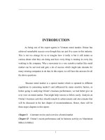
Significant results now implicate cytochrome c as a key mediator of the
apoptotic pathways (Figure 1.1). Many, but not all, inducers of apoptosis
cause the release of cytochrome c from the mitochondria. Also, it is now
assumed that the mitochondrial membrane is the site of action of members
of the Bcl-2 family of pro- and antiapoptotic proteins.
22
It is suggested that
bcl-2,
the mammalian homologue of the
ced-9
gene from
C. elegans
, functions
primarily by inhibiting the release of cytochrome c, whereas proapoptotic
relatives of
bcl-2
may promote this event. The released cytochrome c interacts
with Apaf-1, a positive regulator of apoptosis with homology to the
C. elegans
ced-4
proapoptotic gene. This collaboration results in activation of down-
stream caspases such as caspase 3, which are homologues of the
C. elegans
ced-3
gene. It is the action of these caspases on their substrates that results
in the systematic degradation of key substrates such as nuclear lamins, PARP,
fodrin, protein kinases, and other structural or regulatory proteins. This
process culminates in the organized collapse of the nucleus, membranes, and
cellular organelles. Many neuronal proteins are now recognized as substrates
of caspases, including presenilins and huntingtin.
23,24
The orderly breakdown
of dying cells through the apoptotic mechanisms results in the packaging of
cellular debris into apoptotic bodies which are then cleared by reticuloen-
dothelial cells as well as normal adjacent cells, thus preventing inflammatory
reactions to cell fragments.
The study of existing apoptotic developmental and neurodegenerative
diseases, be they caused by a genetic defect or a triggering environmental
factor, provide us with naturally occurring human models that validate
existing hypotheses in neuronal culture systems and provide new informa-
tion pertinent to basic cell biology.
1.2 Mechanisms of Apoptosis in Neurological Disorders
One theory invoked to explain Alzheimer cases that are apoptosis positive
is that the accumulation of amyloidogenic protein results in excess intracel-
lular calcium, a known trigger for the endonuclease responsible for the DNA
fragmentation seen during the final stages of apoptosis.
25
Oxidative stress
due to defects in energy and/or mitochondrial metabolism contributes to apo-
ptosis in anterior horn cells in amyotrophic lateral sclerosis, in the substantia
© 1999 by CRC Press LLC
FIGURE 1.1
© 1999 by CRC Press LLC
nigra in Parkinson’s disease, and in the penumbral region of infarcts seen
in cerebrovascular disease.
26-29
Excitotoxicity induced via activation of
NMDA receptors and also that induced by application of kainic acid to the
hippocampi has linked apoptosis and hippocampal sclerosis to the occur-
rence of epilepsy.
30
Defects in antiapoptotic genes such as
CLN3
and
NAIP
and
SMN
in the juvenile form of Batten disease and spinal muscular atrophy
type 1, respectively, coexist with massive neuronal loss and have inexorably
coupled these inherited neurodegenerative diseases most intimately with
the occurrence of apoptosis.
31-33
The fact that some sites in the body are immunologically privileged was
recognized over 100 years ago. The brain, like the eye and the testes and
placenta, is an immune-privileged site. Obviously, it is important to protect
the central nervous system and the eye from the ravages of invasive immu-
nopathologic injury.
34
There are multiple hypothetical mechanisms sur-
rounding the concept of immune privilege. One such theory is based on the
Fas–Fas ligand interaction. The expression of Fas is high in the cornea, in
photoreceptors, and in neurons, whereas expression of Fas-L is high in
endothelium. The strong Fas–Fas ligand interaction provides a tight “apop-
totic” vise curtailing the entry of activated macrophages, lymphokines, and
other growth-supporting factors into the sanctuary of the eye or brain. The
existence of conditions such as a defect in an antiapoptotic gene, oxidative
stress, or the presence of a toxic element that engages the apoptotic process,
tips the balance in the direction of neuronal or photoreceptor death in the
brain and eye, but remains phenotypically silent in other tissues.
1.3 Methodologies in Apoptosis
A long list of techniques exists that facilitates the study of apoptosis in
neurobiology and other disciplines. The second part of this book covers
many techniques that the authors have found useful. These include TUNEL
staining and other staining techniques that capitalize on the morphologic
changes of the nucleus and biochemical changes occurring in the cell and
nucleus during apoptosis (see Chapter 8). Also, they include cell viability
assays, electron microscopy (Chapter 9), flow cytometry (Chapter 10), mea-
surement of ceramide and sphingolipids (Chapter 12), and the use of viral
vectors to introduce genes of interest into cells (Chapter 13). The production
of proapoptotic or antiapoptotic gene knockout and/or specific gene over-
expressing mice, as well as the creation of mice with tissue-specific gene
expression have aided in the elucidation of apoptotic pathways and mech-
anisms as we know them.
35
Once the role of a gene or the protein it codes
for has been ensconced as significant, homologous genes and/or interacting
proteins can be fished out using traditional library screening or yeast two-
and three-hybrid systems.
36
© 1999 by CRC Press LLC
Ultimately, the choreography of neuronal apoptotic pathways will become
more complex, detailed, and specific. A better understanding of molecular
mechanisms in apoptotic pathways will make it possible to design effective
drugs targeting defined subsets of neurons at precise points in development
or adult life.
References
1. Hamburger V and Levi-Montalcini R. Proliferation differentiation and degen-
eration of the spinal ganglia of the chick embryo under normal and experi-
mental conditions.
J. Exp. Zool.
111:457, 1949.
2. Kerr JFR, Wyllie AH, and Currie AR. Apoptosis, a basic biological phenomenon
with wide-ranging implications in tissue kinetics.
Br. J. Cancer
26:239, 1972.
3. El-Deiry WS, Tokino T, Velsulesko VE, Levy DB, Parsons R, Trent JM, Lin D,
Mercer WE, Kinzler KW, and Vogelstein B. WAF1, a potential mediator of p53
tumor suppression.
Cell
75:817, 1993.
4. Ibanez CE, Ebendal T, Barbany G, Murray-Rust J, Blundell TL, and Persson H.
Disruption of the low affinity receptor-binding site in NGF allows neuronal sur-
vival and differentiation by binding to the trk gene product.
Cell
69:329, 1992.
5. Lerner TJ, Boustany RM, Anderson JW et al. and the International Batten Dis-
ease Consortium. Isolation of a novel gene underlying Batten disease, CLN3.
Cell
82:949, 1995.
6. Lane SC, Jolly RD, Schmechel DE, Alroy J, and R-M Boustany. Apoptosis as
the mechanism of neurodegeneration in Batten disease.
J. Neurochem.
67:677-683, 1996.
7. Burke JR, Wingfield MS, Lewis KE, et al. The Haw River syndrome: dentatoru-
bropallidoluysian atrophy in an African-American family.
Nat. Genet.
7:521-524,
1994.
8. Gelbard HA, Boustany R-M, and Schor NF. Apoptosis in development and
disease of the central nervous system.
Pediatric Neurol.
16(2):93-97, 1997.
9. Everall IP, Luthbert PJ, and Lantos PL. Neuronal loss in the frontal cortex in
HIV infection.
Lancet
337:1119, 1991.
10. Gamard CJ, Dhbaibo GS, Lie B, Obeid LM, and Hannun YA. Selective involve-
ment of ceramide in cytokine–induced apoptosis.
J.Biol.Chem.
272(26):16474,
1997.
11. Ellis RE, Yuan JY, and Horwitz HR. Mechanisms and functions of cell death.
Annu. Rev. Cell. Biol.
7:663, 1991.
12. Wang XW and Harris CC. p53 tumor-supressor gene: clues to molecular car-
cinogenesis.
J. Cell. Physiol.
173(2): 247, 1997.
13. Bayaert R and Fiers W. Molecular mechanisms of TNF-induced cytotoxicity,
FEBS Lett.
340:9, 1994.
14. Yonehara S., Ischii A and Yonehara M. A cell-killing monoclonal antibody (anti-
fas) to a cell surface antigen co-downregulated with the receptor of tumor
necrosis factor.
J. Exp. Med.
169:1747-1756, 1989.
15. Obeid LM, Linardic CM, Karolak LA, and Hannun YA. Programmed cell death
induced by ceramide.
Science
259:1769, 1993.
© 1999 by CRC Press LLC
16. Reed JC. Double identity for proteins of the Bcl-2 family.
Nature
387:773, 1997.
17. Hochhauser D. Modulation of chemosensitivity through altered expression in
cancer.
Anti-Cancer Drugs
8(10):903, 1997.
18. Nicholson DW, and Thornberry NA. Caspases: killer proteases.
Trends Biochem.
Sci.
22:229, 1997.
19. ZouH, Henzel WJ, Liu X, Lutschg A, and Wang X. Apaf-1, a human protein
homologous to C. elegans ced-4, participates in cytochrome c dependent acti-
vation of caspase-3.
Cell
90:405, 1997.
20. Hu S., Vincenz C, Ni J, Gentz R, and Dixit VM:1-FLICE, a novel inhibitor of
TNFR-1 and CD-95-induced apoptosis.
J. Biol. Chem.
272:17255, 1997.
21. Marchetti P, Casteldo M, Susin SA, Zamzami N, Hirsch T, Macho A, Haeffner
A, Hirsh F, Geuskins M. and Kroemer G. Mitochondrial permeability transition
is a central coordinating event of apoptosis.
J. Exp. Med.
184:1155, 1996.
22. Yang J, Liu X, Bhalla K, Kim CN, Ibrado AM, Cai J, Peng T-I, Jones DP, and
Wang X. Prevention of apoptosis by Bcl-2: Release of cytochrome c from mito-
chondria blocked.
Science
275:1129, 1997.
23. Goldberg YP, Nicholson DW, Rasper DM, Kalchman MA, Koide HB, Graham
AK, Bromm M, Kazimi-Esfarjani P, Thornberry NA, Vaillancourt JP, and Haydn
MA. Cleavage of huntingtin by apopain, a proapoptotic cysteine protease, is
modified by the polyglutamine tract.
Nat. Genet.
13(4):380, 1996.
24. Loetsher H., Deutschle U, Brackhaus M, Reinhardt D, Nelboek P, Mous J,
Grunberg J, Haass C, and Jacobson H. Pesenilins are processed by caspase type
proteases.
J. Biol. Chem.
272:20655, 1997.
25. Forloni G, Chiesa R, Smirolda S et al. Apoptosis mediated neurotoxicity in-
duced by application of beta-amyloid fragment 25-35.
Neuroreport
4:523, 1993.
26. Dipasquale B, Marini AM, and Youle R. Apoptosis induced by 1-methyl-4-phe-
nylpyridinium in neurons.
Biochem. Biophys. Res. Commun.
1181:1442, 1991.
27. Rabizadeh S, Gralla EB, Borchelt DR, Gwinn R, Valentine JS, Sisdia S, Wong
O, Lee M, Hahn H, and Bredeson DE. Mutations associated with ALS convert
SOD from an antiapoptotic gene to a proapoptotic gene: studies in yeast and
cancer cells.
Proc. Natl. Acad. Sci. U.S.A.
92:3024, 1995.
28. Mills EM, Gunasekar PG, Pavlakovic G, and Isam GE. Cyanide-induced apo-
ptosis and oxidative stress in differentiated PC-12 cells.
J. Neurochem.
67:1039,
1996.
29. Okamoto M, Matsumoto M, Ohtsuki T, Taguchi M, Kyanagihara T, Kamada T.
Internucleosomal DNA cleavage involved in ischemia-induced neuronal death.
Biochem. Biophys. Res. Commun.
196:1356, 1993.
30. Pollard H, Cantagrel S, Charriault-Marlangue C, Moreau J, and Yezekiel BA.
Apoptosis associated DNA fragmentation in epileptic brain damage.
Neurore-
port
5:1053, 1994.
31. Puranam K, Qian W-H, Nikbakht K, Venable M, Obeid L, Hannun Y, and
Boustany R-M. Upregulation of Bcl-2 and elevation of ceramide in Batten
Disease.
Neuropediatrics
28:37, 1997.
32. Puranam K, Qian W-H, Nikbakht K, Guo W-X, and Boustany R. CLN3 defines
a novel antiapoptotic pathway operative in neurodegeneration and mediated
via ceramide,
Cell Death and Diffeientiation
, in press.
33. Iwahashi H, Eguchi Y, Yasuhara N, Hanafusa T, Matsuzawa Y, and Tsujimoto
Y. Synergistic antiapoptotic activity between Bcl-2 and SMN implicated in
spinal muscular atrophy.
Nature
390:413, 1997.
© 1999 by CRC Press LLC
34. Griffith TS, Brunner T, Fletcher SM, Green D, and Ferguson TA. Fas-ligand
induced apoptosis as a mechanism of immune privilege.
Science
270:1189, 1995.
35. Zanjani HS, Vogel MW, Delhaye-Bouchaud N, Martinou JC, and Mariani J.
Increased cerebellar Purkinje cell numbers in mice overexpressing a human
bcl-2 transgene.
J. Compar. Neurol.
374(3):332, 1996.
36. Zhou H, and Reed JC. Heterodimerization-independent functions of cell death
regulatory proteins Bax and Bcl-2 in yeast and mammalian cells.
J. Biol. Chem.
272(50): 31482, 1997.
© 1999 by CRC Press LLC
2
Cell Death in the Developing Nervous System
Vinodh Narayanan
CONTENTS
2.1 Introduction
2.2 Target-Independent Cell Death
2.3 Target-Dependent Cell Death and the Discovery
of Trophic Factors
2.4 Cell Death Sensory and Sympathetic Ganglia
2.5 Cell Death in Motoneurons
2.6 Summary
References
2.1 Introduction
In the context of neural development, programmed cell death refers to the
naturally occurring
cell death seen at various stages of development in
almost all neural populations. This term is not entirely synonymous with
“apoptosis,” which refers to a particular cell death mechanism that is trig-
gered both in developmental cell death and in disease or injury. There are
thus many triggers that may initiate the cell death program. Programmed
cell death results in the elimination of cells that are not needed, without
injury to neighboring cells and without an inflammatory response. This
ranges from the removal of extraneous cells that were generated as part of
a lineage, abnormal cells, cells that were produced in excess, cells that did
not succeed in establishing a proper interaction with other cells, cells that
were dependent on a hormone or factor that is not available anymore, or
cells that had a role only at a particular developmental stage. The eventual
form of the nervous system (morphogenesis) is a result of a balance between
© 1999 by CRC Press LLC
the early processes of proliferation and regression, followed by cell growth
and maturation.
Apoptotic cell death has been observed in many different cell types, and
is relevant to the study of many human diseases. Commonly cited examples
of apoptotic cell death that result in the loss of particular tissues include the
elimination of the tail from developing vertebrate embryos (frogs and
humans) and the elimination of webs between the digits of developing
embryos.
1
The dramatic changes that occur during metamorphosis in
amphibia and insects are accompanied by apoptotic cell death in tissues that
are not required in the adult organism.
Naturally occurring cell death as a phenomenon in neural development
has been known for almost a century.
2
However, the systematic and quan-
titative study of neuronal death began with the work of Viktor Hamburger
and Rita Levi-Montalcini. Their work not only quantified cell death in dif-
ferent cell populations, but led to a now generally accepted hypothesis about
the role of target tissue and led to the discovery and characterization of
neurotrophic factors. We shall review here their original work and that of
other neurobiologists, and summarize the current ideas as they pertain to
neural histogenesis.
2.2 Target-Independent Cell Death
One form of programmed cell death is an intrinsically programmed genetic
cell death that is best exemplified by the developing nematode,
Caenorhabditis
elegans
. In this organism, cell identity, cell location, and function are entirely
determined by cell lineage. Of the approximately 1090 cells that are gener-
ated by cell division during development of the adult hermaphrodite, about
131 undergo programmed cell death.
3
It is known precisely which cells in
the developing organism (and at what point in their lineage) are destined
to die, this being one of the terminally differentiated states. In
C. elegans,
there are regional differences in the patterns of programmed cell death, and
cell death appears to function primarily to generate regional diversity,
1
per-
haps by eliminating certain sublineages.
4
Genetic studies have led to the
identification of mutations that affect programmed cell death, and to the
cloning of genes (
ced-3
and
ced-4
) that are
necessary
for
5-7
and genes (
ced-9
)
that
inhibit
cell death.
8,9
Although the determination of cell fate purely by lineage is not found in
more complex organisms, the cellular mechanisms that mediate cell death
in
C. elegans
and vertebrates share common features. The
ced-3
gene from
C. elegans
has been cloned and was found to encode a homologue of human
and murine interleukin-1
β
-converting enzyme (ICE).
7
Conversely, expres-
sion of the murine ICE gene product in cultured mammalian cells causes
© 1999 by CRC Press LLC
apoptotic cell death,
10
indicating a conservation of the molecular mechanisms
of programmed cell death through evolution. ICE is a member of a family
of proteases which activates its substrate by proteolytic cleavage and initiates
a cascade of events leading eventually to apoptotic cell death. The
ced-9
gene
product is a structural and functional homologue of the mammalian
bcl-2
protooncogene, which is known to suppress apoptosis in a variety of model
systems.
9
Thus, programmed cell death in
C. elegans
provided an example
of cell death as a predetermined outcome as a function of lineage, and led
to the discovery of the molecular mechanisms mediating apoptosis that are
functional in more complex organisms.
A form of target-independent (but not strictly predetermined) cell death
has also been observed in vertebrate nervous systems, in the embryonic
cerebral cortex, and even earlier in the developing neural tube. There is a
fairly widespread and uniform appearance of cells undergoing apoptosis in
the embryonic murine cerebral cortex. The peak of this apoptotic cell death
occurs around E14–16, and virtually no dying cells are seen at E10 or in the
adult.
11
Although many dying cells were observed in regions which con-
tained postmitotic neurons (marginal zone, cortical plate, and intermediate
zone), the majority of dying cells were within the proliferative zones.
11
Some
large neurons undergoing apoptosis in the border area between the subplate
and cortical plate were thought to be subplate neurons.
12
The reason for the
observed rate of cell death (average of about 50%) in the proliferative zones
of the embryonic cortex is not clear, but it is interesting that the period of
maximal cell death (E12–E16) corresponds roughly to the neuronogenetic
interval, that time period during which all the terminally postmitotic neu-
rons are generated.
13
Results similar to the above have also been observed in human fetuses.
Apoptotic cells were found in the ventricular zone at the 12th week of
gestation, reaching a peak by the 21st week of gestation.
14
In the oldest fetuses
studied (23 weeks), apoptotic cells were found primarily in the deep portions
of the subplate. As was the case in murine embryos, programmed cell death
in the human embryonic cortex was most prominent in the proliferative
zones. Whether these represent differentiated cells or undifferentiated cells
was not clear. It has been hypothesized that the apoptotic cells represent
postmitotic cells that are uncommitted to reach an appropriate position in
the cortical plate, and are thus eliminated before migration.
14
However, the
precise characteristics that determine whether a cell in the proliferative zone
survives or dies remain unknown. Among the postmitotic regions of the
developing cortex, most of the observed cell death occurs within the sub-
plate, a transient population of neurons that occupies the layer between the
proliferative zone and the cortical plate.
12
Almost all of these cells are elim-
inated early in postnatal life by apoptosis.
15
Another example of cell death occurring at a very early stage of neural
development, before the stage of neuron-target contact, is that seen in the
developing neural tube. In the chick embryo, between the 8- and 12-somite
© 1999 by CRC Press LLC
stages, many dead cells are seen, concentrated in the neural folds.
16
In order
to determine the function of apoptotic cell death in neural tube closure, chick
embryos were cultured at the 8-somite stage, and allowed to develop to the
13-somite stage. In these embryos, the neural tube was completely closed
between somites 1 and 8, similar to what is observed
in vivo
. When these
embryos were cultured in the presence of specific protease (caspase) inhib-
itors, programmed cell death and neural tube closure were blocked.
16
These
results suggest that programmed cell death is required for neural tube clo-
sure, although it is not clear what aspect of neural tube closure depends on
apoptosis.
2.3 Target-Dependent Cell Death and the Discovery
of Trophic Factors
The studies of Hamburger and Levi-Montalcini on the role of targets in
neural development began with the observation that reduction of a periph-
eral field (limb) resulted in a size reduction of the innervating primary nerve
center. Hamburger and Levi-Montalcini studied the mechanisms by which
such changes were brought about.
17
They reported on the effect of reduction
and augmentation of target size on the development of the spinal ganglia
of the chick, examining the rate of proliferation, differentiation, and degen-
eration. The occurrence of cell degeneration in the spinal ganglia during
normal development was noted, and it was observed that there was a distinct
topographic pattern of dying cells within the spinal ganglia. It occurred (in
normal embryos) most extensively in the cervical and thoracic regions, and
was minimal in the brachial and lumbosacral segments. However, limb bud
removal caused an extensive cell degeneration within the brachial ganglia
(wing bud) or lumbosacral ganglia (hind limb bud). Hamburger proposed
that the mechanisms behind cell death in normal and experimental (limb
bud extirpation) embryos were the same, and that the enlarged target offered
by the developing limb (wing or hind limb) prevented the cell degeneration
in the corresponding sensory spinal ganglia. A stated hypothesis was that
either synaptic contact with the target or a trophic substance produced in
the target area was necessary for cell survival, and that competition for these
trophic interactions was what determined whether or not a cell survived or
died.
Following these seminal observations, cell death has been noted in many
different neuronal populations, and may be a universal developmental phe-
nomenon. Examples include the spinal ganglia and motoneurons, the cranial
nerve nuclei, the optic tectum, the retina, and the cerebellum (see Table 1,
Reference 18). The phenomenon of cell death is not limited to neurons, but