Unique role of ionic liquid bminBF4 during curcumin–surfactant association and micellization of cationic, anionic and non ionic surfactant solutions
Bạn đang xem bản rút gọn của tài liệu. Xem và tải ngay bản đầy đủ của tài liệu tại đây (591.18 KB, 6 trang )
Spectrochimica
Acta
Part
A
79 (2011) 1823–
1828
Contents
lists
available
at
ScienceDirect
Spectrochimica
Acta
Part
A:
Molecular
and
Biomolecular
Spectroscopy
j
ourna
l
ho
me
page:
www.elsevier.com/locate/saa
Unique
role
of
ionic
liquid
[bmin][BF
4
]
during
curcumin–surfactant
association
and
micellization
of
cationic,
anionic
and
non-ionic
surfactant
solutions
Digambara
Patra
∗
,
Christelle
Barakat
Department
of
Chemistry,
Faculty
of
Arts
and
Sciences,
American
University
of
Beirut,
P.O.
Box:
11-0236,
Riad
El
Solh,
Beirut,
1107-2020,
Lebanon
a
r
t
i
c
l
e
i
n
f
o
Article
history:
Received
30
September
2010
Received
in
revised
form
17
May
2011
Accepted
24
May
2011
Keywords:
Curcumin
Hydrophilic
ionic
liquid
Micelle
Surfactant
Spectroscopy
a
b
s
t
r
a
c
t
Hydrophilic
ionic
liquid,
1-butyl-3-methylimidazolium
tetrafluoroburate,
modified
the
properties
of
aqueous
surfactant
solutions
associated
with
curcumin.
Because
of
potential
pharmaceutical
applications
as
an
antioxidant,
anti-inflammatory
and
anti-carcinogenic
agent,
curcumin
has
received
ample
attention
as
potential
drug.
The
interaction
of
curcumin
with
various
charged
aqueous
surfactant
solutions
showed
it
exists
in
deprotonated
enol
form
in
surfactant
solutions.
The
nitro
and
hydroxyl
groups
of
o-nitrophenol
interact
with
the
carbonyl
and
hydroxyl
groups
of
the
enol
form
of
curcumin
by
forming
ground
state
complex
through
hydrogen
bonds
and
offered
interesting
information
about
the
nature
of
the
interac-
tions
between
the
aqueous
surfactant
solutions
and
curcumin
depending
on
charge
of
head
group
of
the
surfactant.
IL[bmin][BF
4
]
encouraged
early
formation
of
micelle
in
case
of
cationic
and
anionic
aqueous
surfactant
solutions,
but
slightly
prolonged
micelle
formation
in
the
case
of
neutral
aqueous
surfactant
solution.
However,
for
curcumin
IL
[bmin][BF
4
]
favored
strong
association
(7-fold
increase)
with
neutral
surfactant
solution,
marginally
supported
association
with
anionic
surfactant
solution
and
discouraged
(∼2-fold
decrease)
association
with
cationic
surfactant
solution.
© 2011 Elsevier B.V. All rights reserved.
1.
Introduction
Micellar
systems
of
aqueous
origin
have
immense
technological
applications
as
flow
field
regulators,
solubilizing
and
emulsify-
ing
agents,
membrane
mimetic
media,
nanoreactors
for
enzymatic
reaction
and
drug
delivery
system
[1–8].
It
is
anticipated
that
curcumin,
1,7-bis(4-hydroxy-3-methoxyphenyl)-1,6-heptadiene-
3,5-dione,
may
find
applications
as
a
novel
drug
in
the
near
future
to
control
various
diseases,
including
inflammatory
dis-
orders,
carcinogenesis
and
oxidative
stress-induced
pathogenesis
[9–12].
Curcumin
has
drawn
intense
interest
recently
due
to
its
potential
pharmaceutical
importance
[13–24].
However,
curcumin
is
very
poorly
soluble
in
water
by
reducing
its
effectiveness
as
a
drug.
Therefore,
various
methods
are
being
developed
to
make
cur-
cumin
better
soluble
and
enhance
effectiveness
of
the
drug
during
its
delivery
[16].
Physiochemical
properties
of
an
aqueous
surfactant
solution
depend
on
the
identity
of
the
surfactant.
The
aqueous
solution
of
a
surfactant
at
a
given
concentration
posses
more
or
less
fixed
physiochemical
properties
that
are
difficult
to
modulate.
Other
than
changing
temperature
and
pressure,
the
usual
way
to
mod-
ify
the
physiochemical
properties
of
a
given
surfactant
solution
is
to
use
external
additives,
such
as
cosolvents,
cosurfactants,
∗
Corresponding
author.
Tel.:
+961
1350
000x3985;
fax:
+961
1365217.
address:
(D.
Patra).
electrolytes,
non-polar
organics,
polar
organics,
etc.
Ionic
liquids
(ILs)
are
solvents
composed
entirely
of
ions
and
composed
of
poorly
coordinating
ions
and
can
therefore
be
highly
polar
yet
non-coordinating
[25–27].
These
are
immiscible
with
a
number
of
organic
solvents
and
provide
non-aqueous
polar
alternatives
for
two
phase
systems.
They
are
of
particular
interest
because
of
their
environmentally
friendly
nature,
their
exciting
features
and
their
economical
convenience
[28–35].
The
unusual
properties
of
ILs
demonstrate
a
unique
role
in
altering
the
properties
of
aqueous
surfactant
solutions
such
as
aggregation
number
[3,4].
The
effec-
tiveness
of
this
modification
of
aqueous
surfactant
solutions
by
IL
may
largely
depend
on
the
kind
and
extent
of
interaction/s
between
cation/anion
of
the
IL
and
the
head
group
of
the
surfactant
[4].
How-
ever,
hydrophobic
effect
of
IL
with
surfactant
molecule
might
play
a
role.
In
addition
we
hypothesize
that
IL
may
drive
the
associa-
tion
of
the
drug
molecule
towards
better
solubilization
in
micellar
system
(which
is
very
important
during
drug
delivery)
as
per
the
head
group
of
the
surfactant
charge
and
physiochemical
properties
of
the
drug
molecule.
In
order
to
understand
the
better
insight
of
the
role
of
these
interactions
of
IL
during
solubilization
of
poorly
water
soluble
drug
such
as
curcumin
in
micellar
systems
and
micellization,
we
extend
the
study
of
interaction
of
IL
and
surfactant
solutions
[4]
further
to
systems
composed
of
various
(positive
and
negative)
charged
and
uncharged
surfactant
solutions,
curcumin
and
an
IL
(1-butyl-3-methylimidazolium
tetrafluoroburate,
[bmin][BF
4
]).
The
association
of
curcumin
with
various
charged
surfactant
1386-1425/$
–
see
front
matter ©
2011 Elsevier B.V. All rights reserved.
doi:10.1016/j.saa.2011.05.064
1824 D.
Patra,
C.
Barakat
/
Spectrochimica
Acta
Part
A
79 (2011) 1823–
1828
solutions
and
fluorescence
quenching
of
curcumin
by
o-
nitrophenol
in
different
surfactant
solutions
may
explore
the
kind
of
interaction
between
curcumin
and
various
charged/uncharged
surfactant
solutions
without
IL.
Due
to
cation/anion
of
the
IL,
it
may
remarkably
alter
the
interaction
of
curcumin
and
surfactant
solu-
tions
based
on
the
charge
of
the
head
group
of
the
surfactant
and
deprotonated
form
of
curcumin,
therefore
impact
drug–surfactant
association.
Comparative
study
of
various
charged/uncharged
sur-
factant
molecules
may
conclude
importance
of
hydrophobic
effect
of
IL
during
micellization.
2.
Materials
and
methods
2.1.
Materials
The
surfactants
cetyl
trimethyl
ammonium
bromide
(CTAB),
sodium
dodecyl
sulfate
(SDS)
and
Triton
X-100
(TX100)
were
obtained
from
Acros
Organics
and
were
dissolved
in
different
volumes
of
double
distilled
water
for
the
preparation
of
several
con-
centrations
of
surfactant
solutions.
The
stock
solutions
consisted
of
10
mM
CTAB,
100
mM
SDS
and
10
mM
TX100.
Curcumin
was
also
obtained
from
Acros
Organics
and
was
used
without
further
purifi-
cation.
To
prepare
the
stock
solution,
curcumin
was
dissolved
in
spectroscopic
grade
acetonitrile
(Acros
Organics)
so
that
the
final
concentration
of
acetonitrile
in
the
surfactant
solutions
remained
less
than
1%
(v/v).
1-Butyl-3-methylimidazolium
tetrafluoroburate,
[bmin][BF4]
was
obtained
from
Fluka
and
o-nitrophenol
was
a
Merck
Schuchardt
product.
The
solvents
were
used
without
further
purification.
2.2.
Spectroscopic
measurements
The
absorption
spectra
in
various
solvents
and
in
cationic
CTAB,
anionic
SDS,
and
neutral
TX100
were
recorded
at
room
temperature
using
a
JASCO
V-570
UV–VIS–NIR
Spectrophotometer.
Fluores-
cence
measurements
were
done
on
a
JOBIN
YVON
Horiba
Fluorolog
3
spectrofluorometer.
The
excitation
source
was
a
100
W
Xenon
lamp.
The
detector
used
was
R-928
operating
at
a
voltage
of
950
V.
The
excitation
and
emission
slits
width
were
5
nm.
The
spectral
data
were
collected
using
Fluorescence
software
and
data
analysis
was
made
using
OrginPro
6.0
software.
3.
Results
and
discussion
3.1.
Curcumin–surfactant
interaction
in
absence
of
IL
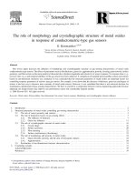
Generally,
curcumin
showed
a
strong
and
intense
absorption
band
in
the
350–480
nm
wavelength
region
in
all
the
investi-
gated
surfactant
solutions.
Representative
absorption
spectra
of
curcumin
in
various
concentrations
of
TX100
solutions
are
depicted
in
Fig.
1.
The
interaction
between
curcumin
and
micelles
can
be
described
as:
C
+
S
K
b
CS
where
C
is
curcumin;
S
is
the
surfactant
(CTAB,
SDS
or
TX100);
CS
is
the
curcumin–surfactant
complex;
and
K
b
is
the
association
constant.
The
concentration
of
the
micellized
surfactant
is
given
by:
S
m
=
S
s
−
cmc
where
S
s
is
the
surfactant
concentration.
700600500400300
0.0
0.1
0.2
0.3
0.4
0.5
0.6
0.7
0.8
0.9
1.0
7-9
6
5
4
3
2
1
Curcu
min with [TX100]
Absorbance
Wavelength
(nm
)
(1)
0.0
2 mM
(2)
0.0
4 mM
(3)
0.1
mM
(4)
0.2
mM
(5)
0.4
mM
(6)
0.6
mM
(7)
1.0
mM
(8)
1.2
mM
(9)
1.4
mM
Fig.
1.
Absorption
spectra
of
curcumin
in
various
aqueous
TX100
concentrations.
Table
1
Association
rate
constants
of
curcumin
with
various
aqueous
surfactant
solutions
in
the
absence
and
presence
of
ionic
liquid.
Sample cmc
used
for
calculation
(mM)
K
b
SDS
7.3
6193
M
−1
CTAB
0.8
20,467
M
−1
TX100
0.2
11,555
M
−1
SDS
+
IL
(1%,
v/v)
0.95
6315
M
−1
CTAB
+
IL
(1%,
v/v) 0.1
10,227
M
−1
TX100
+
IL
(1%,
v/v)
0.4
82,737
M
−1
The
association
constants
can
be
determined
[6,36–39]
as:
C
T
S
m
l
A
=
S
m
ε
s
−
ε
0
+
1
K
gb
(ε
s
−
ε
0
)
where
l
is
the
optical
path
length,
ε
m
is
the
molar
excitation
coeffi-
cient
of
curcumin
fully
bound
to
micelles,
ε
0
is
the
molar
excitation
coefficient
of
curcumin
in
the
solvent,
C
T
is
the
total
curcumin
con-
centration
and
A
=
A
− A
0
where
A
is
the
absorbance
of
curcumin
in
the
presence
of
surfactant
solution
and
A
0
is
the
absorbance
of
curcumin
in
the
absence
of
micelle/surfactant.
Using
Scott’s
plots
[6,36–39],
the
association
constants
of
CTAB,
SDS
and
TX100
were
determined
to
be
20,467
M
−1
,
6193
M
−1
and
11,555
M
−1
(Table
1),
respectively.
It
should
be
noted
that
the
crtical
micellar
concentration
(cmc)
for
the
calculation
of
association
con-
stants
for
various
micelle
was
estimated
by
fluorescence
method
as
explained
later
on.
It
is
observed
that,
K
b
CTAB
>
K
b
TX100
>
K
b
SDS
.
These
results
implied
that
the
different
micelles
have
different
affinities
for
curcumin.
Cationic
CTAB
is
bound
to
curcumin
with
the
highest
affinity,
followed
by
neutral
TX100
and
then
anionic
SDS.
This
could
be
due
to
the
electrostatic
interactions
between
cur-
cumin
and
the
positive
charge
on
the
head
group
of
CTAB
present
in
the
Stern
layer
of
the
micelle,
thus
indicating
that
curcumin
at
the
given
conditions
is
mainly
found
in
its
deprotonated
anionic
forms
[40]
(see
Supplement
1).
In
the
case
of
SDS,
the
repulsion
between
deprotonated
enol
(anionic)
forms
of
curcumin
and
the
negative
charge
on
the
head
group
of
SDS
present
in
the
Stern
layer
of
the
micelle
make
a
weaker
interaction,
hence
decreasing
the
associa-
tion
rate
constant.
However,
given
that
the
head
group
of
TX100
is
nonionic,
the
value
of
the
association
rate
constant
for
TX100
was
in
between
that
of
CTAB
and
SDS.
3.2.
Critical
micellar
concentration
determination
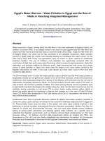
Fluorescence
excitation
and
emission
spectra
of
curcumin
with
various
concentrations
of
surfactant
noted
that
the
fluorescence
D.
Patra,
C.
Barakat
/
Spectrochimica
Acta
Part
A
79 (2011) 1823–
1828 1825
300 35
040
045
0
500 55
060
065
070
0
0.0
4.0x10
6
8.0x10
6
1.2x10
7
1.6x10
7
2.0x10
7
10
8-9
Curc
umin
with [TX100
]
(1) No
TX10
0
(2) 0.02
mM
(3) 0.04
mM
(4) 0.06
mM
(5) 0.1 mM
(6) 0.2 mM
(7) 0.6 mM
(8) 0.8 mM
(9) 1.0 mM
(10
) 1.2
mM
(11
) 1.4
mM
(12
) 1.6
mM
Fluorescence Intensity (a.u)
Wavelength
(nm
)
0.0
4.0x10
6
8.0x10
6
1.2x10
7
1.6x10
7
2.0x10
7
2.4x10
7
2.8x10
7
11-12
11-12
10
8-9
7
7
6
6
1-5
1-5
Fig.
2.
Fluorescence
excitation
and
emission
spectra
of
curcumin
in
various
aqueous
TX100
concentrations.
intensity
of
the
emission
and
excitation
spectra
of
curcumin
in
TX100
(shown
in
Fig.
2)
and
SDS
(not
shown)
increased
as
the
concentration
of
the
surfactant
was
increased.
However,
the
flu-
orescence
spectra
of
CTAB
exhibited
a
different
behavior
(not
shown).
The
fluorescence
intensity
initially
decreased
until
it
reached
0.5
mM
of
CTAB
and
once
the
cmc
was
reached,
the
intensity
started
increasing
with
concentration.
A
red
shift
was
also
observed
after
the
cmc
for
CTAB.
The
Stokes’
shift
of
curcumin
in
various
concen-
trations
of
CTAB,
SDS
and
TX100
was
determined
as
the
difference
between
absorption
and
emission
maxima
obtained
from
the
cor-
rected
spectra
on
the
wavenumber
scale
[41,42].
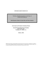
The
plot
of
Stokes’
shift
versus
surfactant
concentration
offered
three
different
kinds
of
change,
respectively,
for
cationic
(CTAB),
anionic
(SDS)
and
neu-
tral
(TX100)
surfactant
solutions.
In
the
case
of
CTAB,
the
value
of
Stokes’
shift
rarely
changed
before
the
cmc.
A
big
jump
of
5000
cm
−1
was
observed
around
the
cmc
and
after
the
cmc
it
remained
more
or
less
unaltered.
The
cmc
of
CTAB
was
estimated
by
finding
the
midpoint
of
the
tangent
joining
the
two
lines,
as
shown
in
Fig.
3A.
For
SDS,
Stokes’
shift
of
curcumin
for
different
surfactant
con-
centrations
varied
differently,
it
initially
decreased
till
the
cmc
was
reached.
Above
the
cmc,
it
marginally
increased.
By
extrapolating
these
two
linear
equations,
before
and
after
the
cmc,
with
respec-
tive
negative
and
positive
slopes,
a
minimum
intersecting
point
was
obtained
to
calculate
the
cmc
(Fig.
3B).
Stokes’
shift
of
curucmin
increased
with
TX100
concentration
until
cmc
was
attained
and
then
it
decreased
dramatically.
In
this
case
the
maximum
value
of
Stokes’s
shift
was
used
to
estimate
cmc
as
marked
in
Fig.
3C.
The
cmc
values
estimated
using
Stokes’
shift
of
curcumin
is
summarized
in
Table
2,
the
values
obtained
without
IL
are
similar
to
the
reported
values
[4,5,43]
establishing
the
reliability
of
the
method.
The
differ-
Table
2
cmc
values
of
aqueous
CTAB,
SDS
and
TX100
solutions
in
the
presence
and
absence
of
ionic
liquid.
Sample
cmc
Curcumin
(cm
−1
)
Pyrene
I
I
/I
III
a
Reported
b
SDS 7.3
mM
7.0
mM
6.0–8.0
mM
CTAB
0.8
mM
–
0.26
mM
TX100
0.2
mM
0.25–0.5
mM
0.9
mM
SDS
+
IL
(1%,
v/v)
0.95
mM
1
mM
(2%,
v/v)
–
CTAB
+
IL
(1%,
v/v)
0.1
mM
–
–
TX100
+
IL
(1%,
v/v) 0.4
mM
0.5–1.0
mM
(2%,
v/v)
–
a
From
Refs.
[3,4].
b
From
Ref.
[43].
0.0000 0.000
5
0.0010 0.001
5
0.0020
5000
6000
7000
8000
9000
1000
0
1100
0
1200
0
A
Stokes' shift (cm
-1
)
[CTAB]
CTAB
0.0000
0.0005
0.0010
0.0015
0.0020
35000
40000
45000
50000
55000
60000
65000
70000
cmc of CTAB
+ IL
cmc of
CT
AB
CTAB
+ IL
0.000
0.005
0.010
0.015
0.020
4000
4200
4400
4600
4800
5000
5200
5400
B
cmc of SDS + IL
Stokes' shift (cm
-1
)
[SDS]
SDS
0.00
0
0.005
0.010
0.015
0.020
18000
18200
18400
18600
18800
19000
19200
19400
19600
19800
cmc of S
DS
SDS + IL
0.000
0
0.000
3
0.0006
0.000
9
0.00
12
0.001
5
0.00
18
3000
3500
4000
4500
5000
5500
6000
C
Stokes' shift (cm
-1
)
[TX10
0]
TX100
0.00
00
0.000
3
0.00
06
0.000
9
0.0012
0.001
5
0.0018
0
1000
2000
3000
4000
5000
cmc of Tx10
0 + I
L
cmc of TX100
TX100 + IL
Fig.
3.
Variation
of
Stokes’
shift
of
curcumin
in
different
concentrations
of
aqueous
CTAB
(A),
SDS
(B)
and
TX100
(C)
in
the
absence
and
presence
of
IL.
ent
trends
of
Stokes’s
shift
for
various
surfactants
could
be
due
to
the
various
kinds
of
interactions
between
the
charged/uncharged
head
groups
of
the
surfactants
and
the
deprotonated
forms
of
cur-
cumin.
3.3.
Quenching
study
by
o-nitrophenol
o-Nitrophenol
can
strongly
quench
the
fluorescence
of
cur-
cumin
by
forming
a
ground
state
complex
through
hydrogen
bonding
[24]
as
given
in
Scheme
1.
However,
the
extent
to
which
it
quenches
may
highly
depend
on
the
conditions
of
the
medium
in
which
curcumin
and
o-nitrophenol
1826 D.
Patra,
C.
Barakat
/
Spectrochimica
Acta
Part
A
79 (2011) 1823–
1828
HO
O
O
H
3
CO
OCH
3
OH
H
H
O
O
-
N
O
Formation
cyclic groun
d stat
e compl
ex of curc
umin with
o-nitroph
enol
Scheme
1.
Ground
state
complex
formation
of
curcumin
with
o-nitrophenol
causing
fluorescence
quenching
of
curcumin
by
o-nitrophenol.
can
interact
and
hence,
on
the
nature
of
the
surfactants.
The
position
of
the
functional
groups
in
o-nitrophenol
and
the
geom-
etry
of
the
molecule
predict
the
location
of
o-nitrophenol
in
the
micelle
[44].
The
benzene
ring
of
the
phenol
is
pushed
towards
the
hydrocarbon
core
and
the
polar
functional
groups
remain
in
the
hydrophilic
layer
of
the
micelle
[44].
Given
that
the
stoichiometric
ratio
of
o-nitrophenol
to
curcumin
is
1:1,
the
nitro
and
hydroxyl
groups
of
the
quencher
interact
with
the
carbonyl
and
hydroxyl
groups
of
the
enol
form
of
curcumin
by
means
of
strong
hydrogen
bonds
[24].
This
associated
complex,
which
is
formed
in
the
ground
state,
greatly
quenches
the
fluorescence
of
curcumin
through
the
following
process:
curcumin* + o-nitrophe
nol [curcumin- o-nitrophenol
]* [curcumin- o-nitrophenol
]
curcumin + o-nitrophenol
[curcumin- o-nitrophenol
] [curcumin- o-nitrophenol
]*
hν
a
hν
a
hν
fl
hν
fl
Using
the
Stern
Volmer
equation
[45]
the
quenching
rate
constant
K
sv
of
curcumin
and
the
quencher,
o-nitrophenol,
was
determined
as
I
0
f
I
f
=
1
+
K
sv
[oNP]
I
0
f
I
f
=
1
+
k
q
0
[oNP]
where
K
sv
is
the
Stern
Volmer
rate
constant,
I
0
f
is
the
fluorescence
intensity
without
the
quencher,
I
f
is
the
fluorescence
intensity
with
the
quencher,
k
q
is
the
quencher
rate
coefficient,
0
is
the
fluores-
cence
lifetime
of
curcumin
without
the
presence
of
the
quencher
and
[oNP]
is
the
concentration
of
o-nitrophenol.
Fig.
4
illustrates
the
fluorescence
spectra
of
curcumin
in
the
presence
of
SDS
with-
out
and
with
various
concentrations
of
o-nitrophenol.
The
insert
in
Fig.
4
presents
the
Stern
Volmer
plot
[45]
for
curcumin
in
presence
of
various
concentration
of
o-nitrophenol.
The
fluorescence
spectra
of
curcumin
in
water,
CTAB
and
TX100
without
and
with
various
concentrations
of
o-nitrophenol
along
with
their
respective
Stern
Volmer
plots
showed
similar
trends
(not
shown).
The
estimated
values
of
K
sv
and
k
q
for
fluorescence
quench-
ing
of
curcumin
by
o-nitrophenol
in
water
and
various
micellar
media
is
determined
as
per
the
Stern
Volmer
equation
[45]
and
given
in
Table
3.
The
quenching
rate
constant
of
curcumin
by
o-
nitrophenol
in
water
was
determined
to
be
449
M
−1
in
comparison
to
3973
M
−1
in
cationic
CTAB.
The
high
quenching
rate
of
CTAB
is
due
to
the
stabilizing
electrostatic
interactions
between
the
pos-
itively
charged
head
groups
of
the
micelles
and
the
negatively
charged
enolic
curcumin
(see
Supplement
1).
This
attractive
inter-
action
facilitates
the
penetration
of
curcumin
in
the
Stern
layer
of
the
micelle
and
hence
the
formation
of
the
complex
[CUR–NP].
In
the
case
of
anionic
SDS,
a
decrease
in
the
quenching
rate
constant
was
found
relative
to
that
of
water.
This
change
can
be
linked
to
Fig.
4.
Fluorescence
emission
spectra
of
curcumin
in
SDS
in
the
presence
of
various
concentration
of
o-nitrophenol.
The
fluorescence
intensity
decreases
with
increase
in
o-nitrophenol
concentration.
Insert
shows
Stern
Volmer
plot
for
the
determina-
tion
of
the
quenching
rate
constant
K
sv
.
D.
Patra,
C.
Barakat
/
Spectrochimica
Acta
Part
A
79 (2011) 1823–
1828 1827
Table
3
Quenching
rate
constants
of
curcumin
by
o-nitrophenol
in
water,
CTAB,
SDS
and
TX100
surfactant
solutions.
Sample K
sv
(M
−1
)
k
q
((
0av
=
2.366
ns)
Water
449
1.9
×
10
11
M
−1
s
−1
CTAB 3973
1.7
×
10
12
M
−1
s
−1
SDS
367
1.6
×
10
11
M
−1
s
−1
TX100
550
2.3
×
10
11
M
−1
s
−1
the
repulsion
between
the
negatively
charged
head
groups
of
the
micelle
and
the
negative
charge
on
the
deprotonated
curcumin,
thus
destabilizing
the
complex
[CUR–NP].
In
the
case
of
neutral
TX100,
a
slight
increase
in
the
quenching
rate
was
observed
relative
to
that
of
water.
The
neutrality
of
this
surfactant
does
not
change
the
physical
properties
of
the
solvent
but
helps
in
bringing
together
o-nitrophenol
and
curcumin
due
to
hydrophobic
interactions.
3.4.
Effect
of
ionic
liquid
[bmin][BF
4
]
on
drug–surfactant
association
The
properties
of
various
aqueous
surfactant
solutions
were
modified
by
a
common
and
popular
hydrophilic
1-butyl-3-
methylimidilazolium
tetrafluoroborate,
[bmin][BF4].
For
modify-
ing
properties
of
aqueous
surfactant
solution,
the
IL
concentration
1%
(v/v)
was
chosen
from
the
literature
[4,5].
The
absorption
(see
Fig.
5)
and
fluorescence
excitation
and
emission
(see
Fig.
5)
spec-
tra
of
curcumin
in
various
surfactant
concentrations
in
presence
of
IL
showed
the
absorbance
or
fluorescence
intensity
of
curcumin
700600500400300
0.0
0.5
1.0
1.5
2.0
2.5
3.0
7-8
6
5
4
3
2
Absorbance
Wavelength (nm)
(1
) NO TX100
(2
) 0.02
mM
(3
) 0.06
mM
(4
) 0.2 mM
(5
) 0.4 mM
(6
) 0.6 mM
(7
) 0.8 mM
(8
) 1.0 mM
Curcum
in plus
IL
with
[T
X10
0]
1
700650600550500450400350300
0.0
4.0x10
6
8.0x10
6
1.2x10
7
1.6x10
7
2.0x10
7
8
8
7
7
6
6
4-5
Fluorescence Intensity (a.u)
Wave
leng
th (nm
)
0
1x10
6
2x10
6
3x10
6
4x10
6
5x10
6
6x10
6
7x10
6
8x10
6
9x10
6
Curcumin plus IL with TX100
10
10
9
9
4-5
1-3
1-3
(1) No TX100
(2) 0.02 mM
(3) 0.04 mM
(4) 0.06 mM
(5) 0.1 mM
(6) 0.2 mM
(7) 0.4 mM
(8) 0.8 mM
(9) 1.0 mM
(10
) 1.6 mM
Fig.
5.
Absorption
and
fluorescence
(excitation
and
emission)
spectra
of
curcumin
in
various
aqueous
TX100
concentrations
in
the
presence
of
IL.
in
CTAB,
SDS
and
TX100,
increased
with
surfactant
concentration.
The
association
constants
for
the
three
surfactants
with
curcumin
in
the
presence
of
IL
were
determined
as
explained
earlier
and
given
in
Table
1.
The
association
constant
of
CTAB
in
the
presence
of
IL
decreased
significantly
relative
to
CTAB
without
IL.
Though
the
short
hydrophobic
effect
of
the
tail
may
encourage
the
IL
to
locate
around
the
Stern
layer
of
the
micelle,
the
positive
charged
head
group
would
repulse
with
the
similar
charged
head
groups
of
CTAB.
Finally
both
CTAB
and
IL
will
compete
to
bind
with
deprotonated
form
of
curcumin.
This
competition
could
account
for
the
decrease
in
the
associa-
tion
constant
of
curcumin
with
CTAB.
However,
in
the
case
of
SDS
in
the
presence
of
IL,
an
increase
of
the
association
rate
constant
was
observed
compared
to
SDS
without
IL.
In
the
absence
of
IL,
there
is
repulsion
between
the
negative
charge
of
the
head
group
(sul-
fate
ion)
of
SDS
and
the
negative
charge
of
the
deprotonated
form
of
curcumin.
When
IL
is
added,
its
positive
charge
head
group
will
act
as
a
stabilizer
between
negatively
charged
SDS
and
negatively
charged
curcumin
(deprotonated
form),
thus
facilitating
the
asso-
ciation
of
curcumin
with
SDS.
On
the
other
hand,
the
association
rate
constant
of
curcumin
with
TX100
increased
significantly
in
the
presence
of
IL.
A
possible
explanation
would
be
the
induction
of
hydrogen
bonding
and
dipole–dipole
forces
by
the
positive
charge
of
the
head
group
of
the
IL
with
TX100
[4],
assisting
interaction
or
strong
association
of
curcumin
with
neutral
surfactant
solution.
3.5.
Effect
of
ionic
liquid
[bmin][BF
4
]
on
micellization
As
discussed
earlier,
the
cmc
of
various
aqueous
surfactant
solu-
tions
was
evaluated
based
on
the
change
in
Stokes’
shift
(see
Fig.
3)
of
curcumin
in
the
presence
of
1%
(v/v)
IL.
Variation
of
Stokes’
shift
with
surfactant
concentration
for
CTAB
with
and
without
IL
showed
similar
trends.
It
could
therefore
be
implied
that
there
is
no
new
kind
of
favorable
interaction
between
the
IL
and
CTAB.
However,
similar
plots
for
SDS
with
and
without
IL
gave
two
different
trends
indicating
that
the
interaction
of
curcumin
with
SDS
in
the
pres-
ence
and
absence
of
IL
are
not
similar.
As
shown
earlier,
in
the
absence
of
IL,
the
Stokes’
shift
of
curcumin
increased
with
increase
in
SDS
concentrations
until
cmc
was
reached.
However,
when
IL
was
present,
Stokes’
shift
continued
to
decrease,
but
at
a
much
smaller
rate,
with
increasing
SDS
concentration.
This
trend
could
imply
that
in
the
case
of
SDS,
there
could
be
a
favorable
interac-
tion
that
stabilizes
the
micelles
in
the
presence
of
IL.
For
TX100,
variation
of
Stokes’
shift
with
surfactant
concentration
showed
dif-
ferent
trends
in
the
presence
and
absence
of
IL.
Without
IL,
there
was
a
big
increase
in
Stokes’
shift
of
curcumin
after
the
cmc
was
reached
whereas
in
the
presence
of
IL,
there
was
a
notable
decrease
of
Stokes’
shift
after
the
cmc.
This
implies
that
the
interactions
of
TX100
solutions
in
the
presence
and
absence
of
IL
are
of
different
nature.
It
was
found
that
cmc
of
CTAB
decreased
when
1%
(v/v)
IL
was
added
(Table
2).
This
decrease
indicates
that
in
the
pres-
ence
of
the
hydrophilic
IL,
the
formation
of
micelles
is
favored
at
relatively
lower
concentrations.
A
possible
reason
for
this
observa-
tion
would
be
the
favorable
hydrophobic
interaction
of
the
carbon
chains
of
both
CTAB
and
[bmin][BF4]
as
well
as
the
cumulative
electrostatic
interaction
among
CTAB,
curcumin
and
[bmin][BF4].
Thus,
both
the
electrostatic
interaction
and
the
tendency
of
the
hydrophobic
chains
to
come
together
further
encourage
the
for-
mation
of
micelles
and
hence
lowers
the
cmc.
Similarly,
the
cmc
of
SDS
decreased
significantly
in
the
presence
of
IL
(Table
3).
The
lowering
of
the
cmc
of
SDS
in
the
presence
of
IL
was
also
reported
earlier
[3]
and
this
could
be
attributed
to
both
the
hydrophobic
effect
and
the
attraction
between
the
anionic
SDS
and
the
positively
charged
IL.
The
cmc
of
TX100
in
IL
increases
from
0.2
mM
to
0.4
mM
by
Stokes’
shift
measurement.
Along
with
an
aryl
and
an
eight
car-
bon
hydrophobic
chain
(C
8
H
17
),
TX100
has
100
monomoric
units
1828 D.
Patra,
C.
Barakat
/
Spectrochimica
Acta
Part
A
79 (2011) 1823–
1828
containing
an
oxygen
atom
(ether
group).
The
head
of
TX100
con-
tains
a
–OH
group
that
interacts
directly
with
the
head
group
of
IL
via
hydrogen
bonding
and
dipole–dipole
interactions
[4].
If
the
micellar
formation
of
TX100
had
to
be
favorable
in
the
presence
of
IL,
then
the
immediately
available
etheric
monomeric
group
of
TX100
(after
the
–OH
group)
must
interact
with
the
immediately
available
hydrophobic
tail
of
IL
(after
the
polar
head
group).
How-
ever,
the
short
hydrophobic
tail
of
IL
and
the
polar
monomeric
chain
of
TX100
make
this
interaction
unfavorable
at
low
concentrations.
Thus,
to
form
micelles,
the
etheric
chains
of
TX100
must
overcome
the
hydrophobic
effect
induced
by
the
tail
of
the
IL.
This
causes
the
cmc
of
TX100
to
increase
in
the
presence
of
IL.
4.
Conclusion
The
association
of
dye/drug
molecule
with
surfactant
solutions
depends
on
the
charge
of
the
head
group
of
the
surfactant
and
physiochemical
properties
of
the
dye
[36–39].
The
present
binding
study
of
curcumin
with
various
surfactant
solutions
and
quenching
of
curcumin
by
o-nitrophenol
clearly
predict
electrostatic
inter-
action
of
head
group
of
surfactant
molecule
and
deprotonated
form
of
curcumin,
while
curcumin
having
greatest
affinity
for
cationic
than
non-ionic
and
finally
anionic
surfactant
solution.
The
observation
that
the
changes
of
association
of
drug
like
cur-
cumin
with
surfactant
solutions
are
dramatic
in
the
presence
of
IL
[bmin][BF
4
]
compared
to
without
IL
[bmin][BF
4
]
presents
clear
evi-
dence
the
importance
of
IL
[bmin][BF
4
]
in
modulating
association
of
curcumin
with
surfactant
solutions.
The
interaction
involving
non-ionic
TX100
surfactant
appear
to
have
more
dramatic
effect
on
the
association
of
curcumin-surfactant
solutions
compared
to
that
involving
cationic
CTAB
and
then
anionic
SDS
surfactant
due
to
interactions
of
IL
[bmin][BF
4
],
curcumin
and
head
group
of
the
surfactant.
Though
the
major
reason
for
alternation
of
aggregation
number
by
IL
[bmin][BF
4
]
[3,4]
is
due
to
electrostatic
interactions
between
head
group
of
the
surfactant
and
anion
[46]
or
cation
[47]
of
the
IL
[bmin][BF
4
],
our
results
showing
early
formation
of
micelle
irrespective
of
cationic
or
anionic
aqueous
surfactant
solutions
and
delay
in
micelle
formation
in
the
case
of
neutral
aqueous
surfactant
solution
suggest
hydrophobic
interaction
of
IL
[bmin][BF
4
]
do
play
a
crucial
role.
These
findings
will
further
enhance
potential
appli-
cation
of
IL
as
a
modulator
in
solubilization
in
the
micellar
system,
association
of
drug–surfactant
during
drug
delivery,
micellization
and
chemistry.
Acknowledgements
Financial
support
provided
by
Lebanese
National
Council
for
Scientific
Research
(LNCSR)
and
American
University
of
Beirut,
Lebanon
through
the
University
Research
Board
(URB)
and
Long-
term
Faculty
Development
grant
to
carry
out
this
work
is
greatly
acknowledged.
Appendix
A.
Supplementary
data
Supplementary
data
associated
with
this
article
can
be
found,
in
the
online
version,
at
doi:10.1016/j.saa.2011.05.064.
References
[1] J.H.
Fendler,
Membrane
Mimetic
Chemistry:
Characterisation
and
Applications
of
Micelles,
Microemulsions,
Monolayers,
Vesicles
and
Host–Guest
Systems,
Wiley,
New
York,
1983.
[2] Y.
Moroi,
Micelles:
Theoretical
and
Applied
Aspects,
Springer,
New
York,
1992.
[3] K.
Behera,
S.
Pandey,
J.
Phys.
Chem.
B
111
(2007)
13307.
[4]
K.
Behera,
M.D.
Pandey,
M.
Porel,
S.J.
Pandey,
J.
Chem.
Phys.
127
(2007)
184501.
[5] R.
Humphry-Baker,
M.
Grätzel,
Y.
Moroi,
Langmuir
22
(2006)
11205.
[6] S.
Göktürk,
M.
Tuncay,
Spectrochim.
Acta
Part
A
59
(2003)
1857.
[7] C.
Jungnickel,
J.
Łuczak,
J.
Ranke,
J.F.
Fernández,
A.
Müller,
J.
Thöming,
J.
Colloid
Surf.
A
316
(2008)
278.
[8] M.
Aoudia,
M.A.J.
Rodgers,
Langmuir
22
(2006)
9175.
[9] I.
Chattopadhyay,
K.
Biswas,
U.
Bandyopadhyayn,
R.K.
Banerjee,
Curr.
Sci.
87
(2004)
44.
[10]
O.
Sharma,
Biochem.
Pharmacol.
25
(1976)
1811.
[11]
K.C.
Srivastava,
A.V.S.
Bordia,
Leuk.
Essent.
Fatty
Acids
52
(1995)
223.
[12]
Y.M.
Sun,
H.Y.
Zhang,
D.Z.
Chen,
C.B.
Liu,
Org.
Lett.
4
(2002)
2909.
[13]
A.
Barik,
K.I.
Priyadarsini,
H.
Mohan,
Photochem.
Photobiol.
77
(2003)
597.
[14] W.
Feng,
W.
Xia,
W.
Fei,
S.
Liu,
Z.
Jia,
J.
Yang,
J.
Fluorescence
16
(2006)
53.
[15]
A.
Barik,
B.
Mishra,
A.
Kunwar,
K.I.
Priyadarsini,
Chem.
Phys.
Lett.
436
(2007)
239.
[16] N.W.
Clifford,
K.S.
Iyer,
C.L.
Raston,
J.
Mater.
Chem.
18
(2008)
162.
[17]
S.
Bisht,
G.
Feldmann,
S.
Soni,
R.
Ravi,
C.
Karikar,
A.
Maitra,
J.
Nanobiotechnol.
5
(2007)
1.
[18]
M.H.M.
Leung,
H.
Colangelo,
T.W.
Kee,
Langmuir
24
(2008)
5672.
[19] M.A.
Rankin,
B.D.
Wagner,
Supramol.
Chem.
16
(2004)
513.
[20]
K.N.
Baglole,
P.G.
Boland,
B.D.
Wagner,
J.
Photochem.
Photobiol.
A
173
(2005)
230.
[21]
S.V.
Jovanovic,
C.W.
Boone,
S.
Steenken,
A.
Trinoga,
R.B.
Kaskey,
J.
Am.
Chem.
Soc.
123
(2001)
3064.
[22]
P.K.
Vemula,
J.
Li,
G.
John,
J.
Am.
Chem.
Soc.
128
(2006)
8932.
[23]
S.V.
Jovanovic,
S.
Steenken,
C.W.
Boone,
M.G.
Simic,
J.
Am.
Chem.
Soc.
121
(1999)
9677.
[24] Y.
Wang,
K M.
Wang,
G L.
Shen,
R Q.
Yu,
Talanta
44
(1997)
1319.
[25]
K.R.
Seddon,
Green
Chem.
4
(2002)
G25.
[26]
T.
Welton,
Chem.
Rev.
99
(1999)
2071.
[27]
P.
Wasserscheid,
T.
Welton,
Ionic
Liquids
in
Synthesis,
Wiley-VCH,
New
York,
2003.
[28] A.M.
Funston,
T.A.
Fadeeva,
J.F.
Wishart,
E.W.
Castner
Jr.,
J.
Phys.
Chem.
B
111
(2007)
4963.
[29]
A.
Paul,
A.
Samanta,
J.
Phys.
Chem.
B
111
(2007)
1957.
[30] K.
Iwata,
M.
Kakita,
H.
Hamaguchi,
J.
Phys.
Chem.
B
111
(2007)
4914.
[31] N.
Li,
Q.
Cao,
Y.
Gao,
J.
Zhang,
L.
Zheng,
X.
Bai,
B.
Dong,
Z.
Li,
M.
Zhao,
L.
Yu,
ChemPhysChem
8
(2007)
2211.
[32]
J.L.
Anderson,
V.
Pino,
E.C.
Hagberg,
V.V.
Sheares,
D.W.
Armstrong,
Chem.
Com-
mun.
(2003)
2444.
[33] Z.
Miskolczy,
K.
Sebök-Nagy,
L.
Biczók,
S.
Göktürk,
Chem.
Phys.
Lett.
400
(2004)
296.
[34]
K.A.
Fletcher,
S.
Pandey,
J.
Phys.
Chem.
B
107
(2003)
13532.
[35] K.A.
Fletcher,
S.
Pandey,
Langmuir
20
(2004)
33.
[36]
A.M.
Wiosetek-Reske,
S.
Wysocki,
Spectrochim.
Acta
Part
A
64
(2006)
1118.
[37]
P.
Pal,
H.
Zeng,
G.
Drocher,
D.
Girard,
R.
Giasson,
L.
Blanchard,
L.
Gaboury,
L.
Villeneuve,
J.
Photochem.
Photobiol.
A
98
(1996)
65.
[38]
M.
Sarkar,
S.
Poddar,
Spectrochim.
Acta
Part
A
55
(1999)
1737.
[39]
S.
Göktürk,
J.
Photochem.
Photobiol.
A
169
(2005)
115.
[40]
H.H.
Tonnesen,
J.Z.
Karlsen,
Lebensm.
Unters.
Forsch.
180
(1985)
402.
[41] J.A.
Degheili,
R.M.
Al-Moustafa,
D.
Patra,
B.R.
Kaafarani,
J.
Phys.
Chem.
A
113
(2009)
1244.
[42]
R.M.
Al-Moustafa,
J.A.
Degheili,
D.
Patra,
B.R.
Kaafarani,
J.
Phys.
Chem.
A
113
(2009)
1235.
[43]
N.C.
Maiti,
M.M.G.
Krishna,
P.J.
Britto,
N.J.
Periasamy,
J.
Phys.
Chem.
B
101
(1997)
11051.
[44]
R.
Chaghi,
L C.
De
Menorval,
C.
Charnay,
G.
Darrien,
J.
Zajac,
J.
Colloid
Interface
Sci.
326
(2008)
227.
[45]
J.R.
Lakowicz,
Principles
of
Fluorescence
Spectroscopy,
Kluwer
Academic,
Plenum
Publishers,
New
York,
1999.
[46]
K.
Behera,
S.
Pandey,
J.
Colloid
Interface
Sci.
331
(2009)
196.
[47]
K.
Behera,
H.
Om,
S.
Pandey,
J.
Phys.
Chem.
B
113
(2009)
786.


![Structures and electronic properties of si nanowires grown along the [1 1 0] direction role of surface reconstruction](https://media.store123doc.com/images/document/14/rc/td/medium_tdu1394959072.jpg)