Molecular Biology Problem Solver 36 pot
Bạn đang xem bản rút gọn của tài liệu. Xem và tải ngay bản đầy đủ của tài liệu tại đây (80.96 KB, 10 trang )
Reagents
Impure reagents—from gel components to buffer salts, stains,
and dyes—can create problems similar to impure water. Gels will
not be reproducible, resolution may be poor, and background
staining may be substantial. For reproducible results and good
resolution, always use the purest components available, elec-
trophoresis grade.
WHICH GEL SHOULD YOU USE? SDS-PAGE, NATIVE
PAGE OR ISOELECTRIC FOCUSING?
The strategy you choose depends on your goal, of course. If
you want to determine the molecular weight of your protein,
use SDS-PAGE. If you want to measure the isoelectric point of
your protein, choose isoelectric focusing (IEF). For proteomics
work, use 2-D electrophoresis (IEF followed by SDS-PAGE).
Native PAGE is used to assay enzyme activity, or other biologi-
cal activity, for example, during a purification procedure. Each
kind of protein PAGE has issues to consider, and these issues
are addressed in the next section. Improving gel resolution is
addressed in a separate section below.
Will Your SDS Gel Accurately Indicate the Molecular
Weight of Your Proteins?
Estimation of the molecular weight of the protein of interest,
accurate to within 2000 to 5000 daltons, requires the protein
band(s) to run within the middle two-thirds of the gel. This is
illustrated in the graph of the log of the molecular weight of a
set of standard proteins vs.the relative mobility of each one (Figure
12.4). Note that the proteins with a relative mobility below 0.3 or
above 0.7 fall off the linear portion of the curve. Thus the most
accurate molecular weight values are obtained when the relative
mobility of the protein of interest is between 0.3 and 0.7. This
means that if your protein doesn’t enter the gel very well, you must
change the gel %T before you can get a good molecular weight
value. The sample may require a different (better) solubilization
procedure also. (See comments on sample preparation, below.)
Should You Use a Straight % Gel or a Gradient Gel?
If you want to resolve proteins that are within a few thousand
daltons of each other in molecular weight, then use a straight
percent gel (the same concentration of acrylamide throughout the
gel). To get baseline resolution for such proteins, that is, to get
clear, unstained space between bands, you may need to use a
Electrophoresis 345
longer gel. Mini gels have 6 to 8cm resolution space. Large gels
have 12 to 20 cm space. The closer the bands are in molecular
weight, the longer the gel must be.
A gradient gel is used to resolve a larger molecular weight range
than a straight percent gel. A 10% gel resolves proteins from
15 to 100 kDa, while a 4% to 20% gradient gel resolves proteins
from 6 to 300 kDa, although the restriction about good molecular
weight determination discussed above still holds. Accurate mole-
cular weights can be determined with gradient gels (Podulso and
Rodbard, 1980).
What Issues Are Relevant for Isoelectric Focusing?
Isoelectric focusing (IEF) measures the isoelectric point, or pI,
of a protein. The main problem for IEF is sample solubility, seen
as streaking or in-lane background on the stained IEF gel, or
horizontal streaking on a 2-D gel. Sample solubilization should be
optimized for each new sample; searching the scientific literature
to identify protocols used for similar samples is a good starting
point. Information on sample preparation is included below in the
discussion about improving resolution.
At present there are two kinds of IEF gels in use: gels formed
with carrier ampholytes, and gels formed with acrylamido buffers,
known as IPG gels (immobilized pH gradient gels).
346 Booz
4
4.2
4.4
4.6
4.8
5
5.2
5.4
R
f
Log MW
200 KD
116.3 KD
97.4 KD
66.2 KD
45 KD
31 KD
21.5 KD
14.4 KD
Log MW vs. Relative Mobility
10%T Gel
0.4
0.6 0.8 1.21
0
0.2
Figure 12.4 Log of the molecular weight (in daltons) of a protein versus the relative
mobility. Reproduced with permission from Bio-Rad Laboratories.
The two kinds of gels suffer from problems specific to each
kind of gel. For gels formed with carrier ampholytes, the main
problem is cathodic drift, the movement of the pH gradient off the
basic part of the IEF gel with time. With cathodic drift, the pH gra-
dient gradually drifts off the basic side of the gel, forming a plateau
in the center of the pH gradient. Cathodic drift occurs after long
focusing times.The drift is controlled by determining the optimum
time of focusing in volt-hours, and then always, reproducibly,
focusing your gels for the determined number of volt-hours. The
optimum time of focusing is determined by performing a time
course, setting up identical gels, and then taking them down one by
one as time passes, and determining from the results when the pro-
teins have reached the optimum resolution. Gels formed with
carrier ampholytes are also limited in the amount of protein that
can be focused, since with an overloaded gel, the gradient will
deform before all the protein has moved to its pI.
Cathodic drift is completely avoided by the use of IPG gels for
isoelectric focusing. The pH gradient is cast into the polyacry-
lamide gel, which is supported by a plastic backing. There is
no cathodic drift because the pH gradient is fixed during the
gel-casting step, rather than formed during the first part of the
electrophoresis, as with carrier ampholyte gels.
There are major additional advantages to IPG gels: they are
much more reproducible than carrier ampholyte gels, and they can
focus much more protein than carrier ampholyte gels, up to 5 mg
or more, because the fixed pH gradient cannot be overbuffered as
above, and because electrophoresis can be carried out at much
higher voltage potentials (up to 10,000 volts) and for much longer
volt-hours (up to 100,000 volt-hours for 17–18 cm IPG gels). Pro-
teins isolated using 2-D electrophoresis can be sequenced or ana-
lyzed by mass sprectrometry, and thus identified. The problems
with IPG strips are still being identified. One problem for 2-D
electrophoresis seems to be the loss of some hydrophobic (mem-
brane) proteins during transfer of the proteins from the IPG strip
to the SDS-PAGE gel (Adessi et al., 1997; Molloy, 2000).Very low
and very high molecular weight proteins may also be problematic,
as well as basic proteins. Procedures to avoid these problems must
be worked out for each sample.
How Can You Resolve Proteins between Approximately
300 and 1000 kDa?
We suggest you use a composite gel for very large proteins.
Composite gels are made of 1% acrylamide and 1% low melt
agarose. The agarose makes the acrylamide strong enough to
Electrophoresis 347
handle, and the acrylamide makes the pores in the agarose gel
small enough to resolve proteins above about 300 kDa. Compos-
ite gels are tricky to pour, as the gel cassette must be warmed to
about 40°C, and the gel mixture must be cooled to just above
the agarose gelling point before pouring. The mixture must be
introduced into the gel casette within a few seconds of adding the
catalysts, as acrylamide polymerization takes place within one or
two minutes at elevated temperatures. Andrews (1986) has a
general procedure for composite gels.
Another option for very large proteins is the use of PAGE with
some additive that may enlarge the pore size and thus permit the
separation of very large proteins. We have not tested this option,
and thus have no recommendations, but Righetti et al. (1992) have
used PEG with a standard 5%T gel to form much larger pores
than normal.
WHAT ISSUES ARE CRITICAL FOR SUCCESSFUL
NATIVE PAGE?
Sample Solubility
Native PAGE is performed under conditions that don’t dena-
ture proteins or reduce their sulfhydryl groups. Solubilizing
samples for native PAGE is especially challenging because most
nondenaturing detergents do not solubilize complex samples well,
and the unsolubilized proteins stick on the gel origin and bleed in,
causing in-lane background.
Location of Band of Interest
Sample proteins move in a native gel as a function of their
charge as well as their mass and conformation, and because of
this, the location of the protein band of interest may be difficult
to determine. For instance, in some buffer systems, BSA, at 64 kDa,
will move in front of soybean trypsin inhibitor, at 17kDa (Garfin,
2000). The easiest way to detect the protein of interest is to deter-
mine its location by Western blotting. Alternatively, the protein’s
location can be monitored by enzyme activity or bioassay, which
usually requires elution from the gel. Elution is discussed below.
How Can You Be Sure That Your Proteins Have Sufficient
Negative Charge to Migrate Well into a Native PAGE Gel?
To determine this, it is useful to have some idea of the pI of the
protein of interest. The pH of the buffer should be at least 2 pH
units more basic than the pI of the protein of interest. An alter-
348 Booz
native is to use an acidic buffer system, and reverse the polarity
of the electrodes. This works well for very basic proteins.
Buffer Systems for Native PAGE
Buffer systems for native PAGE are either continuous or dis-
continuous. Discontinuous buffer systems focus the protein bands
into thin fine lines in the stacking gel, and these systems are
preferred because they provide superior resolution and sample
volumes can be larger and more dilute. In a discontinuous buffer
system, the buffers in the separating gel and stacking gel, and the
upper and lower tank buffers, may all be different in concentra-
tion, molecular species, and pH.The reader should initially try the
standard Laemmli SDS-PAGE buffer system without the SDS
and reducing agent. That buffer system is relatively basic, so most
proteins will be negatively charged and run toward the anode. If
this is not successful for your protein, consult Chrambach and
Jovin (1983), who have published a set of discontinuous buffer
systems covering the whole range of pH, for additional discontin-
uous buffer systems.
Continuous buffer systems have the same buffer throughout the
gel, sample and running buffer. Continuous buffer systems can be
found in McLellan (1982). Continuous buffer systems are easier
to use. For protein gels, the choice between continuous and
discontinuous buffer systems is usually made on the basis of
what works, and the pI of the protein(s) of interest.
Nucleic acid gels, both PAGE and agarose gels, use the same
buffer in all parts of the system: in the gel, in the sample and in
the running buffer (urea, which is uncharged, may be omitted from
the running buffer). The pH, type of buffer, and buffer concen-
tration are the same throughout the system in most methods of
nucleic acid electrophoresis. This makes the gels easy to pour and
to run.
The disadvantage of a continuous buffer system is that the
samples must be low volume, because the bands in such a system
will be as tall or thick as the height of the sample in the well, in
a vertical and horizontal slab gel. This is true of both protein or
nucleic acid samples.
WHAT CAN GO WRONG WITH THE PERFORMANCE
OF A DISCONTINUOUS BUFFER SYSTEM?
In protein electrophoresis, the Laemmli buffer system used for
SDS-PAGE has four different buffers, all different in pH, compo-
Electrophoresis 349
sition, and concentration. Of course, the main voltage potential
across the whole gel drives the proteins into and through the gel.
However, the differences in buffer pH and concentration set up
small voltage potentials within the cell voltage potential. These
small voltage potentials form across areas in a lane where the
number of ions is lower than elsewhere in the lane, causing the
mobility of the macromolecules to increase or decrease, depend-
ing on the voltage potential in that specific location in the
lane. This is the basis of the “stacking condition” (Hames and
Rickwood, 1981).
If the discontinuous buffer protocol is not carried out properly,
the small voltage potentials can occur in the wrong places, causing
the protein bands to spread out sideways into the next lane, or
causing the lane to narrow into a vertical streak of unresolved
protein. Thus it is important to make up the buffers for a discon-
tinuous buffer system properly. For instance, in the Laemmli
buffer system, the resolving gel buffer is TRIS, pH 8.8 (some
authors use pH 8.9). TRIS base is dissolved, and pH’d to the
correct value with 6N HCl. If the pH is made too low, and base is
added to correct the error, then the total ionic strength of the sep-
arating gel buffer will be too high, and the lanes in the gel will
narrow. Or, if the pH is too high (not enough HCl), the bands will
broaden and smear. (A TRIS-based separating gel buffer takes
about 30 minutes to pH correctly. It is best to proceed slowly so
that the buffer is made correctly.)
WHAT BUFFER SYSTEM SHOULD YOU USE FOR
PEPTIDE ELECTROPHORESIS?
The most favored buffer system currently is that described by
Schägger and von Jagow (1987). This discontinuous buffer system
uses much higher concentrations of buffer salts, but the ratios of
the salts are balanced. So the movement of the small proteins
(peptides) is slowed, and they are separated behind the dye
front. The results with this buffer system are excellent, and it has
been widely used for several years for peptides and proteins up
to 100 kDa.
POWER ISSUES
Macromolecules move through a polyacrylamide or agarose
gel because they carry a charge at the pH of the buffer used in the
350 Booz
system, and the voltage potential put across the cell by the power
supply drives them through the gel. This is the effect of the main
voltage potential, set by the power supply.
Constant Current or Constant Voltage—When and Why?
The choice of constant current or constant voltage depends
on the buffer system, and especially on the size of the gel.
Historically constant voltage was used because constant current
power supplies were not available. However, currently available
programmable power supplies, with constant voltage, constant
current, or constant power options, permit any power protocol to
be used as needed.
Generally speaking, constant current provides better resolu-
tion because the heat in the cell can be controlled more precisely
(The higher the current, the higher the heat, and the poorer is the
resolution, due to diffusion of the bands.) However, constant
current runs will take longer than constant voltage runs (Table
12.2).
Electrophoresis 351
Table 12.2 Use of Power Supply Parameters
Size of cell or
inter–electrode
Procedure distance Buffer System Power Parameter
SDS-PAGE Mini cell: gel Discontinuous Constant voltage used
6–8 cm long routinely; better
resolution with
constant current
SDS-PAGE Large cell: gel Discontinuous Constant current
16–20 cm long required; use of
constant voltage
degrades resolution
significantly in the
bottom
–
1
3
of the gel
Native PAGE Large or mini Discontinuous Constant current
cell required; use of
constant voltage
degrades resolution
significantly in the
bottom
–
1
3
of the gel
Native PAGE Large or mini Continuous Constant voltage (no
cell advantage to constant
current; cooling
recommended for
good resolution)
Note: Recommended power conditions can vary among manufacturers.
Why Are Nucleic Acids Almost Always Separated via
Constant Voltage?
Nucleic acids are usually separated with a continuous buffer
system (the same buffer everywhere). Under these conditions, the
runs take the same time with constant voltage as with any other
parameter held constant, and the resolution is not improved
using another parameter as constant. This is usually true for both
agarose and polyacrylamide gel electrophoresis.
The use of continuous buffers in nucleic acid electrophoresis
makes the gels easy to pour and to run. As with protein separa-
tion, small sample sizes must be utilized within continuous buffer
systems, particularly when using vertical systems, to prevent bands
from overlapping.
Why Are Sequencing Gels Electrophoresed under
Constant Power?
Sequencing gels are run under constant voltage or constant
power, at a temperature between 50 and 55°C. If constant voltage
is used, then the voltage must be changed during the run, after
the desired temperature is reached. If constant power is used,
the power can be set, and the voltage and current will adjust as
the run proceeds, maintaining the elevated temperature required
for good band resolution. Elevated temperature and the urea in
the sequencing gel maintain the DNA in a denatured condition.
Should You Run Two Sequencing Cells off the Same Power
Supply under Constant Power?
If the power supply can draw enough current (power) to ac-
commodate two sequencing cells, one might conclude that two
sequencing gels could be run off the same power supply. Don’t do
this! If something happened to one cell, for instance, if the buffer
level fell below the level of the gel so that the circuit in that cell
was interrupted, then the other cell would carry the power needed
for two. The buffer in the second cell would boil away, and the cell
would likely catch fire. In practice, it is very difficult to get each
cell to carry exactly the same current load through the entire run.
When the current loads differ, a vicious cycle/runaway condition
can arise, where one cell requires more current to maintain the
voltage, causing the power supply to increase its output, but the
second cell, because of its lower resistance, receives the additional
power. It just isn’t safe to run two sequencing cells on one power
supply under constant power.
It is acceptable to run two sequencing cells under constant
voltage from the same power supply, as long as the power supply
352 Booz
can provide the needed current. It is urgently recommended that
you remain in the room while the run is proceeding, in case a
problem occurs.
IMPROVING RESOLUTION AND CLARITY
OF PROTEIN GELS
How Can You Generate Reproducible Gels with Perfect
Bands Every Time?
High-quality, reproducible results are generated by using pure,
electrophoresis grade chemicals and electrophoresis grade water,
by preparing solutions the same way every time and with exact
measurement of volumes, by correctly polymerizing your gels
the same way every time as discussed above, and by preparing
the samples so that they enter the gel completely, without con-
taminating components that can degrade the resolution. The most
important factors for good band resolution and clarity are correct
sample preparation and the amount of protein loaded onto the
gel, and they are discussed in greater detail below. Finally, the
detection procedure must be followed carefully, with attention to
detail and elapsed time.
SAMPLE PREPARATION—PROBLEMS WITH
PROTEIN SAMPLES
Some samples require exceptional patience and work to deter-
mine an optimal preparation protocol. Beyond what follows, a lit-
erature search for procedures that worked for proteins similar to
yours is recommended.
What Procedures and Strategies Should Be Used to
Optimize Protein Sample Preparation?
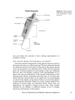
Consider the cellular location of your protein of interest, and
attempt to eliminate contaminating materials at the earliest stages
of the purification. If it is a nuclear binding protein, first isolate the
nuclei from your sample, usually with differential centrifugation,
and then isolate the proteins from the nuclei. If it is a mitochondr-
ial protein, use differential centrifugation to isolate mitochondria
(spin the cell lysate at 3000 ¥ g to remove nuclei, then at 10,000 ¥ g
to bring down mitochondria). If the protein is membrane bound,
use a step gradient of sucrose or other centrifugation medium to
isolate the specific membrane of interest. For soluble proteins, spin
the cell lysate at 100,000 ¥ g to remove all cellular membranes and
Electrophoresis 353
use the supernatant. Note that nucleic acids are very sticky; they
can cause proteins to aggregate together with a loss of elec-
trophoretic resolution. If you have smearing in your sample, add
1 mg/ml of DNase and RNase to remove the nucleic acids.
Is the Problem Caused by Sample Preparation or by
the Electrophoresis?
If a nonprestained standard runs well in a gel, producing sharply
defined, well-shaped bands, then any problems in the sample lanes
lie in sample preparation or in the sample buffer. For this reason
we urge you to run a standard on every gel.
Is the Problem Caused by the Sample or
the Sample Buffer?
For lyophilized standards, make fresh standard buffer. Some-
times it is difficult to determine whether the problem is in the
sample or the sample buffer. Run the standard both with and
without the sample buffer to determine this. It is best to prepare
the sample buffer without reducing agent—dithiothreitol (DTT),
beta-mercaptoethanol (BME), or dithioerythritol (DTE)—freeze
it into aliquots, and add the reducing agent to the aliquot before
use. All these reducing agents evaporate readily from aqueous
solution. Adding the reducing agent fresh for each use means the
reducing agent will always be fresh and in full strength.
Buffer components may separate out during freezing, especially
urea, glycerol, and detergents. Aliquots of sample buffer must be
mixed thoroughly after thawing, to make sure the buffer is a
homogeneous solution.
How Do You Choose a Detergent for IEF or Native PAGE?
Triton X-100 is often used to keep proteins soluble during IEF
or native PAGE, but it may solubilize only 70% of the protein in
a cell (Ames and Nikaido, 1976). SDS is the best solubilizer, but
it cannot be used for IEF because it imparts a negative charge to
the proteins. During the IEF, it is stripped off the proteins by the
voltage potential, and the formerly soluble proteins precipitate in
the IEF gel, resulting in a broad smear. Of course, SDS cannot be
used in native PAGE because it denatures proteins very effec-
tively. Some authors state that SDS may be used in combination
with other detergents at 0.1% or less. It may help solubilize some
proteins when used this way (Molloy, 2000). However, this is not
recommended, as the protein loads must remain low, and other
problems may result (Molloy, 2000).
354 Booz