Development of Enzyme Inhibitors as Drugs pptx
Bạn đang xem bản rút gọn của tài liệu. Xem và tải ngay bản đầy đủ của tài liệu tại đây (2.04 MB, 130 trang )
5
Development of Enzyme
Inhibitors as Drugs
H. John Smith and Claire Simons
CONTENTS
5.1Introduction
5.1.1Basic Concepts
5.1.1.1Substrate (Agonist) Accumulation or Preservation
5.1.1.2Decrease in Metabolite Production
5.2Rational Selection of Suitable Target Enzyme and Inhibitor
5.2.1Target Enzyme
5.2.2Types of Inhibitor for Selected Target Enzyme
5.2.2.1Reversible Inhibitors
5.2.2.2Irreversible Inhibitors
5.3Selectivity and Toxicity
5.4Rational Approach to the Design of Enzyme Inhibitors
5.4.1Lead Inhibitor Discovery
5.4.1.1Modification of the Lead
5.4.2Design from a Knowledge of the Catalytic Mechanism
5.4.2.1Examples
5.4.3Molecular Modeling
5.4.3.1 Crystal Structure of Enzyme or Enzyme–Inhibitor
Complex Available
5.4.3.2 Prediction of 3-D Structure of Enzyme by Other
Means
5.5Development of a Drug Candidate from the Bench to the Marketplace
5.5.1Oral Absorption
5.5.2Metabolism
5.5.2.1Examples
5.5.3Toxicity
5.5.3.1Examples
5.5.4Stereochemistry
5.5.4.1Optical Stereoisomerism
5.5.5Drug Resistance
Further Reading
5.6Enzyme Inhibitor Examples for the Treatment of Breast Cancer
L.W. Lawrence Woo
5.6.1Introduction
5.6.2Endocrine Therapy
© 2005 by CRC Press
5.6.3Aromatase
5.6.4Inhibition of Aromatase as Endocrine Therapy
5.6.4.1Nonsteroidal Aromatase Inhibitors (NSAIs)
5.6.4.2Steroidal Aromatase Inhibitors
5.6.4.3Computer-Aided Drug Design of Aromatase Inhibitors
5.6.5Steroid Sulfatase
5.6.5.1The Enzyme and Breast Cancer
5.6.6Inhibition of Steroid Sulfatase as Endocrine Therapy
5.6.6.1Steroidal STS Inhibitors
5.6.6.2Nonsteroidal STS Inhibitors
5.6.6.3Mechanism of Action for STS and STS Inhibitors
5.6.7Future Directions
Acknowledgment
Further Reading
5.7Enzyme Inhibitor Examples for the Treatment of Prostate Tumor
Samer Haidar and Rolf W. Hartmann
5.7.15a-Reductase and Androgen-Dependent Diseases
5.7.2Inhibitors of 5a-Reductase
5.7.3Prostate Cancer and CYP 17
5.7.4Inhibitors of CYP 17
Further Reading
5.8Thrombin Inhibitor Examples
Torsten Steinmetzer
5.8.1Introduction
5.8.2First Electrophilic Substrate Analog Inhibitors
5.8.3Nonelectrophilic Thrombin Inhibitors
5.8.3.1H-
DPhe-Pro-Agmatine Analogs
5.8.3.2Secondary Amides of Sulfonylated Arginine
5.8.3.3Benzamidine Derivatives of the NAPAP Type
5.8.3.4Nonpeptidic Thrombin Inhibitors
5.8.4Bivalent Inhibitors
Further Reading
5.9HIV-1 Protease Drug Development Examples
Paul J. Ala and Chong-Hwan Chang
5.9.1Introduction
5.9.2Lead Discovery
5.9.2.1Mechanism of Action
5.9.2.2HIV-1 Protease Cleavage Sites
5.9.2.3Structural Information
5.9.3Lead Optimization
5.9.4Drug Resistance
Further Reading
5.10Metalloproteinase–Collagenase Inhibitor Examples
Claudiu T. Supuran and Andrea Scozzafava
5.10.1Introduction
5.10.2Metalloproteinases
5.10.3Inhibition
References
© 2005 by CRC Press
5.1 INTRODUCTION
The majority of drugs used clinically exert their action in one of two ways: (1) by
interfering with a component (agonist) in the body, preventing interaction with its
site of action (receptor), i.e., receptor antagonist, or (2) by interfering with an enzyme
normally essential for the well-being of the body or involved in bacterial or parasitic
or fungal growth causing disease and infectious states, where the removal of its
activity by treatment is necessary, i.e., enzyme inhibitors. In recent years, the pro-
portion of current drugs described as enzyme inhibitors has increased, and this
chapter gives an account of the steps taken for designing and developing such
inhibitors — from identification of the target enzyme to be blocked in a particular
disease or infection to the marketplace.
As has been described in previous chapters, enzymes catalyze the reactions of
their substrates by initial formation of a complex (ES) between the enzyme (E) and
the substrate (S) at the active site of the enzyme. This complex then breaks down,
either directly or through intermediary stages, to give the product (P) of the reaction
with regeneration of the enzyme (Equation 5.1 and Equation 5.2):
(5.1)
(5.2)
where k
cat
is the overall rate constant for decomposition of ES into products; k
2
and
k
3
are the respective rate constants for formation and breakdown of the intermediate
E¢ [i.e., k
cat
= k
2
k
3
/(k
2
+ k
3
)].
Chemical agents known as inhibitors modify the ability of an enzyme to catalyze
the reaction of its substrates, a term that is usually restricted to chemical agents,
other modifiers of enzyme activity such as pH, ultraviolet light, high salt concen-
trations, organic solvents, and heat being known as denaturizing agents.
5.1.1 BASIC CONCEPTS
The body contains several thousand different enzymes, each catalyzing a reaction
of a single substrate or group of substrates. An array of enzymes is involved in a
metabolic pathway each catalyzing a specific step in the pathway up to final metab-
olite production (Equation 5.3). These actions are integrated and controlled in various
ways to produce a coherent pattern governed by the requirements of the cell. Alter-
natively, the enzyme may not be part of a pathway and operates in a single-step
reaction (AB).
(5.3)
ES
ES
E products
k
cat
+æÆæ+
enzyme-substrate
complex
ES ES
E
PEP
k
k
+æÆæ
¢
+æÆæ+
2
3
12
intermediate
ABC
EE
E
E
n12
3
æÆææÆææÆææÆæK metabolite
© 2005 by CRC Press
The use of enzyme inhibitors as drugs is based on the rationale that inhibition
of a suitably selected target enzyme leads first to an accumulation of the substrates
and, second, to a corresponding decrease in concentration of the metabolites; one
of these features leads to a useful clinical response.
5.1.1.1 Substrate (Agonist) Accumulation or Preservation
Where the substrate gives a required response (i.e., agonist), inhibition of its metab-
olizing enzyme leads to accumulation of the intact substrate and accentuation of
that response. Several examples follow:
Accumulation of the neurotransmitter acetylcholine (5.1) by inhibition of the
metabolizing enzyme acetylcholinesterase using neostigmine (5.2) is used for the
treatment of myasthenia gravis and glaucoma (Equation 5.4).
(5.4)
Anticholinesterases, e.g., donepezil (5.3), rivastigmine (5.4), and galantamine
(5.5), capable of penetrating the blood–brain barrier and thereby exerting an effect
on the central nervous system, are used in the treatment of Alzheimer’s disease for
increasing cognitive functions.
Inhibitors have been used (see Equation 5.5) as codrugs to protect an adminis-
tered drug with the required action from the effects of a metabolizing enzyme.
Inhibition of the metabolizing target enzyme permits higher plasma levels of the
CH CO CH CH N CH CH CO H
HOCH CH N CH
acetylcholinesterase
3222 33 32
22 33
+
+
æÆæææææ +()
() ( )5.1
© 2005 by CRC Press
administered drug to persist, thus prolonging its biological half-life and either pre-
serving its effect or resulting in less frequent administration.
Clavulanic acid (5.6), an inhibitor of certain b-lactamase enzymes produced by
bacteria for protection purposes, when administered in conjunction with a b-lacta-
mase-sensitive penicillin, preserves the antibacterial action of the penicillin towards
these bacteria.
(5.5)
Parkinson’s disease is due to degeneration in the basal ganglia, which leads to
reduction in dopamine levels that control muscle tension. Effective treatment for
considerable periods involves administration of the drug L-dopa (5.7), which is
decarboxylated after passage into the brain by a central acting amino acid decar-
boxylase (AADC).
Because L-dopa is readily metabolized by peripheral AADCs (see Figure 5.1),
it is administered with a peripheral AADC inhibitor, i.e., benzserazide (5.8) and
carbidopa (5.9) (which cannot penetrate the brain), to decrease this metabolism and
reduce the necessary administered dose.
FIGURE 5.1 Peripheral and central metabolism of L-Dopa (5.7).
Drug
or
agonist Codrug (inhibitor)
Inert product(s)
metabolizing enzyme
æÆææææææ
≠
Blood-Brain
Barrier
Basal Ganglia
Plasma
central
AADC
L-Dopa L-Dopa
(5.7)
Dopamine
COMT
AADC
3-methoxydopa
Dopamine
© 2005 by CRC Press
A further adjuvant to the above combinations is a catechol-O-methyltransferase
(COMT) inhibitor. COMT peripherally converts L-dopa to 3-methoxydopa with loss
of potency. Entacapone (5.10) (COMTESS) is the inhibitor currently available for
this purpose; tolcapone (5.11) (Tasma), previously used, led in a few instances to
fatal hepatic effects and has been discontinued in the U.K.
5.1.1.2 Decrease in Metabolite Production
When the metabolite has an action judged to be clinically undesirable or too pro-
nounced, inhibition of a relevant enzyme reduces its concentration with a decreased
(desired) response.
Allopurinol is an inhibitor of xanthine oxidase and is used for the treatment of
gout. Inhibition of the enzyme reduces the formation of uric acid from the purines
xanthine and hypoxanthine, from the external precursors; otherwise, the uric acid
deposits and produces irritation in the joints (Equation 5.6).
(5.6)
In the above example, an enzyme acting in isolation was targeted, but additional
strategies may be used with enzyme inhibitors to produce an overall satisfactory
clinical response.
(1) Where the target enzyme is part of a biosynthetic pathway consisting of a
sequence of enzymes with their specific substrates and coenzymes (Equation 5.7),
inhibition of a carefully selected target enzyme in the pathway (see Section 5.2.1)
would lead to prevention of overall production of a metabolite that either clinically
gives an unrequired response or is essential to bacterial or cancerous growth.
Xanthine
Allopurinol
xanthine
oxidase
æÆæææ
≠
uric acid
© 2005 by CRC Press
(5.7)
(2) Sequential chemotherapy involves the use of two inhibitors simultaneously
on a metabolic chain (Equation 5.8) with the aim of achieving a greater therapeutic
effect than by application of either alone.
(5.8)
This situation arises when dosage with a single inhibitor is limited by host
toxicity or resistant bacterial strains have emerged. The best-known combination is
the antibacterial mixture cotrimoxazole, consisting of trimethoprim (5.12) (dihydro-
folate reductase [DHFR] inhibitor) and the sulfonamide sulfamethoxazole (5.13)
(dihydropteroate synthetase inhibitor), although the usefulness of the latter in the
combination has been queried.
(3) A rare example of metabolic pathway inhibition is shown in Equation 5.9 in
which inhibition of an enzyme occasionally leads to formation of a “dead-end”
complex between the enzyme, coenzyme, and inhibitor, rather than straightforward
interaction between the inhibitor and the enzyme. 5-Fluorouracil (5.14) inhibits
thymidylate synthetase to form a dead-end complex with the enzyme and coenzyme,
tetrahydrofolate, thus preventing bacterial growth (Equation 5.9).
(5.9)
Cofactor Z E
2
Z¢ Inhibitor
+ Inhibitor (Dead-end complex)
Topoisomerases I and II are nuclear enzymes that catalyze the concerted breaking
and rejoining of DNA strands to produce the necessary topological and conforma-
tional changes in DNA critical for many cellular processes such as replication,
recombination, and transcription. The antitumor drugs doxorubicin (5.15) and amsa-
A
inhibitor
BCD E
E
E
E
E
n
1
2
3
æÆæ
≠
æÆææÆæºæÆæ (metabolite)
A
inhibitor
BCD
inhibitor
E
E
E
E
E
n1
2
3
12
æÆæ
≠
æÆææÆæº
æÆæ
≠
(metabolite)
ABCDE
EE
E
E
12
3
4
æÆææÆææÆææÆæ (metabolite)
© 2005 by CRC Press
crine (5.16) exert their action by binding to the enzyme-(broken)DNA complex in
a nonproductive ternary dead-end complex.
5.2 RATIONAL SELECTION OF SUITABLE TARGET
ENZYME AND INHIBITOR
5.2.1 T
ARGET ENZYME
Selection of a suitable target enzyme for a particular disease or infection may be
aided by (1) fortuitous discovery of the side effects noted for an existing drug being
used for another purpose where its main target enzyme is known, (2) drugs intro-
duced into therapy after detection of biological activity in screening experiments in
the anticancer and antibacterial setting where the target enzyme was subsequently
searched for and found, (3) examination of the biochemical pathways involved either
in the normal physiological functioning of the cellular processes that may have been
affected in the disease or growth requirements of the bacterial or parasitic infections
and requirements for viral multiplication and spread.
Drugs in current use for one therapeutic purpose have occasionally exhibited
side effects indicative of potential usefulness for another, subsequent work estab-
lishing that the newly discovered drug effect is due to inhibition of a particular
enzyme. Although the drug may possess minimal therapeutic usefulness in its newly
found role, it does constitute an important “lead” compound for the development of
analogs with improved clinical characteristics.
© 2005 by CRC Press
The use of sulfanilamide (5.17) as an antibacterial drug was associated with
acidosis in the body due to its inhibition of renal carbonic anhydrase (CA). This
observation led to the development of the currently used potent inhibitor acetazola-
mide (5.18) as an antiglaucoma agent and subsequently the important chlorothiazide
group of diuretics [e.g., chlorothiazide (5.19) and methylchlorothiazide (5.20)]
although these have a different mode of action. Further developments with carbonic
anhydrase have shown the presence of 14 isoenzyme forms of CA and that CA IX
in particular aids hypoxia (oxygen deficiency) and thus growth in solid cancerous
tumors by creating an acidic environment; specific inhibitors of CA would add to
the anticancer armory.
The anticonvulsant aminoglutethimide (5.112) was withdrawn from the market
due to inhibition of steroidogenesis (steroid hormone synthesis) and an insufficiency
of 11b-hydroxy steroids. Aminoglutethimide, in conjunction with supplementary
hydrocortisone, is now in clinical use for the treatment of estrogen-dependent breast
cancer in postmenopausal women due to its ability to inhibit aromatase, the terminal
enzyme in the pathway, which is responsible for the production of estrogens from
androstenedione. Other much more potent aromatase inhibitors free of depressive
side effects have subsequently been developed (see Section 5.6 examples).
Iproniazid (5.21), initially used as a drug in the treatment of tuberculosis, was
observed to be a central nervous system stimulant due to a mild inhibitory effect on
MAO. This observation, with eventual identification of the enzyme target, led to the
discovery of more potent inhibitors of MAO, such as phenelzine (5.22), tranyl-
cypromine (5.23), selegiline ((-)-deprenyl) (5.24), and chlorgyline (5.25).
© 2005 by CRC Press
Many drugs introduced into therapy following detection of biological activity
by cell culture or microbiological screening experiments have subsequently been
shown to exert their action by inhibiting a specific enzyme in the tumor cell culture
or parasite. This knowledge has helped in the development of clinically more useful
drugs by limiting screening tests to involve only the isolated pure or partially purified
target enzyme concerned and thus introducing a more rapid screening protocol.
A priori examination of the biochemical or physiological processes responsible for
a disease or condition in which these are known or can be guessed at, may point to a
suitable target enzyme in its biochemical environment, the inhibition of which would
rationally be expected to lead to alleviation or removal of the disease or condition.
Inhibitors of the noradrenaline biosynthetic pathway were intended to decrease
production of noradrenaline at the nerve–capillary junction in hypertensive patients,
with an associated reduction in blood pressure. The selected target enzyme, aromatic
amino acid decarboxylase (AADC), catalyzes the conversion of dopa to dopamine
in the second step of the biosynthesis of noradrenaline from tyrosine (Figure 5.2).
Many reversible inhibitors, although active in vitro against this enzyme, fail to lower
noradrenaline production in vivo; however, in an isolated scenario, they may slow
down decarboxylation of dopa in peripheral tissues. Irreversible inhibitors of AADC
successfully lower noradrenaline levels (see Section 5.2.2.2). A possible explanation
for the inability of the AADC inhibitors to produce a satisfactory response in a
metabolic chain is as follows.
In a metabolic chain of reactions with closely packed enzymes in a steady state
(see Equation 5.10) in which the initial substrate (A) does not undergo a change in
concentration as a consequence of changes effected elsewhere in the chain, any type
of reversible inhibitor that inhibits the first step of the chain effectively blocks that
sequence of reactions.
(5.10)
Inhibitors acting at later points in the chain of closely bound enzymes may not
block the metabolic pathway. If the reaction B Æ C (Equation 5.10) is considered,
FIGURE 5.2 Conversion of dopa to dopamine by the action of AADC.
A B C D metabolite
EE
E
E
1
1
2
2
3
3
4
4
æÆææÆææÆææÆæ
uu u u
© 2005 by CRC Press
competitive inhibition of E
2
initially decreases the rate of formation of C, but
eventually the original velocity (n
2
) of the step is attained as the concentration of B
rises due to the difference between its rates of formation and consumption. However,
these changes relating to an increase in concentration of B may have secondary
effects on the chain due to product inhibition (B on E
1
) or product reversal (A ¨ B);
either of these effects can reduce n
1
, thus leading to a slowing down of the overall
pathway, i.e., here, inhibition of the second enzyme has a successful outcome. There
is a general misconception that the overall rate in a linear chain can be depressed
only by inhibiting the rate-limiting reaction, i.e., the one with the lowest velocity at
saturation with its substrate. Because individual enzymes cannot be saturated with
their substrates, the overall rate is determined largely by the concentration of the
initial substrate, so that the first enzyme will often be rate limiting, irrespective of
its potential rate due to a low concentration of its substrate.
A knowledge of the structure, life cycle, and replication of the human immun-
odeficiency virus (HIV) has led to the development of inhibitors of the virally
encoded protease essential for maturation of the virus and hence production and
spread. Aspects of this work are discussed in Section 5.9.
5.2.2 TYPES OF INHIBITOR FOR SELECTED TARGET ENZYME
As described in detail in Chapter 4, enzyme-inhibiting processes may be divided
into two main classes, reversible and irreversible, depending upon the manner in
which the inhibitor (or inhibitor residue) is attached to the enzyme.
5.2.2.1 Reversible Inhibitors
Reversible inhibitors may be competitive, noncompetitive, or uncompetitive, depend-
ing upon their point of entry into the enzyme–substrate reaction scheme. In either
case, the inhibitor is bound to the enzyme through a suitable combination of forces;
van der Waal’s, electrostatic, hydrogen bonding, and hydrophobic (see Chapter 2).
The extent of the binding is determined by the equilibrium constant K
I
for breakdown
of the EI or EIS complex for classical inhibitors. However, on rare occasions a
covalent bond may be formed with an active site residue, as in the case of a
hemiacetal or hemiketal bond with the catalytic serine in serine proteases with a
polypeptide aldehyde or ketone-based inhibitor, but the EI complex readily dissoci-
ates back into free enzyme and inhibitor as the free inhibitor concentration falls due
to dilution, excretion, metabolism, etc.
Competitive inhibitors, as their name suggests, compete with the substrate for
the active site of the enzyme, and by forming an inactive enzyme–inhibitor complex,
decrease the rate of catalysis by the enzyme of the substrate (Equation 5.11):
(5.11)
ES
IK
EI
ES E P
I
K
k
S
+
≠Ø
æÆææÆæ+
inactive enzyme-
inhibitor complex
2
© 2005 by CRC Press
The Michaelis–Menten equation for the rate (n) of an enzyme-catalyzed reaction
in the presence of an inhibitor is given by
(5.12)
where it is seen that, in the presence of the inhibitor, the extent to which the reaction
is slowed is dependent on the inhibitor concentration [I] and the dissociation con-
stant, K
i
, for the EI complex. A small value for K
i
(ª10
6
to 10
8
M) indicates strong
binding of the inhibitor to the enzyme. With this type of inhibitor, the inhibition
may be overcome, for a fixed inhibitor concentration, by increasing the substrate
concentration as seen in Equation 5.12. With competitive inhibition, only substrate
binding, i.e., K
m
, is affected because the inhibitor competes with the substrate for
the same binding site, i.e., K
m¢
= K
m
(1 + [I]/K
I
). Determination of K
I
has been
previously described in Chapter 4 and is a parameter for comparing the potency of
inhibitors because it is independent of substrate and inhibitor concentration.
Noncompetitive inhibitors combine with the enzyme–substrate complex and
prevent the breakdown of the complex to products (Equation 5.13).
(5.13)
These inhibitors do not compete with the substrate for the active site; they only
change the V
max
parameter for the reaction. The binding strength of the inhibitor to
either E or ES is identical so that there is a single value for K
I
. The kinetics for this
type of inhibitor are given by
(5.14)
The extent of the inhibition by a fixed concentration of inhibitor is not reversed
by increasing the substrate concentration (in contrast to competitive inhibition)
because substrate and inhibitor bind at different sites.
Uncompetitive inhibition is the third type of reversible inhibitor and is rare in
single-substrate catalysis.
(5.15)
u=
++
Ê
Ë
Á
ˆ
¯
˜
V
K
S
I
K
m
I
max
[]
[]
11
ES
EI S
ES
EIS
EP
I
K
I
K
I
I
II
+
≠Ø
+
Ø≠
æÆæ+
+
-+
-
u=
+
Ê
Ë
Á
ˆ
¯
˜
◊
+
=
+
+
V
I
K
S
SK
V
IK
KS
I
m
I
m
max
max
[]
[]
[]
([]/)
(/[])
1
1
1
ES ES
EIS
EP
K
I
I
I
+
Ø≠
æÆæ+
+
-
© 2005 by CRC Press
This type of inhibitor binds only to the enzyme–substrate complex; perhaps
substrate binding produces a conformation change in the enzyme, which reveals an
inhibitor-binding site. The modified Michaelis–Menten equation is shown in Equa-
tion 5.16 where it could be seen that both K
m
and V
max
are modified.
(5.16)
Noncompetitive and uncompetitive inhibitors are uncommon and have only
recently made their appearance in drug discovery studies as a result of random
screening of chemical libraries by the pharmaceutical industry for inhibitors of a
new drug target. These types of inhibitors are impossible to design (without a lead)
because the characteristics of their binding sites in the ES complex are not comple-
mentary to the structure of the known substrate on which the great majority of
competitive inhibitors can be readily modeled.
As previously discussed in Chapter 4, occasionally the Lineweaver–Burk plot,
used to determine the inhibitor type, shows a pattern that can lie between either (1)
competitive and noncompetitive inhibition, or (2) noncompetitive and uncompetitive
inhibition. This form of inhibition is termed mixed inhibition and arises because the
inhibitor binds to both E and the ES complex but with different binding constants
(K
i
and K
I
).
There are two special types of competitive inhibitors that bind very strongly to
the target enzyme; the transition-state analog and tight-binding inhibitor.
A transition-state analog is a stable compound that resembles in structure the
substrate portion of the enzymic transition state for chemical change; it differs in
this respect from the transition-state structure formed after reaction between, for
example, a serine moiety at the active site of a serine protease and a peptidyl ketone
inhibitor, i.e., the oxyanion-containing tetrahedral intermediate (see Chapter 3).
An organic reaction between two types of molecules is considered to proceed
through a high-energy-activated complex known as the transition state, which is
formed by the collision of molecules with greater kinetic energy than the majority
present in the reaction. The energy required for the formation of the transition state
is the activation energy for the reaction and is the barrier to the reaction occurring
spontaneously. The transition state for the reaction between hydroxyl ion and methyl
iodide involves both the commencement of formation of a C–OH bond and the
breaking of the C–I bond; it may break down to give either the components from
which it was formed or the products of the reaction. Enzymes catalyze organic
reactions by lowering the activation energy for the reaction, and one view is that they
accomplish this by straining or distorting the bound substrate towards the transition
state (see Chapter 2). Equation 5.17 shows a single-substrate enzymatic reaction and
the corresponding nonenzymatic reaction in which ES
π
and S
π¢
represent the transition
u=
+
Ê
Ë
Á
ˆ
¯
˜
+
+
Ê
Ë
Á
ˆ
¯
˜
=
¢
+
¢
V
I
K
K
S
I
K
V
K
S
I
m
I
max
max
max
[]
[]
[]
[]
1
1
1
1
© 2005 by CRC Press
states for the enzymatic and nonenzymatic reaction, respectively, and K
N
π
and K
E
π
are equilibrium constants, respectively, for their formation. K
s
is the association
constant for formation of ES from E and S, and K
T
is the association constant for
the hypothetical reaction involving the binding of S
π¢
to E. Analysis of the relationships
between these equilibrium constants shows that K
T
K
N
π
= K
s
K
E
π
. Because the equi-
librium constant for a reaction is equal to the rate constant mutiplied by h/kT, where
h is Planck’s constant and k is the Boltzmann’s constant, K
T
= K
s
(k
E
/k
N
), where k
E
and k
N
are the first-order rate constants for breakdown of the ES complex and the
nonenzymatic reaction, respectively. Because the ratio k
E
/k
N
is usually of the order
10
10
or greater, it follows that K
T
>> K
s
. This means that the transition state S
π¢
is
considered to bind to the enzyme at least 10
10
times more tightly than the substrate.
(5.17)
A transition-state analog is a stable compound that structurally resembles the
substrate portion of the unstable transition state of an enzymatic reaction. Because
the bond-breaking and bond-making mechanism of the enzyme-catalyzed and non-
enzymatic reactions are similar, the analog resembles S
π¢
and has an enormous
affinity for the enzyme, and binds more tightly than the substrate. It would not be
possible to design a stable compound that mimics the transition state closely because
the transition state itself is unstable by possessing partially broken and partially
made covalent bonds. Even crude transition-state analogs of substrate reactions
would be expected to be sufficiently tightly bound to the enzyme to be excellent
reversible inhibitors, an expectation that has been borne out.
As will be seen in Section 5.4.2.1.3, the design of a transition-state analog for
a specific enzyme requires knowledge of the mechanism of the enzymatic reaction.
Fortunately, the main structural features of the transition states for the majority of
enzymatic reactions are either known or can be predicted with some confidence.
Tight-binding inhibitors bind tightly to the enzyme either noncovalently or
covalently and are released very slowly from the enzyme because of the tight
interaction. The slow binding is a time-dependent process and is believed to be due
either to an enforced conformational change in the enzyme structure or reversible
covalent bond formation or, more probably, simply the very low inhibitor concen-
tration used during measurement to allow observation of a residual activity. The
drugs coformycin, methotrexate, and allopurinol belong to this class and are useful
drugs. Tight binding, in which the dissociation from the complex takes days, is not
distinguishable in effect from covalent bonding, and this type of inhibitor may be
classed as an irreversible inhibitor.
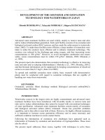
5.2.2.1.1 Parameters for Determining Relative Inhibitory Potency
In the initial screening of inhibitors, it is convenient to compare potencies within
tested compounds as percentage inhibition. However, the relationship between the
ES
ES
ES
ES
EP
EP
K
S
K
K
K
T
N
N
+
Ø≠
¢
+
Ø≠
æÆæ
æÆæ
+
Ø≠
π
π
¢
π
π
© 2005 by CRC Press
percentage inhibition of an enzyme and inhibitor concentration ([S] = constant) is
not linear, and the relative concentration to inhibit enzyme activity by 50, 90, and
99% under standard conditions increases logarithmically, i.e., 10
0
, 10
1
, and 10
2
,
respectively (see Table 5.1). In screening tests, although the potencies of different
inhibitors may appear similar within the range 90 to 95% inhibition, their potencies
may be very different when IC
50
values from further experiments are compared.
Expression of potency as an IC
50
value (concentration of inhibitor required to
inhibit enzyme activity by 50%) is a convenient measure of potency within a
laboratory, but this value should be used with care when comparing interlaboratory
results for competitive inhibition because it is dependent on the concentration of
substrate used (Equation 5.18), which may vary between laboratories.
(5.18)
For noncompetitive and uncompetitive inhibitors, IC
50
= K
I
and is independent
of substrate concentration. K
I
is independent of substrate and inhibitor concentrations
for all classes of reversible inhibitors.
5.2.2.2 Irreversible Inhibitors
Compounds producing irreversible enzyme inhibition fall into two groups: active
site–directed (affinity labeling) inhibitors and mechanism-based inactivators (k
cat
inhibitors, suicide substrates).
5.2.2.2.1 Active Site-Directed Irreversible Inhibitors
These resemble the substrate sufficiently to form a reversible enzyme–inhibitor
complex, analogous to the enzyme–substrate complex, within which reaction occurs
TABLE 5.1
Relationship between Percentage Competitive
Reversible Inhibition of an Enzyme and Relative
Inhibitor Concentration
Percentage Inhibition Relative Inhibitor Concentrations
10 0.1
50 1
a
67 3
76 5
90 10
99.01 100
99.90 1000
99.99 10000
a
IC
50
value
IC
50
1=+
Ê
Ë
Á
ˆ
¯
˜
K
S
K
I
m
© 2005 by CRC Press
between functional groups of the inhibitor (e.g., –COCH
2
Cl, –COCHN
2
,
–OCONHR, –SO
2
F) and enzyme (–SH, =N–, –NHR, –OH). A stable covalent bond
is formed with irreversible inhibition of the enzyme. Active site-directed irreversible
inhibitors are designed to exhibit specificity towards their target enzymes because
they are structurally modeled on the specific substrate of the enzyme concerned.
These inhibitors are termed affinity-labeling agents when used to probe the nature
of the functional groups present in the active site.
Irreversible inhibitors progressively reduce enzyme activity with time, and the
reaction follows pseudo-first-order kinetics as described in Chapter 4. The biochem-
ical environment of the enzyme (see Section 5.2.1) is unimportant so that any step
in a biosynthetic pathway may be inhibited with decrease in overall metabolite
production. However, because these compounds belong to a group that mainly
consists of alkylating and acylating agents, they have not been developed as drugs
as they would be expected to react with a range of tissue constituents containing
amino or thiol groups besides the target enzyme, with potentially serious side effects.
However, they have been used successfully as affinity labeling agents.
5.2.2.2.2 Mechanism-Based Enzyme Inactivators
Many irreversible inhibitors of certain enzymes have previously been recognized,
among which the range of electrophilic centers normally associated with active site-
directed irreversible inhibitors, e.g., –COCH
2
Cl, –COCHN
2
, –OCONHR, –SO
2
F,
were absent, and therefore the means by which they inhibited the enzyme was
unclear. The action of these inhibitors has, in more recent years, become understand-
able because they have been categorized as mechanism-based enzyme inactivators.
Mechanism-based enzyme inactivators bind to the enzyme through the K
s
parameter
to form a complex and are modified by the enzyme in such a way as to generate a
reactive group that irreversibly inhibits the enzyme by forming a covalent bond with
a functional group present at the active site. Occasionally, catalysis leads not to a
reactive species but an enzyme–intermediate complex that is partitioned away from
the catalytic pathway to a more stable complex by bond rearrangement (e.g., b-
lactamase inhibitors).
These inhibitors are substrates of the enzyme, as suggested by their alternative
name k
cat
inhibitors, where, as explained earlier, k
cat
is the overall rate constant for
the decomposition of the enzyme–substrate complex in an enzyme-catalyzed reac-
tion. Mechanism-based inactivators do not generate a reactive electrophilic center
until acted upon by the target enzyme. Reaction may then occur with a nucleophile
on the enzyme surface, or alternatively the species may be released and either react
with an external nucleophile or decompose (Equation 5.19)
(5.19)
The biochemical environment of the target enzyme is unimportant (see Section
5.2.1). For example, in the noradrenaline biosynthetic pathway, a-monofluorometh-
EI EI
EI
EP
EI
k
k
k
k
k
+æÆæ
Ø
+
æÆæ-
-
+
+
+
+
1
1
2
3
4
*
© 2005 by CRC Press
yldopa (5.26), a mechanism-based inactivator of AADC, produces a metabolite that
irreversibly inhibits and decreases the level of the enzyme by >99%. This leads to
a near-complete depletion of catecholamine levels in brain, heart, and kidney, despite
the occurrence of the enzyme in the second step of the noradrenaline biosynthetic
pathway, as discussed earlier.
Mechanism-based inactivators do not possess a biologically reactive functional
group until after they have been modified by the target enzyme and, consequently,
would be expected to demonstrate high specificity of action and low incidence of
adverse reactions. It is these features that have encouraged their active application
in inhibitor design studies.
5.2.2.2.3 Parameters for Determining Relative Potency of
Irreversible Inhibitors
Previously, for reversible inhibitors, the potency of an inhibitor was shown to be
reflected in the K
I
value, which is characteristic of the inhibitor and independent of
inhibitor concentration. Similarly, the potency of an irreversible inhibitor is given
by the binding and kinetic rate constants, both of which are independent of inhibitor
concentration (Equation 5.20). This allows a precise comparison of the relative
potency of inhibitors, which is necessary in the design and development of more
effective inhibitors of an enzyme.
(5.20)
complex inhibited
enzyme
K
I
and k
+2
can be determined by the method previously described in Chapter 4,
and inhibitor potency can be related to the ratio k
+2
/K
I
for comparative purposes.
The potency increases as binding increases (K
I
decreases) and the rate constant for
the reaction increases (k
+2
increases). In a similar manner, the binding and reaction
rate constant for a mechanism-based enzyme inactivator can be determined, but
another aspect, the partition ratio, enters into the efficiency of these agents.
The ratio of the rate constants, i.e., k
+4
/k
+3
, gives the partition ratio (r) for the
reaction, and when this approaches zero, the mechanism-based inactivation will
proceed with a single turnover of the inhibitor, where the noncovalent enzyme–inhib-
itor complex (EI) is transformed into an activated species (EI*) that then irreversibly
inhibits the enzyme (Equation 5.21).
(5.21)
EI EI EI
K
k
I
+æÆæ-
+
()()
2
EI EI EI EI
K
k
k
I
+æÆææÆæ-
+
+
2
3
*
© 2005 by CRC Press
For mechanism-based inactivators, the turnover rate of the enzyme is important
because of enzyme resynthesis, and this rate may be 10
3
to 10
5
times slower than
for natural substrates. The partition ratio of the reaction should ideally be close to
zero, in which case every turnover results in inhibition; the reactive electrophilic
species, by not being free to react with other molecules in the biological media, has
a high degree of specificity for its target enzyme and exhibits low toxicity.
5.3 SELECTIVITY AND TOXICITY
Inhibitors used in therapy must show a high degree of selectivity towards the target
enzyme. The term selectivity is preferred to specificity because the latter is unob-
tainable within a group of closely related enzymes.
Inhibition of closely related enzymes with different biological roles (e.g., trypsin-
like enzymes such as thrombin, plasmin, and kallikrein), or reaction with constituents
essential for normal functioning of the body (e.g., DNA glutathione, liver P450-
metabolizing enzymes) could lead to serious side effects. An inhibitor being devel-
oped for potential clinical use is put through a spectrum of in vitro tests against
other realistic potential enzyme targets to ascertain that it is suitably selective towards
the intended target. An inhibitor with high potency, e.g., IC
50
= 5 nm, would be
screened at 1 mM against other targets, and a small percentage inhibition would rate
as a demonstration of acceptable selectivity. The aromatase inhibitor fadrozole (5.27)
at higher doses than likely to be achieved clinically showed inhibition of the 18-
hydroxyase in the steroidogenesis pathway, which could affect aldosterone produc-
tion in the clinical setting. With the further developed compound letrozole (5.28)
the observed selectivity between the two enzymes noted with fadrozole (10-fold)
was widened by at least an order (100-fold).
Where the target enzyme is common to the host’s normal cells as well as to
cancerous or parasitic cells, chemotherapy can be successful when host and parasitic
cells contain different isoenzymes, e.g., dihydrofolate reductase (DHFR), with those
of the parasite being more susceptible to carefully designed inhibitors.
On very rare occasions, the target enzyme may be absent from the host cell.
Sulfonamides [e.g., sulfamethoxazole (5.13)] are toxic to bacterial cells by inhibiting
dihydropteroate synthetase, an enzyme on the biosynthetic pathway to folic acid
essential for bacterial growth. The host cell is unaffected because it utilizes pre-
formed folic acid from the diet, which the susceptible bacteria is unable to do.
© 2005 by CRC Press
Another example relates to the carbonic anhydrase (CA) isoforms CA IX and
CA XII that predominate in cancer cells (but are absent in normal cells) and are
concerned in maintaining an acidic environment leading to hypoxia essential for
solid tumor growth. Inhibitors of CA IX and XII, as antitumor agents, would need
to be very selective because up to 12 other CA isoforms are known in man and are
also involved in the interconversion between CO
2
and HCO
3
–
, critical for many
physiological processes (especially, CA I, CA II, and CA IV).
Normal and cancerous cells contain the same form of the target enzyme, DHFR,
but the faster rate of growth of tumor cells makes them more susceptible to the
effects of an inhibitor. Although side effects occur, these are acceptable due to the
life-threatening nature of the disease.
5.4 RATIONAL APPROACH TO THE DESIGN OF
ENZYME INHIBITORS
Once the target enzyme has been identified, then usually a lead inhibitor has previ-
ously been reported, or can be predicted from studies with related enzymes, or has
appeared, in more recent years, from the rapid screening methods now available of
industrial chemical collections (libraries). The design process is then initially con-
cerned with optimizing the potency and selectivity of action of the inhibitor to the
target enzyme using in vitro biochemical tests; nowadays, this has the advantage
that the pure enzyme from recombinant DNA technology may be available for such
studies. Candidate drugs are then examined by in vivo animal studies for oral
absorption, stability to the body’s metabolizing enzymes, and toxic side effects.
Because many candidates may fall at this stage, the whole design cycle recommences
because further design is then necessary to maintain desirable features and eliminate
undesirable features from the in vivo profile. Because an in vivo profile in animal
studies is not directly translatable to the human situation, studies with human vol-
unteers are also required before a drug enters clinical trials.
5.4.1 LEAD INHIBITOR DISCOVERY
A lead inhibitor is usually a compound of low potency and selectivity whose structure
can be used as a scaffold for structural modification to other compounds whose
potency and selectivity are enhanced, together with many other desirable features,
thereby leading to a compound that may eventually reach the marketplace. Discovery
of a lead inhibitor for a new target enzyme may arise in conjunction with the
discovery of the target enzyme (see Section 5.2.1) through (1) discovery of side
effects noted for an existing drug (used for another purpose with a different target)
where the side effect is the clinical effect required and expected to be shown on
inhibition of the new proposed target enzyme, (2) compounds showing low potency
in pharmacological, antibacterial, antiparasitic, or antiviral effects from screening
experiments in which subsequent examination has revealed that it acts on the pro-
posed target enzyme.
© 2005 by CRC Press
5.4.1.1 Modification of the Lead
The design of a novel inhibitor of a new target enzyme takes into account a com-
bination of several different design approaches based on (1) modification of the
structural scaffold of a lead inhibitor, if this is available, (2) a knowledge of the
substrate and the mechanism of the catalytic reaction and perhaps a lead inhibitor
(which may be from a closely related enzyme with a known structure), and (3) use
of computer-assisted molecular graphics (molecular modeling).
Once a lead inhibitor has been identified, from whatever combination of the
design aspects described above, a process of optimization of its potency and selec-
tivity using in vitro tests is undertaken. This process involves chemical manipulation
of the lead, and although many general structural approaches are available, an
appropriate strategy will suggest itself for a particular lead. The principal concepts
followed are generalized as follows: (1) Replace existing groups with groups com-
parable in size (volume) and charge, an approach well known in medicinal chemistry
for receptor agonists as well as enzyme inhibitors as “isosteric replacement” (see
Section 5.4.1.1.1). (2) Because the binding of peptide substrates to enzymes occurs
through a hydrogen-bonding network between hydrogen bond acceptors (R
2
C=O,
–O–, R
3
N:) and donors (=NH, bound H
2
O, –OH) along the two surfaces (as for
a–helix and b–pleated sheet structures in proteins), as well as the interaction of
oppositely charged groups (COO
–
, R
3
NH
+
), increasing the potential hydrogen-bond-
ing groups in the inhibitor could increase potency if they are correctly positioned.
(3) Many enzymes have a hydrophobic (water-repelling) cavity containing alkyl or
aromatic or heterocyclic residues as part of the active site for binding of peptide
amino acid residues. Introduction of similar residues in the inhibitor if correctly
positioned will improve binding.
However, concepts (2) and (3) will affect the hydrophilic–hydrophobic balance
in the drug, necessary for the penetration of membranes in its transport to the site
where its target is situated (see Section 5.5.1). The size of the hydrophobic cavity
may limit the size of any hydrophobic groups introduced. (4) There is restriction
in the lead compound of extended alkyl and carbon chains because these are not
only susceptible to metabolic hydroxylation by liver-metabolizing P450 enzymes
(see Section 5.5.2) but can also be freely rotated and therefore may not be correctly
positioned for binding in the required position of the enzyme; the energy gained
on binding will be depleted by the energy required to reposition the chain, i.e.,
lower binding energy. Ring formation between flexible chains restricts flexibility
and is a technique frequently used. (5) Remove, where possible, stereochemical
aspects from the inhibitor, i.e., asymmetric carbon centers leading to (R)- and (S)-
isomers and cis or trans (Z/E) isomerization around a C=C double bond. Such
stereochemistry complicates drug development because activity usually resides with
one isomer (see Section 5.5.4).
Potent, selective drug candidates from in vitro studies may fail in an in vivo
animal model and require further structural manipulation to improve their in vivo
profile when the whole of the synthetic and in vitro testing cycle recommences.
Even then, for human testing there is usually a backup compound available to replace
the main candidate drug, should it fail.
© 2005 by CRC Press
5.4.1.1.1 Isosterism
A receptor agonist or antagonist, or enzyme inhibitor, has a skeleton complementary
to the respective target protein site so as to present binding groups in the correct
orientation to complementary sites on the protein for specific hydrogen bond, ionic,
dipolar, and nonspecific hydrophobic interactions. Isosteric replacement of groups
or atoms in the skeleton was an attempt in the very early days of drug design to
maintain the structurally specific requirements of a drug before much was known
concerning drug–target-protein interactions. The concept is that isosteric modifica-
tion is the replacement of an atom, or group of atoms, in a molecule by another
group with similar electronic and steric configurations. Isosterism was an attempt
to apply to molecules or molecular fragments the premise that similarities to prop-
erties of elements within vertical groups of the Periodic Table were due to identical
valence electronic configurations. Thus, two molecular fragments containing an
identical numbered arrangement of electrons should have similar properties, and
were termed isosteres. Further, the early recognition that benzene and thiophene
were alike in many of their physical properties led to the term ring equivalent to
describe the interchanging of –CH=CH– and –S–, which distinguishes their struc-
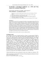
tures. Table 5.2 shows isosterically related atoms or groups (where the presence of
hydrogen atoms is ignored).
The outer electrons (numbers in parentheses) are calculated as follows: For –N=,
the lone pair is unshared, and the other 6 electrons of the three bonds are shared
giving a total of (2 + 6/2) = 5 outer electrons.
The isosterism concept has been applied with great success in the past and is
invariably referred to in lead manipulations in the current development of drugs, but
needs to be applied with caution. Preservation of the template for correct orientation
of specific atom or group bonding (i.e., the pharmacophore) by isosteric substitution
should not be confused with (1) substitution of nonspecific functions that are hydro-
phobic and bind to suitably adjacent hydrophobic amino acid residue side chains on
the target protein but outside the pharmacophore, thus increasing binding and
potency, or (2) hydrophobic or hydrophilic functions introduced to adjust the balance
of such functions in the molecule to improve membrane penetration or introduce
metabolic stability. The isosteric concept does not distinguish between the structur-
ally specific requirements of a drug, and its nonspecific requirements outside the
pharmacophore area for improving inherent drug potency and its ability to reach its
target protein.
TABLE 5.2
Isosterically Related Atoms and Groups
Electronic Configurations 2(4) 2(5) 2(6) 2(7)
=C= –N= –O– –F
–CH= –NH– –OH
–CH
2
– –NH
2
–CH
3
© 2005 by CRC Press
Furthermore, the concept of isosteric replacement does not take into account
ionization, and the –NH
2
>N– function replacements mentioned here are weak bases
of the phenylamine type where protonation does not occur at physiological pH.
5.4.1.1.1.1 Replacement of Univalent Atoms or Groups
The analogous groups considered here –F (or –Cl), –OH, –NH
2
, and –CH
3
are
seen in Table 5.2, and such replacements have been successfully used in the
development of hypoglycemic agents — development of inhibitory activity to
dihydrofolate reductase when the –OH group of folic acid (5.29) was replaced by
–NH
2
[aminopterin (5.30)], 6 –OH of hypoxanthine (5.31), and guanine (5.32)
was replaced by –SH to give the anticancer drugs 6-mercaptopurine (5.33) and 6-
thioguanine (5.34), and barbiturates to short-acting thiobarbiturates (–N=C–OH
Æ –N=C–SH).
5.4.1.1.1.2 Replacement of Divalent Atoms or Groups
In esters, the rotation of C–O–C bonds is restricted by resonance (5.35), and aliphatic
esters exist, predominantly, in the cis configuration (5.35) rather than the trans (5.36).
Studies on amides have also revealed similar planar structures and a predominant
configuration (5.37) analogous to the cis ester. Conversion of the ester function in
local anesthetics to amide prolongs their action in the body by preventing esterase
actions; here polarity is maintained.
© 2005 by CRC Press
Interchange of –O–, –S–, –NH–, and –CH
2
– have been used in the development
of several groups of drugs, e.g., antihistamines, anticholinergic spasmolytics, anti-
depressant drugs, and antiulcer drugs.
5.4.1.1.1.3 Interchange of Trivalent Atoms and Groups
The substitution of –CH= by –N= in aromatic rings has been one of the most
successful applications of isosterism. One of the most potent antihistamines,
mepyramine (5.38), has evolved from replacement of a phenyl group in antergan
(5.39) by pyridyl. The p-electron deficiency of the pyridine nucleus enables the
nitrogen electron pair to hydrogen-bond with a water molecule, causing an increase
in hydrophilicity that is significant in determining the high level of biological activity.
This is a nonspecific replacement.
The substitution of a benzene ring by pyridine has also resulted in improved
activity in the tricyclic, antihistaminic, and neuroleptic (major tranquilizing) drugs
with the introduction of isothipendyl (5.40) cf. promethazine (5.41) and prothipendyl
(5.42) cf. promazine (5.43).
Earlier examples of –N=/–CH= substitutions are to be found in the sulfonamide
antibacterials with the development of sulfapyridine (5.44) and sulfadiazine (5.45),
in which the heterocyclic ring confers greater acidity on the sulfonamide group,
leading to the required degree of ionization at physiological pH.
© 2005 by CRC Press
Aromatic ring substitution of –CH=CH– by –S– or –O–, previously referred to
as “ring equivalents,” has been profitable. These are nonspecific bonding moieties.
Replacements in the pyridyl ring of sulfapyridine (5.44) has produced the five-mem-
bered ring structures sulfathiazole (5.46), sulfisoxazole (5.47), and sulfamethizole
(5.48), which are more soluble in urine and less liable to crystallize in the renal tubules.
The reason for increased solubility is that pyridine and pyrimidine analogs confer
increased water solubility compared to the phenyl ring because the nitrogen lone
pair does not participate in the heteroaromatic ring resonance (p-electron-deficient
ring) and is able to hydrogen-bond with water. Alternatively, pyrrole, furan, and
thiophene, ring equivalent analogs of phenyl, are almost insoluble in water because
the N, O, and S lone pairs participate in ring resonance and are not available for
hydrogen-bonding to water. However, replacement of –C= by –N= (thus converting
pyrrole to imidazole, furan to oxazole, and thiophene to thiazole) increases water
solubility as expected, due to introduction of a p-electron-deficient nitrogen into the
ring system. The interchanging of phenyl with sulfur-, oxygen-, and nitrogen-con-
taining heterocyclic rings has also been extensively exploited in the development of
semisynthetic penicillins and cephalosporins with broader spectra of activity and
greater stability towards b-lactamases.
© 2005 by CRC Press
Ring replacement of –N< by –HC< and its subsequent modification to >C= have
resulted in a variety of useful drugs. This is seen in the development of psychother-
apeutics such as chlorprothixene (5.49) and amitriptyline (5.50), and the antiinflam-
matory drug sulindac (clinoril) (5.51). Substitution of the pyridyl amino –N< by
–HC< in the antihistamine mepyramine (5.38) (and 4-methoxybenzyl by 4-chlo-
rophenyl) produces chlorpheniramine (5.52) valued for its short, powerful action
and relative freedom from sedation. Similarly, the indole nucleus of the antiinflam-
matory drug indomethacin (5.53) (a prostaglandin synthetase (COX) inhibitor) has
been modified (>N–C(=O)– Æ >C=CH–) in sulindac (5.51).
5.4.1.1.1.4 Other Isosteric Modifications
Several modifications are summarized here, in which the modified molecule bears a
more general resemblance to the parent and are only termed isosteric for convenience.
Reversal of groups — An ester may be reversed from –OC(=O)R to
–C(=O)–OR, or an amide from –C(=O)NHR to –NHC(=O)R. The dipolar character
and hydrogen-bonding capacity of the molecule is maintained.
Ring opening and closure — Sulfonamide oral hypoglycemic agents arose
directly from the clinical observation that a sulfathiazole derivative (5.54), which
was being used specifically for treating typhoid, lowered the blood sugar almost to
a fatal level. Modifications involving opening of the thiazole ring to give a thiourea
unit in which =S was ultimately replaced by =O yielded carbutamide (5.55a) that
was later replaced by the less toxic tolbutamide (5.55b).
The antitubercular thiosemicarbazones were developed from the observation that
sulfathiadiazole (5.56) possessed weak antitubercular properties and through subse-
quent testing of intermediates in the synthesis of aminothiadiazoles, i.e., the open
chain analog (5.57). The thiosemicarbazone group is also associated with antiviral
activity, and methisazone (5.58) was developed, which prevents smallpox infection
among people who have previously been in contact with fatal cases.
© 2005 by CRC Press