Introduction to Modern Liquid Chromatography, Third Edition part 12 potx
Bạn đang xem bản rút gọn của tài liệu. Xem và tải ngay bản đầy đủ của tài liệu tại đây (165.53 KB, 10 trang )
66 BASIC CONCEPTS AND THE CONTROL OF SEPARATION
present, it will be necessary to add a buffer to the mobile phase so as to control its
pH; pK
a
values for sample compounds, if available, can be useful during method
development (Chapter 7). The molecular weight of the sample may also affect the
choice of separation conditions for initial experiments (Sections 9.1.1, 13.4.1.4).
Samples that contain enantiomers will require the development of a special method
for their separation (Chapter 14).
The separation goals for a sample may not be limited to adequate resolution
and minimum run time. Depending on the equipment that is available for the routine
application of the final method, gradient elution may not be possible (an isocratic
method will therefore be required). Trace analysis, as in the determination of com-
pound impurities, may impose additional requirements on both sample preparation
and the detector. Quantitative analysis calls for some minimum precision (e.g.,
±1–2% for major components and ±10–20% for trace constituents). However,
often only some of the sample components will require separation; for example,
drugs present in blood or urine, or pesticides in water or soil samples.
2.5.4.2 Sample Pretreatment
Prior to injection the sample may require some processing in order to remove com-
ponents that can damage the column or interfere with the separation of compounds
of interest. Sample pretreatment procedures (Chapter 16) involve multiple steps and
use of a wide range of separation media. For this reason these procedures can be
more difficult to develop than the subsequent HPLC separation. When a pretreat-
ment procedure for a similar combination of analyte and sample matrix is available
(either as developed in the same laboratory or as reported in the literature), its use
or adaptation for the sample is often preferred, so complete re-development of a
sample-pretreatment procedure can be avoided (this approach is less likely to be
applicable for HPLC separation per se; see Section 2.5.4.5).
2.5.4.3 Selection of Chromatographic Mode
Reversed-phase chromatography is the default choice for HPLC method develop-
ment. However, depending on the sample, other chromatographic modes may be
preferable. Often times this does not become apparent until initial experiments with
RPC prove unsuccessful. Similarly, if isomeric compounds are present in the sample
and prove difficult to separate by RPC, the use of NPC with unbonded silica as
column packing will often prove more successful. NPC with unbonded silica is also
preferred for preparative-scale separations (Chapter 15).
2.5.4.4 Detector Selection
The variable-wavelength UV detector is usually a first choice when sample compo-
nents have an adequate chromophore. The molecular structures of suspected sample
components may suggest the use of one detector in preference to another (Chapter 4).
Nonspecific detectors based on evaporative light scattering or (less often) refractive
index may be necessary when a sample contains components of unknown structure
that can be missed with UV detection. Mass spectrometric detection (LC-MS) can
be used as a supplement to UV detection because of its versatility in dealing with
many kinds of samples and separation goals. The future use of LC-MS is expected
2.5 RESOLUTION AND METHOD DEVELOPMENT 67
to increase greatly, and mass spectrometers may one day be the detector of choice
for most HLPC applications.
2.5.4.5 Choice of Separation Conditions
The selection of conditions for HPLC separation is discussed in Sections 2.5.1–2.5.3,
and also in later chapters that deal with individual separation modes or special
samples. For most samples, a systematic, trial-and-error approach can be followed,
based on three successive steps. First, mobile-phase strength (%B) is varied until
the right retention range is achieved, for example, 1 ≤ k < 10. Second, different
separation conditions are explored for acceptable selectivity (values of α)and
resolution. The first conditions that should be explored for improved selectivity are
changes in %B (e.g., ±10% B) and temperature (e.g., 30–50
◦
C). If some peaks are
still overlapped and poorly separated, other conditions can be varied to improve
selectivity (as described in later chapters). The third step is to vary column conditions:
column length, particle size, and/or flow rate. A change in column conditions can
provide a moderate increase in the plate number N and resolution, usually at the
expense of a longer separation time (run time). When the sample resolution is better
than necessary (R
s
2) after optimizing selectivity, a reduction in column length
and/or an increase in flow rate can result in a much shorter run time. In many cases,
adequate separation can be achieved within a day or two, based on a small number
of experiments as outlined above.
For methods involving a large number of samples, and where adequate resolu-
tion must be combined with run times that are as short as possible, it can be profitable
to spend more time initially on ‘‘scouting’’ experiments. The experimentation may
be with different columns, different B-solvents, and variations in mobile-phase pH
and temperature. Use of gradient elution during the experiments can help avoid the
need to separately optimize values of %B for each variable studied.
Still another approach is to search the literature for a separation of the same
or similar sample. Trial-and-error modifications of conditions are then followed
until an acceptable separation is achieved. We do not recommend this approach
because possible deficiencies in literature methods can delay subsequent attempts at
achieving a final, acceptable separation. A systematic approach based on starting
conditions suggested in this book will usually require fewer experiments and result in
a better final method. Nevertheless, apart from the selection of separation conditions,
literature separations can be useful for selecting a detector and detection conditions
(Chapter 4) and/or a sample preparation procedure (Chapter 16) for a specific
analyte or sample.
2.5.4.6 Anticipation, Identification, and Solution of Potential Problems
Different problems may be encountered during the development and subsequent rou-
tine use of an HPLC procedure. Most problems (poor peak shape, drifting baselines,
etc.) are immediately obvious and reflect deficiencies in materials, equipment, or
laboratory technique. Chapter 17 (on troubleshooting) provides information on the
likely causes of such problems, as well as means for their solution. Some problems
can be anticipated in advance, allowing experiments to be carried out that will
minimize the likelihood of their occurrence:
68 BASIC CONCEPTS AND THE CONTROL OF SEPARATION
• poor retention of very polar samples
• overlooked peaks
• poor batch-to-batch reproducibility of the column
• non-robust separation conditions
• variations in equipment
A common problem in RPC is poor retention of very polar samples
(Sections 6.6.1, 7.3.4.3). For non-ionized solutes, it may be necessary to switch to
normal-phase chromatography (Chapter 8), which retains polar solutes strongly.
For weakly retained solutes that are ionized, the use of ion-pair chromatography
(Section 7.4) or ion-exchange chromatography (Section 7.5) may be indicated.
Overlooked peaks can arise for two reasons: (1) poor detection sensitivity,
or (2) failure of the chromatographic procedure to separate two adjacent peaks
(overlapping peaks). Poor detection sensitivity often can be dealt with by the
complementary use of a nonspecific detector (Sections 4.11–4.13), which is advisable
when using UV detection for samples whose composition is not fully known at the
start of method development. Overlapped peaks are more likely to be missed when
one peak is much larger than the other. The problem of missing peaks can also
be addressed in part by the use of mass spectrometric detection (Section 4.14),
which is able to deconvolute overlapping peaks. An alternative approach, following
the apparent separation of all peaks in the sample by a ‘‘primary’’ procedure, is
the development of an orthogonal separation (Section 6.3.6.2), whose selectivity is
very different from that of the primary method. An orthogonal separation should
be able to move a missing peak to another part of the chromatogram where it is
more noticeable. Hidden peaks can arise during method development or during
later routine use if an unsuspected component in the new samples under analysis is
overlapped by another peak in the chromatogram.
Poor batch-to-batch reproducibility of the column is today an infrequent
problem. It is more likely to arise for complex samples where the chromatogram
is crowded and many peaks have marginal or barely adequate resolution. Small,
unintended changes in column selectivity (Section 5.4.2) can result in a decrease
in resolution for one or more peaks. A commonly used approach for avoiding
problems due to batch-to-batch variability in column selectivity is as follows: After
the conditions for the final method are selected, several different manufacturing lots
of the column are tested to confirm equivalent performance. Usually all the tested
column lots will provide adequate separation, and this helps eliminate concern
about column reproducibility in the future. Additional means for dealing with the
possibility of varying column selectivity are discussed in Sections 5.4.2 and 6.3.6.1.
Non-robust separation conditions can result in a loss in resolution from
small, inadvertent changes in one or more separation conditions. For example,
small variations in mobile-phase pH are difficult to avoid during normal laboratory
operation, yet they can result in significant changes in resolution when ionizable
compounds are present in the sample (Section 7.3.4). To confirm that the final
method is robust, the effect on resolution of small changes in each separation
condition should be determined (Section 12.2.6). It is usually possible to modify
separation conditions so as to improve method robustness.
2.6 SAMPLE SIZE EFFECTS 69
Possible variations in equipment and their effect on the separation should
also be addressed during method development. The most important requirement
is the development of standard test procedures that will guarantee satisfactory
performance of the equipment (Sections 3.10.1, 3.1.0.2). The holdup or dwell
volume of equipment used for gradient elution often varies from system to system,
and this can lead to failure of the method as a result of consequent changes in relative
retention. Various means for dealing with dwell volume variability are presented in
Section 9.3.8.
2.5.4.7 Method Validation and System Suitability
When the development of an HPLC method is complete, the method is usually
tested to ensure its suitability for the intended purpose, often according to guidelines
issued by a regulatory agency (e.g., FDA and ICH). The precision and accuracy of
the method can be determined by analyzing suitable samples in replicate, based on
specifications for precision, resolution, peak shape, and other factors pertaining to
system suitability.
For methods under the oversight of regulatory agencies, a formal validation
is required, with extensive documentation. Because a failed validation attempt
can require extensive documentation (deviations, investigations, etc.), it is wise
to perform ‘‘pre-validation’’ experiments. Pre-validation is simply a test of some
subset of the validation (e.g., precision, accuracy, linearity, capability of analyzing
a full batch of samples) that demonstrates that the method will successfully pass a
formal validation. Our experience has shown that the extra day or so invested in
pre-validation will result in a much higher percentage of passing validations for an
overall savings in time and money. For a detailed discussion of method validation
and system suitability, see Chapter 12.
2.6 SAMPLE SIZE EFFECTS
As long as the weight and volume of the injected sample are sufficiently small, a
change in sample weight or volume should affect peak height and area, but not
retention times, peak widths, or resolution. For sufficiently large samples, however,
column overload results; peak widths increase and resolution decreases. HPLC assay
procedures are normally carried out with samples whose size is sufficiently small
that retention times and resolution do not vary. If it is suspected that the weight or
volume of the sample may be too large—so as to degrade the separation, the sample
volume can be reduced by half and the separation repeated. If there is no change
in retention or resolution, the original sample size was not too large. When the
purpose of HPLC separation is the purification of a crude product, it is customary
to use a much larger sample (nonlinear separation), so as to maximize the amount
of recovered material (Chapter 15).
For typical separations on columns with lengths of 50 to 250 mm, and an
internal diameter of 4 to 5 mm, the weight of individual compounds in the sample
should be limited to ≤ 50 μg, with a sample volume ≤ 25 μL (when the mobile
phase is used as the injection solvent). For smaller diameter columns, sample size
should be reduced in proportion to the square of column diameter. However, if the
70 BASIC CONCEPTS AND THE CONTROL OF SEPARATION
sample contains ionized solutes, column overload may occur for sample weights
>
1 μg (Sections 7.3.4.2, 15.3.2.1). An understanding of the effects of sample size
on HPLC separation is important for the following reasons:
• to avoid an undesirable change in separation due to a sample size that is too
large
• to increase detection sensitivity for trace analysis, by using the largest
possible sample size
• to maximize the recovered weight of purified product in preparative HPLC
(Chapter 15)
A change in resolution and/or retention that results from the injection of a sample
whose volume or weight is too large is referred to, respectively, as volume overload
and mass overload.
2.6.1 Volume Overload: Effect of Sample Volume on Separation
If the sample (dissolved in the mobile phase) is introduced to the column in a volume
V
s
, and if the baseline volume of a peak for a very small-volume sample is V
p0
,the
peak volume V
p
for a larger sample volume will be [64]
V
p
=
4
3
V
s
2
+ V
p0
2
0.5
(2.27)
Assuming that the concentration of solute in the sample is constant, the effect of the
sample volume V
s
on peak size and shape is illustrated in Figure 2.22 (assumes no
mass overload). As sample volume increases from injections 1 to 4, the peak begins
to widen and then develops a flat top. For Equation (2.27) and the examples of
Figure 2.22, we assume the delivery of an undistorted (i.e., cylindrical) sample plug
to the head of the column; however, the sample volume V
s
typically is increased by
about 50% in the process of being washed from the sample valve.
The peak volume for a small volume of injected sample can be obtained from
V
p0
= WF and Equations (2.7) and(2.10a):
V
p0
≈ 0.002Ld
c
2
N
−0.5
(1 + k) (2.28)
1
3
4
5 67
2
Time (min)
Figure 2.22 Effect of sample volume V
s
on peak width and shape. V
s
/V
c
= 0.3 (peak 1); 3
(peak 2); 5 (peak 3); 15 (peak 4). Computer simulations, courtesy of Geoff Cox, Prochrom
R&D.
2.6 SAMPLE SIZE EFFECTS 71
As long as the sample volume V
s
is < 0.4V
p0
, the increase in peak width and loss
in resolution will be <10% (Eq. 2.27), which is usually acceptable. (Alternatively,
for a loss of resolution < 1%, use V
s
< 0.15V
p0
.) If we assume a flow rate that is
approximately optimum (for h ≈ 2), then H = 2d
p
, N = L/H = L/2d
p
. For a value
of k no smaller than 1, then with Equation (2.28) and k ≥ 1 we have the allowable
sample volume V
s
= 0.4V
p0
given by
V
s
< 0.14L
0.5
d
c
2
d
p
0.5
(V
s
in μL, L and d
c
in mm d
p
in μm) (2.29)
Maximum sample volumes for several different column configurations are as follows:
(150 × 4.6-mm, 5-μm particles) V
s
≤ 80 μL; (100 × 4.6-mm, 3-μm particles) V
s
≤
50 μL; (30 × 4.6-mm, 3-μm particles) V
s
< 30 μL; (30 × 2.1-mm, 1.5-μm particles)
V
s
< 4 μL. If the resolution of early peaks in the chromatogram is not critical, larger
sample volumes can be injected.
A sample may be provided as a solution in a solvent other than the mobile phase.
When the sample solvent is weaker than the mobile phase, larger sample volumes can
be injected without adverse effect on peak width or resolution. Conversely, injection
of the sample dissolved in a solvent stronger than the mobile phase often leads to
broadening and/or distortion of early peaks in the chromatogram [65–67]; the use
of sample solvents that are stronger than the mobile phase is a common mistake
and should be avoided if possible. If it is inconvenient to change the sample solvent,
smaller injection volumes of sample dissolved in a strong solvent can sometimes
be tolerated. A 1:1 dilution of the sample with the weaker A-solvent (e.g., water)
followed by injection of a 2-fold larger sample volume may also prove effective for
minimizing sample-solvent problems while maintaining the same weight of injected
sample (especially for a sample solvent that is
>
50% B). Larger sample volumes and
stronger sample solvents can be used in gradient elution (Chapter 9), because the
sample mixes with the weaker mobile phase (lower %B) at the start of the gradient.
If in doubt, it is always a good idea to inject half and double the desired sample
volume, observe the effect on resolution, and then make adjustments accordingly.
An interesting exception to the conclusions above on the use of a strong
sample solvent has been reported [68, 69], for solutes that elute with moderate
values of k when pure water (or buffer) is used as mobile phase. Strong solvents
such as propanol, tetrahydrofuran, isopropyl acetate, and 4-methyl-2-pentanone can
be used to dissolve the sample in the latter case, without adverse effects on peak
shape or width for solutes that elute before the sample solvent. Occasionally this
observation can be useful for solutes that are not sufficiently soluble in water. For
further information on sample volume and separation, see [70, 71].
2.6.2 Mass Overload: Effect of Sample Weight on Separation
Even when a small volume of the sample is injected, it is possible for the weight
of dissolved sample to overload the column, causing sample peaks to broaden and
change shape. This occurs because the column has a limited capacity to retain
sample molecules; that is, the stationary phase adjacent to a band can become
saturated with the sample. A representation of peak broadening due to column
overload is shown in Figure 2.23. Here a small volume of a sample compound has
been injected repeatedly, varying only the sample concentration (and weight); the
72 BASIC CONCEPTS AND THE CONTROL OF SEPARATION
4
1
1
c
b
a
3
2
Figure 2.23 Effect of sample weight on peak width and shape. Superimposed solute peaks
(1–4) for injections of increasing sample mass; peaks 1 (a–c) are for small sample weights
where peak width is not affected by sample weight.
resulting chromatograms have been superimposed. Injections 1a–c involve small
sample weights, so there is no peak broadening or distortion, and peak height is
proportional to sample weight. The injection of successively larger sample weights
(2, 3, and 4), however, results in the formation of broader peaks with a right-triangle
shape. Sample weights increase in Figure 2.23 in the sequence 1a (smallest weight)
< 1b < 1c 2 < 3 < 4 (largest weight).
As long as the weight of an individual sample component in the injected
sample is not excessive (typically <50 μg for 4.6-mm i.d. columns, less for smaller
i.d. columns), each band moves through the column without being affected by the
presence of other bands in the sample. Consequently it is the weight of individual
compounds in the sample that normally determines column overload and changes in
peak shape, not the total weight of sample. This behavior is illustrated in Figure 2.24.
In Figure 2.24a the sample size is 2.5 mg for the (overloaded) RPC separation of each
of these two solutes (xanthines); this is about 50 times larger than the maximum
solute weight for nonoverloaded separation (and the peaks therefore tail). The same
weight (2.5 mg each) of the two compounds was injected separately (solid line, A
and B
) and as a mixture (dashed line, A and B), and the three chromatograms
(a)(b)(c)
2.5 mg A
2.5 mg B
2.5 mg A
10 mg B
2.5 mg A
25 mg B
A,
A’
B,
B’
A
A’
B,
B’
A
A’
B,
B’
34 5 6 (min) 3 4 5 6 (min) 3 4 5 6 (min)
Figure 2.24 Effect on separation of severe mass overload. Sample consists of β-hydroxyethyl-
theophylline A and 7β-hydroxypropyltheophylline B, either alone or as a mixture. Sample
weights are shown in figure. Conditions: 150 × 4.6-mm column, 5-μm particles; other (iso-
cratic) conditions given in [70]. Peaks labeled A
and B
are for the injection of samples of
pure of A or B; peaks labeled A and B are for the separation of mixtures of A and B. Adapted
from [72].
2.6 SAMPLE SIZE EFFECTS 73
were superimposed. There is little difference in the resulting peaks for A or B in
this moderately overloaded separation, whether the compounds are injected alone
or in mixture with each other; that is, overloading of the column by one compound
does not affect the separation of other peaks in the sample (as long as they are
baseline-resolved). A similar behavior is seen in Figure 2.24b for separations where
the weight of B was increased to 10 mg, although here the retention of peak A is
decreased slightly as a result of the presence of peak B in the combined sample.
When the weight of compound B is increased sufficiently (25 mg in Fig. 2.24c),
however, the separation of peak Aisaffected. One message of Figure 2.24 is that
column overload with resulting peak tailing will not occur until the weight of an
individual compound becomes too large (e.g.,
>
50 μg for a column with an internal
diameter of 4.6 mm)
2.6.3 Avoiding Problems due to Too Large a Sample
When carrying out routine HPLC separations for sample analysis, it is desirable
that values of k, N ,andR
s
remain constant for different compounds being analyzed
by the same procedure. This requirement simplifies both quantitation and peak
identification based on retention time. Constant values of k, N,andR
s
in turn require
sample sizes small enough so that column overload is avoided and separation is not a
function of sample size—usually requiring sample weights and volumes that do not
exceed some limit (as discussed above). Sample volume is normally held constant for
HPLC analysis, so the main concern is then a sample with too large a concentration
of the analyte(s). Again, the value of w
max
is for each compound in the sample, not
for the total sample weight. For example, if no component of the sample comprises
more than 10% of the sample weight, the maximum sample weight will be 10-fold
greater than for a sample that contains only a single solute.
2.6.3.1 Higher Than Expected Sample Concentrations
If the concentration of an analyte changes from sample to sample, mass overload
may result for higher concentration samples, causing loss of resolution and change in
retention time. Such an effect on the separation of the analyte concentration or weight
should be considered for the final HPLC procedure (following method development),
and a maximum analyte concentration or sample weight w
max
should be established
that will avoid problems due to mass overload—including possible overload of the
detector causing nonlinear detection. Samples exceeding this concentration should
be diluted and re-assayed.
2.6.3.2 Trace Analysis
In trace analysis it is desirable to maximize peak height so as to increase signal
to noise. Usually the quantity of a trace analyte injected is too small to overload
the column, but other components of the sample may cause column overload, with
potentially adverse effects on the separation of the analyte. That is, a large enough
injected weight of one compound can affect the separation of a second, adjacent
band. This is illustrated in Figure 2.24. Note that in Figure 2.24a the presence
of compound B in the sample does not affect the separation of compound A.In
Figure 2.24c, on the other hand, where the amount injected of compound B is
74 BASIC CONCEPTS AND THE CONTROL OF SEPARATION
increased 10-fold (to 25 mg), the separation and quantitation of compound A is
markedly affected. For injection of 2.5 mg of A alone, the retention time of A is
3.6 min; for injection of 2.5 mg of A in the presence of 25 mg of B, the retention
time shifts to 3.1 min, the band becomes narrower, and resolution is poor.
When a sample contains excessive amounts of interfering compounds, the best
approach prior to HPLC separation is a sample cleanup to remove the interfering
compounds (Chapter 16). In trace analysis it is advantageous to inject the largest
possible sample volume. When the peak of interest is well resolved from adjacent
peaks and if sufficient sample is available, larger sample volumes can raise peak height
further. If the sample is dissolved in a solvent that is much weaker than the mobile
phase, still larger volumes can be injected with a proportionate increase in peak size
and no additional peak broadening (this approach is especially useful when gradient
elution is used). The latter approach for increasing detection sensitivity assumes that
large volumes of sample are available.
2.7 RELATED TOPICS
Some additional topics of varying importance are as follows:
• column equilibration
• gradient elution
• peak capacity and two-dimensional separation
• peak tracking
• secondary equilibria
• column switching
• retention predictions based on solute structure
These topics are introduced in this section to provide an adequate background for
the more detailed accounts of later chapters.
2.7.1 Column Equilibration
When an HPLC system is turned on and mobile-phase flow has begun, 30 to 60
minutes may be required before the system is ready for use and sample injections
can begin. This equilibration time can be shortened by storing the column in the
mobile phase for subsequent use, but often it is desirable to flush the system and
column at the end of a working day with water, followed by 100% organic solvent
(Section 3.10.2.1). Other changes in mobile phase may be necessary during the
day, such as for switching from one HPLC assay procedure to another, between
repetitive gradient runs, or during method development. After each change in mobile
phase, equilibration of the column may require the passage of 10 or more column
volumes of the new mobile phase before sample injections can resume. The use of
a new column may require an even larger equilibration volume. Whenever there
is a change in mobile phase or column, it should be confirmed that the column
has been equilibrated before resuming sample analysis or method development.
Column equilibration can be checked by injecting replicate samples; when no
2.7 RELATED TOPICS 75
change is observed in successive chromatograms, the column can be assumed
to be equilibrated. For further information on column equilibration, see Sections
7.4.3.2 (ion-pair chromatography), 8.5.2 (normal-phase chromatography), and 9.3.7
(gradient elution).
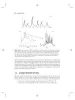
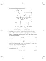
2.7.2 Gradient Elution
Isocratic elution with a fixed mobile-phase composition works well for many samples
and is the simplest form of liquid chromatography. For some samples, however,
no single value of %B can provide a generally satisfactory separation, as illustrated
by the RPC examples of Figure 2.25a, b for the separation of a nine-component
herbicide sample. With 50% acetonitrile/water (Fig. 2.25a), later peaks are very
wide and have inconveniently long retention times. As a result run time is excessive
(140 min), and later peaks are less easily detected (e.g., peak 9 is only 3% as tall
as peak 1). The use of 70% acetonitrile (Fig. 2.25b) partly addresses the latter two
difficulties, but at the same time it introduces another problem: the poor resolution
of peaks 1 to 3. This example illustrates the general elution problem: the inability
of a single isocratic separation to provide adequate separation within a reasonable
run time for samples with a wide range in retention (peaks with very different values
of k).
0 20 40 60 80 100 120 140
Time (min)
5
1
2
3
4
0246810
Time (min)
1
+
2
5
6
89
7
89
50% ACN
70% ACN
(a)(b)
(c)
46
Time (min)
8
76
5
4
321
9
30-85% B in 7 min
100% B
80%
60%
40%
20%
0%
3 4
6
7
Figure 2.25 Illustration of the general elution problem and the need for gradient elution. The
sample is a mixture of herbicides. (a) Isocratic elution using 50% acetonitrile (ACN)-water as
mobile phase; 150 × 4.6-mm C
18
column (5-μm particles), 2.0 mL/min, ambient temperature;
(b)sameas(a), except 70% ACN-water; (c)sameas(a), except gradient elution: 30–85%
ACN in 7 minutes. Computer simulations based on data of [5].