Introduction to Modern Liquid Chromatography, Third Edition part 36 pptx
Bạn đang xem bản rút gọn của tài liệu. Xem và tải ngay bản đầy đủ của tài liệu tại đây (231.46 KB, 10 trang )
306 IONIC SAMPLES: REVERSED-PHASE, ION-PAIR, AND ION-EXCHANGE CHROMATOGRAPHY
0
20
40
60
80
100
HA
B
2 3 4 5 6 7 8 9 10 11 12
50% ionized
k
%
ioniized
pH-4
pH-5
pH-6
pH-7
pH-8
pH-9
pH-10
(a)
(b)
pK
a
HA
pK
a
B
B
B
B
B
+
HA
HA
HA
HA
HA
HA
HA
0 2 4 6 8 10 (min)
B
B
B
t
0
10
8
6
4
2
0
pH
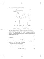
Figure 7.1 Hypothetical illustration of the RPC separation of an acidic compound HA
from a basic compound B as a function of pH. (a) Ionization of HA and B as a function of
mobile-phase pH and effect on k;(b) sample separation as a function of mobile-phase pH;
values of k
0
for HA and B are assumed equal, k
±
= 0, and t
0
= 1.0min.
7.2 ACID –BASE EQUILIBRIA AND REVERSED-PHASE RETENTION 307
and
(bases) F
±
=
1
{1 + (K
a
/[H
+
])}
(7.4b)
When pH = pK
a
(or [H
+
] = K
a
), a compound is half ionized (F
±
= 0.5).
Figure 7.1b shows chromatograms for the separation of HA and B as a function
of mobile-phase pH. As the pH of the mobile phase increases from 4 to 10, the
acid HA (shaded peak) becomes more ionized and less retained, while the base B
eventually becomes less ionized and more retained (for pH = 7, k = 0.1 for each
peak). It can be appreciated from this example that a change in mobile-phase pH
can be a powerful means of controlling relative retention (selectivity) and separation
for samples that contain acids and/or bases. The relative retention of two acids (or
bases) can vary with pH when their values of pK
a
, k
0
, and/or k
±
differ (usually the
case). Some authors have claimed that the separation of partly ionized solutes (e.g.,
where significant amounts of both HA and A
−
are present at the same time) will
necessarily lead to poor peak shape, but there is no empirical or theoretical basis for
this belief .
A similar representation of RPC retention behavior as a function of pH is shown
in Figure 7.2, for the variation of retention time t
R
as a function of mobile-phase pH
for a hypothetical, weakly basic solute with pK
a
= 5.0 (e.g., a substituted aniline or
pyridine). When pH is varied over a sufficiently wide range, solute retention exhibits
a characteristic S-shaped plot as shown; this retention plot mirrors the ionization of
thesampleasinFigure7.1a. At the midpoint of this retention versus pH curve (solid
circle in Fig. 7.2), the mobile-phase pH is equal to the pK
a
value of the solute. The
mobile-phase pH is often chosen in order to control selectivity and resolution. When
the mobile-phase pH ≈ pK
a
for a critical compound or compounds, a change in
pH will provide a maximum change in retention and separation. Thus mobile-phase
pH should fall within region ‘‘II’’ of Figure 7.2 (pH = pK
a
± 1.0),ifwewantto
change selectivity and resolution by varying pH. However, as discussed below, a
mobile-phase pH that allows a greater control over selectivity (i.e., region II) can
mean a less reproducible separation—one of many necessary compromises in HPLC
method development.
When an acid or base is half-ionized, a change in pH of 0.1 unit will result in a
change of k by about 10%. For typical separation conditions, a 10% change in k for
a solute can result in a change in resolution of as much as ±2.5R
s
units, meaning a
possible change in separation from baseline resolution (R
s
>
1.5) to complete overlap
(R
s
= 0). Thus, if a solute is half-ionized, a change in mobile-phase pH by 0.1 unit
can cause a complete loss of resolution. This suggests that mobile-phase pH may need
to be controlled within about 0.02 units for such a separation, which could prove
difficult for many laboratories (see Section 7.3.4.1). In order to avoid pH-related
variations in retention, the mobile-phase pH can be selected to be different from the
pK
a
values of all sample components, by at least ±1.5 pH-units (regions I and III of
Fig. 7.2). As the majority of compounds have pK
a
values
>
4, low-pH separations
(2 ≤ pH ≤ 3) are more likely to be less sensitive to small changes in pH—which
is one reason for beginning method development with a low-pH mobile phase (as
recommended in this chapter). Separations at high pH (≥ 10) can also be used for
this purpose, although special columns are required which are stable under these
conditions (Sections 5.2.5, 5.3.2).
308 IONIC SAMPLES: REVERSED-PHASE, ION-PAIR, AND ION-EXCHANGE CHROMATOGRAPHY
II
pK
a
± 1.0
pK
a
= 5
34 5 67
pH
I
III
Retention time
Figure 7.2 RPC retention as a function of pH. A basic solute with pK
a
= 5.0 is assumed.
A further illustration of the dependence of separation on pH is provided by
Figure 7.3, for several compounds of varying acidity or basicity. Figure 7.3a maps
retention time versus mobile-phase pH for five solutes: compound 1 is salicylic acid
(a relatively strong carboxylic acid), compound 2 is phenobarbital (a weak acid),
compound 3 is phenacetin (a neutral compound in this pH range), compound 4 is
nicotine (a weak base), and compound 5 is methylamphetamine (a strong base).
Figure 7.3b–e shows the corresponding chromatograms for the separation of this
sample as a function of mobile-phase pH. Note, for example, the relative (and
absolute) change in retention for strongly basic compound 5 (shaded peak); as pH
increases, compound 5 becomes less ionized, and more retained.
Several points are worth making about the example of Figure 7.3. First, for
this mixture of acids, bases, and neutrals, a change in pH is a powerful means of
varying relative retention and thereby optimizing resolution. A maximum resolution
of R
s
= 7.2 can be obtained for this sample at pH-8.3 (Fig. 7.3f )inatimeof
28 minutes. Alternatively, baseline separation (R
s
= 2.0) can be obtained in the
shortest time (11 min) at pH-5 (Fig. 7.3c). However, by reducing column length
from 300 to 50 mm for the separation at pH-8.3, and increasing flow rate from 2.0
to 5.0 mL/min, run time can be shortened to 2 minutes (Fig. 7.3g), while maintaining
R
s
≥ 2.0.
Second, this sample contains acids and bases with a wide range in pK
a
values
(see following discussion) and therefore exhibits sizable changes in retention for
small changes in pH throughout the range 3 < pH < 9. Consequently, either a
careful control of mobile pH will be required for the separation of this sample
(Section 7.3.4.1) or conditions must be selected that provide excess resolution
(R
s
2). For example, the separation of Figure 7.3g with pH = 8.3andR
s
= 2.0
could be made more robust by using a 10-cm column (for R
s
= 2.8inaruntimeof
4 min), holding other conditions the same.
Finally, the shape of a plot of retention versus pH for a peak allows a
determination of its sample type (acid, base, or neutral), and a rough estimate of its
pK
a
value. Thus compounds in Figure 7.3a whose retention increases significantly as
7.2 ACID –BASE EQUILIBRIA AND REVERSED-PHASE RETENTION 309
40
30
20
10
0
35 7 9
pH
5 strong base
(… )
4 weak base ( )
3 neutral (
)
2 weak acid ( _ . ._ )
1 strong acid ( ___ )
5.0 8.3
(a)
pH
Retention time (min)
Figure 7.3 Effect of mobile-phase pH on RPC retention as a function of solute type. Sample:
1, salicylic acid; 2, phenobartitone; 3, phenacetin; 4, nicotine; 5, methylamphetamine (shaded
peak). Conditions for separations (a–f ): 300 × 4.0-mm C
18
column (10-μm particles); 40%
methanol-phosphate buffer; ambient temperature; 2.0 mL/min. Flow rate is 5.0 mL/min and
column length is 50-mm in (g). Adapted from [1], with chromatograms (b–g) recreated by
computer.
pH increases are bases (4 and 5), those whose retention decreases with an increase
in pH are acids (1 and 2), and compounds that show little change in retention with
pH (3) are either neutral or are fully ionized over the pH range studied. Compounds
2and4areseentohavepK
a
values of about 8 and 6.5, respectively. While the pK
a
values of compounds 1 and 5 cannot be estimated accurately (a complete retention
vs. pH curve is required), it is safe to say that pK
a
≥ 9 for compound 5, and pK
a
≤ 3
for compound 1.
The relationship between RPC retention and mobile-phase pH is more com-
plicated for amphoteric compounds that contain both acidic and basic groups. This
is illustrated in Figure 7.4 for the retention of two amino acids as a function of
pH. A molecule of each compound contains both an acidic –COOH group and a
basic –NH
2
group. As a result minimum retention is observed at intermediate pH
values, because for 4 < pH < 8 both the carboxyl and amine groups are ionized.
More precisely, the molecule is maximally ionized in this pH range, even though
the net charge is zero (different ionized groups within a molecule—even of different
sign—can each prefer the more polar mobile phase).
7.2.1 Choice of Buffers
Whenever acids or bases are separated, it is necessary to buffer the mobile phase in
order to maintain a constant pH and reproducible retention during the separation.
The use of a pH meter to measure (and control) pH will be less precise when the
mobile phase contains organic solvent because the electrode response tends to drift
310 IONIC SAMPLES: REVERSED-PHASE, ION-PAIR, AND ION-EXCHANGE CHROMATOGRAPHY
pH-3
pH-5
pH-7
pH-9
pH-8.3 (300-mm column)
pH-8.3 (50-mm column)
024681012
Time (min)
0
0
02040
2 4 6 8 10 12 14
246810
Time (min)
Time (min)
Time (min)
Time (min)
4
5
2
1
+
3
1
4
5
2
3
1
2
3
4
5
(b)
(c)
(d)
(e)
(f)
(g)
1
2
3
4
5
1
2
3
4
5
02
Time (min)
020
1
2
3
4
5
Figure 7.3 (Continued)
for organic-water solutions. Consequently we recommend that pH measurements
be carried out for the A-solvent (aqueous buffer) prior to the addition of organic to
form the final mobile phase. The pH of the final mobile phase (including organic
solvent) can then be equated to (or labeled as) that of the A-solvent, although the
actual mobile-phase pH will be somewhat different (Section 7.2.3). This uncertainty
7.2 ACID –BASE EQUILIBRIA AND REVERSED-PHASE RETENTION 311
02468101214
p
H
25
20
15
10
5
k
70
60
50
40
30
20
10
RCHCOO
−
RCHCOO
−
RCHCOOH
NH
3
+
NH
3
+
NH
2
Figure 7.4 Dependence of RPC retention on mobile-phase pH for amphoteric compounds.
Sample: phenylalanine (
•
), leucine (o). Conditions: polystyrene column, 40-mM phosphate
buffer as mobile phase. Adapted from [2].
concerning the final mobile-phase pH is unimportant for the routine application of
RPC. When the A-solvent is prepared in this way, different laboratories should be
able to obtain the same final mobile-phase pH within ±0.04 to 0.05 units [3]. If a
closer control of mobile-phase pH is required, see Section 7.3.4.1. When we refer
to mobile-phase pH in this book, we will generally mean the pH of the A-solvent.
Directions for the preparation of buffer solutions of varied pH and buffer-type are
given in Appendix II.
In selecting a buffer for RPC separation, several buffer properties may prove
relevant:
•pK
a
and buffer capacity
• solubility
• UV absorbance (when UV detection is used)
• volatility (when mass-spectrometric or evaporative light-scattering detection
is used)
• ion-pairing properties
• stability and compatibility with the equipment
The first four buffer properties are usually the most important.
7.2.1.1 Buffer pK
a
and Capacity
‘‘Buffer capacity,’’ or the ability of the buffer to maintain a constant pH, depends on
312 IONIC SAMPLES: REVERSED-PHASE, ION-PAIR, AND ION-EXCHANGE CHROMATOGRAPHY
•pK
a
value of the buffer
• buffer concentration
• pH of the mobile phase
Just as for the ionization of a sample component in Figure 7.1a, the fractional
ionization of the buffer as a function of pH can be expressed by Equations (7.2) or
(7.2a); that is, buffer and solute ionization are identical functions of mobile-phase
pH and pK
a
. Maximum buffering occurs when the concentrations of the two forms
of the buffer (e.g., HA and A
−
) are equal; that is, when the buffer pK
a
equals
the mobile-phase pH. Buffer capacity decreases as values for the buffer pK
a
and
mobile-phase pH become more different. Consequently the first requirement of the
buffer is that its pK
a
value should be within ±1.0 units of the selected mobile-phase
pH (this requirement can be relaxed to ±1.5 unit for higher concentrations of the
buffer). The buffer capacity of the mobile phase is proportional to buffer concentra-
tion, which typically falls within a range of 5 to 25 mM. To minimize the possibility
of inadequate buffering of the sample during RPC separation, it is generally desirable
for the sample to be dissolved in the mobile phase (or buffered to the same pH as the
mobile phase); this practice becomes especially important for lower concentrations
of the mobile-phase buffer, and/or for larger volumes of injected sample.
Table 7.1 provides a list of buffers that can be used in RPC, along with
pertinent properties such as buffer pK
a
and the mobile-phase pH range in which the
buffer is effective. For separations with UV detection, and a mobile-phase pH ≤ 8,
popular buffers include phosphate, trifluoroacetate, acetate, and formate. In addition
ammonium bicarbonate can also be considered. The pK
a
values of ammonia (9.2)
and bicarbonate (10.3) overlap, hence somewhat extending the buffering range of
ammonium bicarbonate (8.2 ≤ pH ≤ 11.3). This buffer is volatile and therefore
compatible with LC-MS; however, when the buffer pH < 8.5, loss of CO
2
(e.g.,
from excessive degassing) may lead to an unintended increase in pH. Because of this
instability it is recommended to prepare fresh ammonium bicarbonate buffers daily.
The remaining discussion of this section (7.2.1.1) is more detailed. The reader
may wish to skip this digression, proceed to Section 7.2.1.2, andreturntothepresent
section as needed.
We can define the ‘‘effective buffer capacity’’ of the mobile phase to mean that
an increase in this quantity will result in fewer problems due to insufficient buffering.
The effective buffer capacity of the mobile phase increases for:
1. a smaller difference between values of the buffer pK
a
and the pH of the
mobile phase (change either the buffer or pH)
2. a greater difference between the mobile-phase pH and the pK
a
of the solute
(for large differences, the solute is either non-ionized or completely ionized;
buffering is then much less important)
3. increased buffer concentration
4. smaller volumes of injected sample
5. a sample whose pH is adjusted to that of the mobile phase
An example of inadequate buffering is provided by the chromatograms of
Figure 7.5 for the solute 3,5-dimethylaniline as a function of mobile-phase pH, using
a 25 mM potassium phosphate buffer. Despite this sizable buffer concentration,
7.2 ACID –BASE EQUILIBRIA AND REVERSED-PHASE RETENTION 313
Table
7.1
Buffers for Use in HPLC Separation
Buffer Acid
a
pK
a
Approximate UV Comments
(25
◦
C) Buffer Range Cutoff
b
Trifluoroacetic acid >2 1.5–2.5 210 nm (0.1 %) Ion-pairing, volatile
Phosphoric acid 2.1 1.5–3.5 <200 nm (10 mM) Limited solubility
Monophosphate 7.2 6.0–8.5 <200 nm (10 mM) Limited solubility
Diphosphate 12.3 11.0–13.5 <200 nm (10 mM) Limited solubility
Citric acid 3.1 2.0–4.5 230 nm (10 mM) Equipment problems
c
Monocitrate 4.7 3.5–6.0 230 nm (10 mM) Equipment problems
c
Dicitrate 5.4 4.0–7.5 230 nm (10 mM) Equipment problems
c
Formic acid 3.8 2.5–5.0 210 nm (10 mM) Volatile
Acetic acid 4.8 3.5–6.0 210 nm (10 mM) Volatile
Carbonic acid 6.4 5.0–7.5 <200 nm (10 mM) Volatile
d
Monocarbonate 10.3 9.0–11.5 <200 nm (10 mM) Volatile
e
Bis-tris propane.HCl
e
6.8 75.5–8.0 215 nm (10 mM) Possibly unstable
f
Bis-tris propane 9.0 7.5–10.5 225 nm (10 mM) Possibly unstable
f
Tris.HCl
g
8.0 7.0–9.5 205 nm (10 mM) Possibly unstable
f
Ammonia.HCl 9.2 8.0–10.5 200 nm (10 mM)
o-Boric acid 9.1 8.0–10.5 200 nm (10 mM)
mono-o-borate 12.7 11.5–14.0 200 nm (10 mM)
1-methylpiperidine.HCl 10.1 9.0–11.5 215 (10 mM) Possibly unstable
f
Triethylamine.HCl 11.0 9.5–12.5 <200 (10 mM) Possibly unstable
f
a
Buffer composed of buffer acid plus ionized acid, such as for phosphoric acid, H
3
PO
4
and H
2
PO
−
4
.
b
Aqueous solutions; absorbance <0.5 AU at wavelengths above cutoff
c
Claimed to attack stainless steel; we have experienced equipment problems associated with the
long-term use of citrate buffer.
d
Use of this buffer is impractical, because of loss of CO
2
from the reservoir.
e
1,3-Bis[tris(hydroxymethyl)methylamino] propane.
f
Ammonium carbonate is unstable at pH-7.0 (loss of CO
2
to atmosphere), but stable at pH-8.5 [11]; amine
buffers tend to oxidize, with a large increase in UV absorbance.
g
Tris(hydroxymethyl)aminomethane.
a slight tailing of the peak is seen in Figure 7.5 for a mobile-phase pH = 3.0.
As the pH increases to 3.5 (Fig. 7.5b) and 4.0 (Fig. 7.5c), the peak progressively
broadens and becomes more distorted (peak fronting)—the result of decreased
buffer capacity. The pK
a
values of solute and buffer are 3.8 and 2.1, respectively,
so as pH increases above 3.0 in this example, the difference between the buffer pK
a
value and mobile-phase pH (factor 1 above) increases, while the difference between
mobile-phase pH and the pK
a
of the solute decreases (factor 2 above). Together, this
results in a decrease of the effective buffer capacity as the mobile-phase pH increases
from 3 to 4—and a progressive deterioration of peak shape. Poor peak shape in
each of the examples of Figure 7.5 could be improved by any of the experimental
factors 3 to 5 on p. 312 (increased buffer concentration, etc.)
314 IONIC SAMPLES: REVERSED-PHASE, ION-PAIR, AND ION-EXCHANGE CHROMATOGRAPHY
pH =
3.0 3.5 4.0
Figure 7.5 Effect of insufficient buffer capacity on peak shape for 3,5-dimethylaniline
as solute. Conditions: column, 250 × 4.6-mm cyano column (5-μm particles); 25%
methanol-buffer, buffer is 50-mM potassium monophosphate; 35
◦
C; 1 mL/min. Adapted
from [4].
When we wish to reduce buffer concentration for any reason (limited buffer
solubility, increased UV absorbance, etc.), we need to optimize other contributions to
buffer capacity (factors 1, 2, 4, or 5 on p. 312); for example, choose a mobile-phase
pH that provides either minimal or maximal ionization of the sample (factor 2). For
further discussion of buffer capacity in RPC, see [5].
7.2.1.2 Other Buffer Properties
The remaining buffer properties listed at the end of Section 7.2.1 should also be
considered when developing a RPC separation for an ionic sample.
Buffer Solubility. Organic buffers are usually adequately soluble in all organic-
water mobile phases (0–100% B), but many inorganic buffers have limited solubility
in mobile phases which are predominantly organic (high %B). Consequently there
is a danger that combining the A- and B-solvents may result in buffer precipitation,
which could lead to blockage of the column or HPLC equipment. If there is any
doubt as to whether a mobile phase might precipitate, the complete solubility of
the buffer in the final mobile phase should be confirmed first (over the intended
pH range), especially when the A- and B-solvents are mixed by the HPLC pumping
system. Thus varying proportions of the A- and B-solvents can be combined manually
in a container and observed over a period of 30 minutes or so. If any cloudiness
develops, or a precipitate is observed for a given mobile-phase composition (%B),
mobile phases of that %B or higher should be excluded or the buffer concentration
should be reduced (see the discussion of [5, 6] for further details). Buffer solubility
is of special concern for separations by gradient elution (Section 9.3.1).
Buffer solubility is affected by several separation conditions [6]. The buffer
counter-ion is one such factor; for acidic buffers such as phosphate, buffer solubility
usually increases as
sodium salt (least soluble) < potassium salt < ammonium salt (most soluble)
Similarly buffer solubility varies with the organic solvent (B-solvent), provided that
comparisons are made for the same %B and pH:
tetrahydrofuran (least soluble) < acetonitrile < methanol (most soluble)
7.2 ACID –BASE EQUILIBRIA AND REVERSED-PHASE RETENTION 315
Buffer solubility is also affected by the relative ionization of the buffer; as the charge
on the buffer ion increases (e.g., HPO
=
4
vs. H
2
PO
−
4
), the buffer becomes less soluble
in high-%B mobile phases. Thus the choice of buffer and other separation conditions
permits a considerable control over buffer solubility. Because most isocratic RPC
separations of ionic samples are carried out with mobile phases of <60% B, however,
buffer solubility is usually not a problem. To a lesser extent this is also true for
gradient elution because, when buffer is added only to the A-solvent (the usual
practice for inorganic buffers), buffer concentration in the mobile phase becomes
inversely proportional to %B (also true for isocratic elution method development).
Buffer solubility is usually only an issue for inorganic buffers, especially
phosphate. One study [6] has reported that potassium phosphate has an
ambient-temperature solubility at pH-7 of 10 mM for either 85% MeOH-water or
75% ACN-water (with higher solubilities at lower %B, and vice versa for higher
%B). At pH-3, a solubility of 10 mM can be achieved with 85% MeOH or 85%
ACN. A phosphate-buffer concentration of 1 to 2 mM, combined with other
favorable choices from the list ‘‘effective buffer capacity’’ of Section 7.2.1.1, should
allow %B values as high as 90% for either methanol or acetonitrile as B-solvent.
Fortunately, %B-values this high are rarely required for ionized compounds, in
which case buffer solubilty may no longer be an issue.
Detector Requirements. The absorbance of the buffer is proportional to buffer
concentration and adds to the absorbance of the water-organic mixture used for
the mobile phase. Table 7.1 provides a rough guide for assessing whether a given
buffer will result in a significant increase in the UV absorbance of the mobile phase.
The influence of buffers on gradient baseline drift is illustrated in Figures 17.7 to
17.10. Additional information on buffer absorbance versus wavelength is provided
in Table I.2 of Appendix I.
Mass spectrometric detection (LC-MS) requires a volatile buffer. Common
choices include trifluoroacetic acid (TFA), acetic acid, formic acid, and their ammo-
nium salts. For the separation of basic compounds at low pH, with volatile buffers
such as formic or acetic acid, it is preferable to select a mobile phase with a higher
ionic strength; that is, a higher buffer concentration and a pH where the buffer
is significantly ionized (i.e., choose a mobile-phase pH that is fairly close to the
pK
a
value of the buffer [7]). Otherwise, even small weights of injected sample can
result in column overload and peak tailing because of the ionic repulsion of retained
molecules of protonated bases BH
+
(Section 15.3.2.1). For example, ammonium
formate can be used as buffer at a pH of 3.5 to 4.0, whereas the use of formic
or acetic acid alone (at a lower pH) provides much less ionization of the buffer.
With reduced buffer ionization, column overload and peak tailing become more
likely. For additional information on the best choice of buffer for MS detection, see
Section 4.14.
Ion-Pairing by the Buffer. So far we have assumed that the sole effect of the
buffer on sample retention and separation is to control mobile-phase pH and sample
ionization. Additionally an ionized buffer X can interact with an ionized solute