Introduction to Modern Liquid Chromatography, Third Edition part 37 docx
Bạn đang xem bản rút gọn của tài liệu. Xem và tải ngay bản đầy đủ của tài liệu tại đây (288.29 KB, 10 trang )
316 IONIC SAMPLES: REVERSED-PHASE, ION-PAIR, AND ION-EXCHANGE CHROMATOGRAPHY
A
−
or BH
+
by ion-pairing:
ionized solute ion pair
(acids) A
−
+ X
+
⇔ A
−
X
+
(7.5)
(bases) BH
+
+ X
−
⇔ BH
+
X
−
(7.5a)
hydrophilic (less retained in RPC) hydrophobic (more retained in RPC)
Buffers that ion-pair are usually organic ions, and they tend to be more hydrophobic
than inorganic ions. For example, trifluoroacetate is a significant ion-pairing agent
(Section 13.4.1.2), whereas phosphate is not. Ion-pairing by inorganic buffers is
usually not significant, although some studies suggest that even phosphate may
undergo weak ion-pairing with protonated bases [8]. For further information on
ion-pairing by the buffer, see Section 7.4.2.1.
Buffer Stability and Equipment Compatibility. Inorganic buffers and carboxylic
acids are usually stable, but amines are prone to oxidation with a consequent
increase in their UV absorbance at wavelengths <250 nm. For example, although
pure triethylamine (TEA) should be transparent at wavelengths ≥ 200 nm, one study
[9] reported that a 1% aqueous solution of TEA (90 mM) has an absorbance
>
2
AU at 220 nm.
Citrate buffers have been claimed to attack stainless steel, whereas other
reports contradict this claim as applied to HPLC equipment [10]. One laboratory
has experienced occasional equipment problems that appeared associated with the
use of citrate buffer [11], but this behavior has not been confirmed. While it appears
likely that citrate concentrations <10 mM can be used without concern, and its
use is convenient for applications where pH is to be varied continuously over the
range 2 < pH < 7, caution is nevertheless advised. Overall, the (real or imagined)
problems in the use of citrate, its relatively high UV cutoff of 230 nm (Table 7.1),
and the availability of favorable alternative buffers (e.g., phosphate plus acetate)
make citrate a seldom-used buffer by most workers.
7.2.1.3 Preferred Buffers
UV detection in the 200 to 220 nm region is often used for samples with low
concentrations of compounds that absorb poorly at higher wavelengths. Phosphate
buffers have been preferred for this application, as long as the mobile-phase pH can be
accommodated within the ranges specified in Table 7.1 (pH ≤ 3.5, 6.0 ≤ pH ≤ 8.5,
or pH ≥ 11.0). Formate and acetate cover the pH range of 2.5 to 6.0, can be used at
wavelengths of 210 nm or higher, and are volatile for LC-MS detection; for LC-MS
detection at low pH with basic solutes, ammonium acetate or formate buffers provide
an increase in ionic strength that allows higher sample concentrations without peak
tailing [12]. Phosphate, acetate, and formate are the most commonly used buffers
for separations with a mobile-phase pH < 8.0.
There is increasing use of higher pH mobile phases (pH
>
8) as a result of the
development of RPC columns that are stable at high pH (Section 5.2.5). Borate and
ammonia have been used to some extent as buffers for high-pH operation, but note
the further discussion of Section 5.8 concerning column stability at high pH. See
7.2 ACID –BASE EQUILIBRIA AND REVERSED-PHASE RETENTION 317
also Appendix II for details on the more convenient preparation of some common
buffers of required pH.
7.2.2 pK
a
asaFunctionofCompoundStructure
When selecting a range of mobile-phase pH values within which to carry out method
development (i.e., optimization of pH), it can be useful to know the approximate
pK
a
values of the various sample components. This information allows the mobile
phase to be restricted within an appropriate range of pH values. For example, a
mobile-phase pH range that varies from pK
a
− 1topK
a
+ 1 (for the sample) is useful
for controlling retention and selectivity by changes in mobile-phase pH. Similarly
a mobile-phase pH outside this range will result in a more robust method that
is less sensitive to small (unintended) changes in pH. Values of pK
a
vary widely
for different organic compounds, but a large number of pK
a
values have been
determined experimentally [13], and additional pK
a
values can be estimated on the
basis of solute molecular structure. One very extensive source of experimental plus
predicted pK
a
values (ACD/pK
a
DB: pK
a
prediction) is available from Advanced
Chemistry Development (Toronto, Canada).
Exact pK
a
values for sample components are not required in RPC method
development, and in many cases the chemical structures of sample components may
not even be known. Values of pK
a
are determined by ionizable acid or base groups
attached to the solute molecule, for example, –COOH, –NH
2
. This means that
estimates of pK
a
can be obtained from literature pK
a
values for compounds of
similar functionality (e.g., benzoic acid, as a representative of aromatic carboxylic
acids). Table 7.2 summarizes pK
a
values in water for some common acid- or
base-substituent groups present in typical sample molecules. It is also possible to
infer values of pK
a
from experimental plots of retention against pH, as for peaks
2and4inFigure7.3a.
7.2.3 Effects of Organic Solvents and Temperature on Mobile-Phase pH
and Sample pK
a
Values
This detailed section is less essential in everyday operation, so the reader may wish
to skip to Section 7.3. Nevertheless, the conclusions of this section are potentially
useful for an accurate interpretation of the relationship between sample retention
and mobile-phase pH.
Literature values of pK
a
for different compounds (as in Table 7.2) are usually
reported for aqueous solutions at near-ambient temperatures. Quite often, however,
RPC separations of ionic samples are carried out at higher temperatures, with mobile
phases that contain varying amounts of organic solvent. Both mobile-phase pH and
values of pK
a
for the sample can be affected by added organic (specifically, the
value of %B) and by temperature. However, a knowledge of the true (mobile-phase)
pH and solute pK
a
values as a function of %B and temperature has little practical
importance, so far as routine RPC assays are concerned; it is only important that %B
and temperature are maintained constant for all runs so that solute ionization and
retention remain unchanged from run to run. In the case of method development,
however, approximate values of pK
a
can be useful for selecting the pH of the
mobile phase (as discussed above). It is useful in this connection to define an
‘‘effective’’ pK
a
value for the solute, which takes into account the effects of %B
318 IONIC SAMPLES: REVERSED-PHASE, ION-PAIR, AND ION-EXCHANGE CHROMATOGRAPHY
Table 7.2
Approximate pK
a
Values for Acidic or Basic Functional Groups (aqueous solutions)
pK
a
Acid Base
Group Aliphatic Aromatic Aliphatic Aromatic
Sulfonic acid, –SO
3
H11
Amino acid, –C(NH
2
)–COOH
a
310
Carboxylic acid, –COOH 5 4
Thiol, –SH 10 7
Purine 3 9
Phenol, –OH 11
Pyrazine 1
Sulfoxide, –SO– 2
Thiazole 2
Amine, –NH
2
,–NR
2
10 5
Pyridine 5
Imidazole 5
Piperazine 10
Note: Values can vary by 1 to 2 pK
a
units or more as a result of adjacent groups in the molecule.
a
See Figure 13.1 for values of individual amino acids.
and temperature on the pK
a
values of both solute and buffer (Tables 7.1 and 7.2).
Effective pK
a
values for the solute can be used with the pH of the buffer (not
the buffer-organic mobile phase) to estimate solute ionization and retention as a
function of pH. An effective pK
a
value is equivalent to the value that can be inferred
from an experimental plot of retention against mobile-phase pH (i.e., buffer pH, as
in Fig. 7.2).
7.2.3.1 Effect of %B on Values of Effective pK
a
for the Solute
The pH of the mobile phase depends on the pK
a
value for the buffer, while a
change in %B will affect pK
a
values of both the buffer and the solute [14–16]. If
the buffer and solute are each acidic (e.g., phosphate buffer and a carboxylic acid
solute), changes in pK
a
with %B will be similar for both solute and buffer—and
hence cancel approximately. For the latter case, effective pK
a
values for the solute
can be assumed equal to the values of Table 7.2. The same will be true when both
the buffer and solute are basic (i.e., effective solute pK
a
values that are similar to
values measured in water at room temperature). However, when the buffer is acidic
and the sample is basic, and vice versa, the apparent change in pK
a
with %B can be
substantial, so that effective pK
a
values for the solute will no longer be the same as
literature values measured in water.
Commonly used RPC buffers are more often acidic than basic (e.g., phosphate,
acetate, formate). For the case of basic solutes and acidic buffers (with methanol as
B-solvent), a decrease in the apparent pK
a
by about 0.03 units can be expected for
7.3 SEPARATION OF IONIC SAMPLES BY REVERSED-PHASE CHROMATOGRAPHY (RPC) 319
each 1% increase in %B (experimental data of [17, 18]). For example, consider the
separation of several substituted anilines, using 25% methanol-phosphate buffer,
as reported in [17]. Effective pK
a
values for these solutes would be expected to be
25 × 0.03 = 0.75 units lower than literature values. For eight different solutes with
literature pK
a
values of 2.7 to 5.3, experimental pK
a
values (as in Fig. 7.2) for this
sample were lower by an average 0.7 ± 0.1 units (1 SD). Similarly, for six substituted
benzoic acids separated with 40% methanol and acetate buffer [17], effective pK
a
values were the same as literature values (±0.1 unit, 1 SD). The effect of added
acetonitrile on effective pK
a
values is similar to that for the addition of methanol.
The effect on relative retention of changes in effective pK
a
values with %B is
equivalent to a change in mobile-phase pH (the ‘‘effective’’ pH of the mobile phase),
which suggests that a change in %B can have a larger effect on relative retention
and selectivity for ionic samples than for neutral samples. This has been observed
for gradient elution as a function of gradient steepness [19, 20], which is equivalent
to a change in %B for isocratic elution (Section 9.1.3).
In the discussion above, the mobile-phase pH is equated to that of the measured
pH of the aqueous buffer, a procedure that we recommend for reasons given in
Section 7.2.1. The pH of the final, water-organic mobile phase could be measured
instead (less conveniently, and less reproducibly), as suggested by some workers
[14–16]. However, possible changes in solute pK
a
values with %B would still
require correction; that is, the use of pH values measured in the mobile phase does
not solve the problem of solute pK
a
values that vary with %B. Again, we strongly
recommend measuring the pH of the buffer, not the final mobile phase.
7.2.3.2 Effect of Temperature on Values of pK
a
Values of pK
a
for both the buffer and sample can vary with temperature [21–25].
For protonated basic solutes, a lowering of effective pK
a
values with increasing
temperature [24] can lead to a decrease in solute ionization and an atypical increase
in retention at higher temperatures. Because of such changes in pK
a
with temperature,
significant changes in relative retention with temperature can also result for ionic
samples—more so than for neutral samples [19, 20]. That is, temperature selectivity
will generally be greater for ionic samples. While the correction of pK
a
values for a
change in temperature should be possible (as above for a change in %B), at present
there are no simple guidelines for this purpose. As temperature is seldom varied
over a very wide range (e.g., 30–50
◦
C is typical), the effect of temperature on solute
pK
a
values will usually be small and can therefore be ignored when using estimated
values of pK
a
for method development.
7.3 SEPARATION OF IONIC SAMPLES BY REVERSED-PHASE
CHROMATOGRAPHY (RPC)
Reversed-phase chromatography (RPC) should be a first choice for the separation
of mixtures of ionizable organic compounds. Method development for the RPC
separation of ionic samples (Section 7.3.3) proceeds in similar fashion as for neutral
samples, with some important differences that are developed in the remainder of this
320 IONIC SAMPLES: REVERSED-PHASE, ION-PAIR, AND ION-EXCHANGE CHROMATOGRAPHY
section. The following information on RPC separation (Sections 7.3.1, 7.3.2) should
be useful for both method development and in troubleshooting routine separations.
7.3.1 Controlling Retention
Following an initial experiment, mobile-phase strength (%B) can be varied to obtain
a desirable retention range (e.g., 1 ≤ k ≤ 10), the same way as for neutral samples
(Sections 2.5.1, 6.2.1). Alternatively (and generally preferable), initial experiments
can be carried out using gradient elution, as discussed in Section 9.3.1. Once a value
of %B has been selected for the reasonable retention of the sample, the next step is
the adjustment of separation selectivity for optimal relative retention and maximum
resolution.
7.3.2 Controlling Selectivity
For the separation of neutral samples, selectivity can be varied by changing solvent
strength (%B), temperature, the B-solvent, or the column. These variables also
affect the separation of ionic samples, usually to a greater extent than for neutral
samples. In addition separation selectivity for ionic samples is strongly affected
by mobile-phase pH, and to a lesser extent by the kind and concentration of the
buffer. In the past, mobile-phase additives for the suppression of silanol activity
(alkylamines, quaternary ammonium compounds) have been added to the mobile
phase, and these additives can further change selectivity. Today, however, the
widespread preference for type-B columns (Section 5.2.2.2) has virtually eliminated
the need for silanol suppression by such additives. Depending on the nature of the
sample, all of these separation conditions may exert a significant effect on relative
retention and resolution, as discussed below. One study [25] ranked the relative
importance of different conditions in affecting the selectivity of RPC separations of
ionic samples as follows:
pH (most important)
>
solvent type ≈ column type
>
%B
>
temperature
buffer concentration and type (least important).
7.3.2.1 Mobile-Phase pH
The ability of a change in mobile-phase pH to affect the relative retention of
ionizable samples is apparent from the example of Figure 7.3. We have noted
that solute retention changes with pH only when the pH of the mobile phase
is within ≈±1.5 units of the pK
a
value of the solute (Fig. 7.2). Consequently, if
mobile-phase pH is to have an effect on separation selectivity, the pH must be similar
to pK
a
values of the sample constituents. Carboxylic acids and amines are the most
commonly encountered examples of ionic solutes; reference to Table 7.2 suggests for
a mobile-phase pH between 2 and 3 that bases (pK
a
≈ 5–10) will be fully ionized,
and acids (pK
a
≈ 5) will be in the neutral form. This is only approximately true,
since it overlooks the effects of %B and temperature on values of pK
a
,aswellas
changes in pK
a
that can result from the presence of different substituents in the
solute molecule. Consequently, while pH selectivity is usually reduced at low pH, it
can still be significant—depending on the sample and the value of %B.
7.3 SEPARATION OF IONIC SAMPLES BY REVERSED-PHASE CHROMATOGRAPHY (RPC) 321
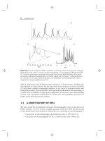
Another example of a change in relative retention with pH is shown in
Figure 7.6 for a mixture of substituted benzoic acids (peaks 1–4) and anilines
(peaks 5–7). As mobile-phase pH is increased from 3.2 to 4.3 (Fig. 7.6a–c), the
retention of acids 1 to 4 decreases, while the retention of bases 5 to 7 (shaded peaks)
increases. For a mobile-phase pH of 4.3 or higher, the acidic compounds 1 to 4 are
mainly in the ionized form and therefore retained weakly; similarly, at higher pH
the basic compounds 5 to 7 are largely non-ionized and more strongly retained. As
a result for a mobile-phase pH
>
4 there is a separation of these acids and bases
into two groups of peaks. An optimum mobile-phase pH = 3.4(Fig.7.6d)provides
acceptable resolution of the sample. However, even at this relatively low pH, the
separation of Figure 7.6d is seen to be somewhat sensitive to small changes in pH;
0 2 4 6 8 10 12 14
5
1
2
6
3
4
7
pH-3.2
5 ≤ k ≤ 15
R
s
= 0.6
pH-3.7
2 ≤ k ≤ 15
R
s
= 0.9
pH-4.3
0.5 ≤ k ≤ 15
R
s
= 0.2
pH-3.4
4 ≤ k ≤ 15
R
s
= 2.7
(a)
(b)
(c)
(d)
024681012
Time (min)
Time (min)
Time (min)
Time (min)
5
1
2
6
3
4
7
02468101214
2
1
3
5
4
6
7
02468101214
2
1
3
4
5
6
+
7
1-4 acids
5-7 bases
Figure 7.6 Effect of mobile-phase pH on the RPC separation of a mixture of acids and
bases. Sample: 1, 2-fluorobenzoic acid; 2, 3-chlorobenzoic acid; 3, 3-nitrobenzoic acid;
4, 3-fluorobenzoic acid; 5, 3,5-dimethylaniline; 6, 4-chloroaniline; 7, 3-chloroaniline. Con-
ditions: 150 × 4.6-mm C
18
column (5-μm particles); mobile phase, 13% acetonitrile-buffer
(buffer is citrate plus phosphate); 2.0 mL/min; 35
◦
C. Peaks for basic compounds 5 to 7 are
shaded. Chromatograms based on data of [19].
322 IONIC SAMPLES: REVERSED-PHASE, ION-PAIR, AND ION-EXCHANGE CHROMATOGRAPHY
the buffer pH should therefore be maintained within ±0.1 pH units of the specified
value for reproducible separation. The presence of partly ionized acids and bases in
the same sample for 3 < pH < 4 is the reason for both changes in relative retention
with pH and the marginally robust nature of the separation of Figure 7.6d.
7.3.2.2 Solvent Strength (%B) and Temperature
In Figure 7.7 the effects of a change in %B and temperature on relative retention
are illustrated for the sample of Figure 7.6, in each case for the same mobile-phase
pH = 3.2. It is apparent that significant changes in relative retention occur as either
temperature or %B is changed. These changes in relative retention can be attributed
to the same factors that are operative for the separation of neutral compounds (e.g.,
Fig. 6.26), plus more important changes in the ‘‘effective’’ mobile-phase pH as a
result of change in either %B or temperature (Section 7.2.3). Consequently a change
in %B or temperature usually has a larger effect on the relative retention (and
resolution) of ionic samples than neutral samples, as noted above. In the examples of
Figure 7.7 we see an increase in the relative retentionofbases5to7(shadedpeaks)
compared with acids 1–4 as either %B or temperature increase (while the absolute
retention of all compounds decreases). This implies that the pK
a
values of the
sample bases have decreased as a result of an increase in either %B or temperature,
which is equivalent to an increase in mobile-phase pH for these basic compounds
Time (min)
13% B, 35
°
C
5 ≤ k ≤ 16
R
s
= 0.6
28% B, 35
°
C
1 ≤ k ≤ 5
R
s
= 0.3
19% B, 49
°
C
3 ≤ k ≤ 9
R
s
= 3.3
28% B, 60
°
C
1 ≤ k ≤ 4
R
s
= 1.0
13% B, 60
°
C
5 ≤ k ≤ 11
R
s
= 0.7
0246810
12 14 0 2 4 6 8 10
Time (min)
Time (min)Time (min)
5
1
2
6
3
4
7
1
5
2
3
6
4
7
(a)
(c)
(b)
(d)
024 024
5
1
2
3
4
+
6
7
5
2
3
4
6
7
02468
Time
(
min
)
5
1
2
3
6
4
7
(e)
Figure 7.7 Effect of mobile-phase strength (%B) and temperature on the separation of a
mixture of acids and bases. Sample: same as in Figure 7.6; conditions also the same, except
pH = 3.2, and values of %B and temperature are noted in the figure. Peaks for basic com-
pounds 5 to 7 are shaded. Chromatograms based on data of [19].
7.3 SEPARATION OF IONIC SAMPLES BY REVERSED-PHASE CHROMATOGRAPHY (RPC) 323
(see the discussion of Section 7.2.3 for the case of an acidic buffer and basic solutes).
A maximum resolution of R
s
= 3.3 is observed in Figure 7.7e for 19% B and 49
◦
C.
The latter optimized separation can be found by trial-and-error changes in %B
or temperature, but a more efficient procedure is the use of computer simulation
(Section 10.2.2).
The remainder of this section represents an alternative way of interpreting the
changes in retention of Figure 7.7. Since it is not essential to an understanding of
the effects of %B and temperature on retention, the reader may prefer to skip to the
following Section 7.3.2.3.
The effects of changes in %B, temperature, or other conditions on relative
retention can be further interpreted in terms of
(acids, bases) k = k
0
(1 − F
±
) + k
±
F
±
(7.4)
Because k
0
k
±
, this relationship can be simplified to
k ≈ k
0
(1 − F
±
), (7.6)
where k
0
is the value of k for the neutral (non-ionized) molecule, and F
±
is the
fractional ionization of the solute for a given mobile-phase pH. An increase in either
temperature or %B will lead to a decrease in values of k
0
for the solute, regardless of
whether it is ionic or neutral. Additionally a change in conditions that also changes
the ‘‘effective’’ mobile-phase pH (and therefore values of F
±
) can have a further
effect on the separation of an ionic sample. Thus in Figure 7.7 an increase in either
%B or temperature appears to increase mobile-phase pH slightly (equivalent to a
decrease in pK
a
values for these solutes)—with a preferential retention of basic
solutes 5 to 7.
7.3.2.3 Solvent Type
A change in solvent type (e.g., methanol replacing acetonitrile) is expected to have
a comparable effect on the relative retention of both ionic and neutral samples. In
addition any change in ‘‘effective’’ pK
a
values as a result of this change in B-solvent
can further affect the relative retention of ionic samples—similar to the case of a
change in %B or temperature. The latter effect should lead to larger changes in
relative retention for ionic as opposed to neutral samples when the B-solvent is
changed. This was observed to be the case in one study [26], where the average
change in values of α for 45 neutral solutes was ±0.04 units for a change in B-solvent
from 50% ACN to 45% ACN + 5% MeOH. The corresponding change in α for 22
ionic compounds was ±0.09 units (twice as large as for neutral solutes). See Sections
6.3.2 and 6.4.1 for a related discussion of the effect of solvent type on the separation
of non-ionic samples.
7.3.2.4 Column Type
Separations of a neutral sample with four columns of different type (different ligands)
were illustrated in Figure 6.14. Similar separations of an ionic sample are shown
in Figure 7.8 for three of the same columns (note that an ‘‘ionic’’ sample may also
contain neutral solutes, as in this example). A comparison of results for these two
324 IONIC SAMPLES: REVERSED-PHASE, ION-PAIR, AND ION-EXCHANGE CHROMATOGRAPHY
Time (min)
0246
Time (min)
Symmetry C18
2 ≤ k ≤ 9 , R
s
= 0.3
Altima HP C18 amide
2 ≤ k ≤ 7, R
s
= 0.5
F
s
= 35
Luna phenyl-hexyl
2 ≤ k ≤ 8, R
s
= 0.4
F
s
= 33
Time (min)
1
5
2
3
4
+
6
7
1
2
+
5
3
4
7
6
1
2
+
3
4
5
6
7
(a)
(b)
(c)
02 4 6 8
6246
Figure 7.8 Separation of an ionic sample as a function of column type. Sample: 1,
5-phenylpentanol; 2,4-n-hexylaniline; 3, toluene; 4, ethylbenzene; 5,4-n-butylbenzoic acid;
6, trans-chalcone; 7, mefenamic acid. Conditions: 150 × 4.6-mm columns (5-μm particles);
50% acetonitrile-buffer (buffer is pH-2.8 phosphate buffer); 35
◦
C, 2.0 mL/min. F
s
values ver-
sus Symmetry C18. Recreated separations based on data of [27, 28].
samples (Fig. 6.14 vs. Fig. 7.8) shows somewhat greater changes in relative retention
for the ionic sample of Figure 7.8; this is expected, as discussed below. We also
see marginal resolution (R
s
< 1 for the least-resolved peak-pair) for each column
in Figure 7.8, similar to the result of Figure 6.14 for the separation of a neutral
sample with these same columns. That is, a change in just the column is not likely
to provide a significant improvement in overall (‘‘critical’’) resolution. However, a
change in column combined with optimized values of other conditions (e.g., %B and
temperature) is much more promising, as shown in Figure 6.27 for the separation of
a neutral sample on these same columns. Differences in selectivity among these three
columns can be described by the column-comparison function F
s
(Section 5.4.2):
Symmetry/Altima, F
s
= 35; Symmetry/Luna, F
s
= 33. In each case these F
s
values
suggest significant differences in column selectivity, although much larger differences
can be achieved with other pairs of columns.
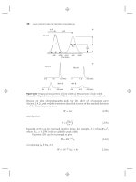
Another example of the effect of the column on selectivity is shown in
Figure 7.9 for the separation of a mixture of five, fully protonated strong
bases (1–5), five partly ionized weak acids (6–10), and a neutral reference
7.3 SEPARATION OF IONIC SAMPLES BY REVERSED-PHASE CHROMATOGRAPHY (RPC) 325
compound (11; shaded peak). In Figure 7.9a,mixturesofeither the strong bases
or weak acids plus neutral compound 11 are separated on each of these three
columns. In Figure 7.9b corresponding separations of samples containing all 11
compounds are shown. The relative retention of the fully protonated strong bases
(1–5) of Figure 7.9 is most affected by values of the ion-exchange capacity C for the
column (Section 5.4.1); larger values of C mean a greater retention of protonated
bases. Values of C at pH-2.8 for these columns are, respectively, −0.47 (Inertsil),
−0.30 (Symmetry), and 0.18 (Discovery). As expected, the relative retention of
basic solutes 1 to 5 increases in proceeding from the Inertsil to the Symmetry to the
Discovery column (note the retention ranges for peaks 1–5 in Fig. 7.9b, indicated at
the top of each chromatogram by arrows). The relative retention of the weak acids
6 to 10 and neutral compound 11 are quite similar on the three columns because
their retention is not affected by values of C.
02
Time (min)
2460
Time (min)
02
Time (min)
024
Time (min)
02
Time (min)
0
2
Time (min)
1
2
3
11
4
5
1
2
11
4
5
11
2
1
3
4
5
6
+
7
11
8
9
10
6
7
11
8
9
10
6
7
11
8
9
10
Strong bases Acids
Inertsil ODS-3
C = −0.47
Inertsil ODS-3
Symmetry C18
C = −0.30
Symmetry C1
8
Discovery C18
C = 0.18
Discovery C18
3
(a)
Figure 7.9 RPC separation of an ionic sample as a function of column type. Sample: (bases)
1, propranolol; 2, prolintane; 3, diphenhydramine; 4, nortriptyline; 5, amitriptyline; (acids) 6,
2-fluorobenzoic acid; 7, 3-cyanobenzoic acid; 8, 2-nitrobenzoic acid; 9, 3-nitrobenzoic acid;
10, 2,6-dimethylbenzoic acid; (neutral compound, shaded) 11, benzylalcohol. Conditions:
150 × 4.6-mm columns (5-μm particles); 30% acetonitrile-buffer (buffer is pH-2.8 phosphate
buffer); 35
◦
C; 2.0 mL/min. (a)Separationofacids6to10orbases1to5,ineachcasewith
neutral-compound 11 added; (b) separation of all 11 compounds. Recreated separations based
on data of [26, 29].