Introduction to Modern Liquid Chromatography, Third Edition part 57 pptx
Bạn đang xem bản rút gọn của tài liệu. Xem và tải ngay bản đầy đủ của tài liệu tại đây (152.31 KB, 10 trang )
516 QUALITATIVE AND QUANTITATIVE ANALYSIS
(comprising all the constituents of the sample excluding the analyte; for example,
blank plasma or the excipients in a pharmaceutical product) or sample diluent (i.e.,
injection solvent). For example, make a known dilution of an over-limit plasma
sample with blank plasma prior to sample pretreatment, or a dilution of a pesticide
formulation with additional injection solvent prior to injection. In applications
where occasional over-limit samples are likely to be encountered, such as preliminary
pharmacokinetic studies, it is wise to include dilution tests as part of the validation
process. For example, spiked samples could be prepared to demonstrate that a
10-fold dilution of an over-limit sample gives the same result as a sample prepared
at 1/10 the concentration. The method then could be written to allow dilution of
any sample, up to 10-fold over-range, into the concentration range approved for
analysis.
Samples that have concentrations below the standard curve may require a
larger mass of injected sample. In some cases, this can be accomplished simply by
injecting a larger sample volume. In other cases, concentration of the sample during
sample pretreatment, or less dilution, can provide a solution. In any event, the
process should be validated so that one has confidence in the quality of the resulting
data. Some methods may be written so that samples with peaks between the LOD
and the LLOQ are reported as below limit of quantification (BLQ), indicating that
analyte is present but in an insufficient amount to quantify. In every case, the means
adopted should be appropriate to the use of the final results.
11.3 QUALITATIVE ANALYSIS
Qualitative measurements are those that identify or help to identify the structure of
an analyte. In general, chromatography is a weak tool for qualitative analysis, but
a well-behaved HPLC system coupled to an appropriate detector can make it much
more suitable. Three approaches are common for qualitative analysis by HPLC:
• retention time
• on-line qualitative analysis
• off-line analysis
Remember that no matter which technique is used, it is much easier to prove that
two peaks are not the same compound than to prove that they are the same. That is,
compounds with closely related structures usually have similar retention times and
UV spectral characteristics, so retention time plus UV spectra may not be sufficient
for peak identification—it may be necessary to use mass spectrometry, NMR, FTIR,
or other techniques to confirm peak identity. Nevertheless, the identification of a
peak is generally easier when its behavior and properties can be shown to be identical
to those of a specific compound or standard—as opposed to a compound that is not
available.
11.3.1 Retention Time
The most common technique for qualitative analysis by HPLC is to compare the
retention time t
R
of the analyte to that of a reference standard (as discussed in
Section 11.2.2, relative retention is often used instead of absolute retention time).
11.3 QUALITATIVE ANALYSIS 517
If all HPLC conditions (mobile-phase composition, temperature, flow, etc.) are kept
constant, the retention time should be constant. Of course, conditions are never
exactly constant, and this results in small variations in retention from run to run
(e.g., ±0.02–0.05 min). If an injected analyte falls within the retention range of the
standard, this supports the conclusion that the standard and analyte peaks are the
same compound. However, retention time is characteristic, but not unique;more
than one compound can have the same retention time.
In order to minimize the possibility of confusion of one compound for another
with the same retention time, efforts should be taken during method development
to ensure that adequate resolution is achieved between the analyte of interest
and any likely interfering substances. The examples of adjacent peaks in Figure
2.17 can serve as a guide for how much resolution is required for this purpose,
which depends considerably on the relative size of two adjacent peaks and how
much they tail. Further considerations are (1) the likelihood that the column plate
number and peak tailing can change over time, (2) relative retention can vary with
inadvertent—usually small—changes in separation condition, and (3) the observed
retention time can differ from the true retention time when two peaks overlap.
Retention also can be influenced by the sample matrix; for example, the retention
time of a pure reference standard may differ slightly from t
R
of the same compound
in plasma. This is one reason why methods for drugs in biological samples should be
calibrated using matrix-based standards [6] (by spiking known concentrations of the
reference standard into blank [drug-free] matrix). Because several factors can create
uncertainty in the use of retention time for confirming peak identity (qualitative
analysis), it is best to limit this technique to methods where a particular analyte is
likely to be present and no other sample components are likely to overlap the analyte
peak.
Another retention-related technique for qualitative analysis is co-injection of a
reference standard. In this case the sample is injected; then the reference standard is
added to the sample and it is injected again. This technique is related to the method
of standard addition (Section 11.4.1.4) used for quantitative analysis. If the peaks
in the two injections have the same retention times, peak widths, and peak shapes
(within normal variability), there is additional evidence to conclude that the two
compounds are identical. On the other hand, if co-injection produces a broader
peak, a distorted peak, or two peaks, it is strong evidence that the reference standard
and analyte are not the same.
Finally, the use of retention-time predictions, literature values for retention
time, or retention times of related substances are never sufficiently accurate to
confirm the identity of a compound— although such estimates may prove useful for
certain purposes (e.g., the combination of a retention estimate with mass spectral
information for peak identification). The use of a retention time alone to identify an
analyte should be limited to comparisons with the retention of a known reference
standard, where the presence of interfering peaks is unlikely.
11.3.2 On-line Qualitative Analysis
Structural elucidation of unknown analytes can be performed with the aid of
HPLC detectors, but rarely is HPLC detection as effective as off-line qualitative
procedures. Several HPLC detectors (e.g., UV, NMR, or IR), provide qualitative
518 QUALITATIVE AND QUANTITATIVE ANALYSIS
spectral information about the sample; other detectors, such as the chemiluminescent
nitrogen detector, laser light-scattering detector, MS, or chiral detectors, generate
more specific and quantitative information about the analyte, such as nitrogen con-
tent, approximate mass, analyte molecular weight, or optical rotation, respectively.
Detectors usually collect information while the sample passes through the detector
flow cell, so the time available for measurement during passage of the peak through
the flow cell is similar in magnitude to the peak width—often only a few seconds. A
further constraint is that the sample is usually very dilute. Stopped-flow operation is
also possible, with the advantage of increasing the time allowed for measurement.
When compared to the same instrumental techniques used in an off-line,
stand-alone application—where analysis time is not limited and analyte concen-
tration often is much greater—the information content of on-line techniques is
consequently reduced. For this reason on-line data may be most useful for proving
that a particular peak is not a specific compound, rather than establishing chemical
structure. However, when a reference standard is available, the combination of
retention time with a single detector response can be sufficient to legally prove the
identity of an analyte. An example of this is the use of LC-MS (or LC-MS/MS) in the
forensic analysis of drugs of abuse. Finally, in many cases the presumed identity of a
peak (e.g., a metabolite of a drug) plus qualitative information from on-line detec-
tion may be sufficient for tentative structural confirmation. When data from several
HPLC detectors (e.g., FTIR, MS, or chemiluminescent nitrogen) are combined, the
structural identity of an analyte can be inferred with greater confidence.
11.3.2.1 UV Detection
The diode-array UV detector (Section 4.4.3), and less commonly the variable-
wavelength UV detector (Section 4.4.2) in the stopped-flow scanning mode, can
generate UV spectra of chromatographic peaks as they pass through the detector
flow cell. UV spectra alone, whether obtained on-line or off-line, rarely have enough
information content to assign an analyte structure. The spectra may be sufficient
to help confirm the presence of a compound suspected to be in a sample, but
the spectral similarity of structurally similar compounds usually prevents any final
conclusion about structure. For example, the UV spectra may be sufficient to confirm
which peak is the active ingredient in a drug dissolution sample, but it would not be
satisfactory to prove the present of a drug of abuse in a forensic situation.
11.3.2.2 LC-MS
The mass spectral detector (Section 4.14), especially in the MS-MS mode, can provide
sufficient spectral information to confirm the identity of a peak. The quadrupole
LC-MS in the MS-MS mode generates data on precursor-to-product ion transitions
that can be used to help elucidate the structure of an unknown, especially when
several different transitions can be obtained from the same analyte. The ion-trap
LC-MS has the capability to generate additional structural information in the MS
n
mode, where product ions may be successively fragmented into smaller product
ions (Section 4.14.2). However, with each successive fragmentation, the sample is
diluted, reducing the quality of the data. The time-of-flight LC-MS also measures
analyte-mass information that can help to provide structural identity. Because the
mass resolution (e.g., m/z ≈ 1) of LC-MS detectors is much lower than stand-alone
MS units, fractional mass differences cannot be used for structural elucidation.
11.3 QUALITATIVE ANALYSIS 519
11.3.2.3 LC-FTIR
The Fourier transform infrared detector (Section 4.15.1) is used most commonly by
trapping and evaporating aliquots of the column effluent, followed by spectroscopic
measurements. The LC-FTIR can generate valuable structural information (e.g.,
Fig. 4.34) that can be used to determine or confirm the chemical structure of a
chromatographic peak.
11.3.2.4 LC-NMR
The nuclear-magnetic-resonance LC detector (Section 4.15.2) in the flow-through or
stopped-flow mode can provide valuable structural information about a peak (e.g.,
Fig. 4.35). For
1
H NMR, deuterated solvents are required for the mobile phase, or
the mobile phase must be evaporated—this can restrict the scope of application of
LC-NMR.
11.3.2.5 Chemiluminescence Nitrogen Detector (CLND)
The CLND (Section 4.9) responds to the nitrogen content of the analyte. Because the
detector response is proportional to the molar content of nitrogen in the sample, the
detector can be calibrated with compounds of known nitrogen content. The molar
nitrogen content of an analyte can be determined from the detector response and the
mass of analyte injected (e.g., Fig. 4.20). Although this information is not sufficient
to determine molecular structure, it can aid in structural analysis.
11.3.2.6 Laser Light-Scattering Detector (LLSD)
The LLSD (Section 4.12.3) can assign an approximate molecular weight to a
macromolecular analyte, without the need for a reference standard of the analyte.
This capability can be sufficient to distinguish between monomeric and dimeric
forms of an analyte (e.g., Fig. 4.26). However, the accuracy of LLSD for determining
analyte molecular weight is far below that of LC-MS.
11.3.2.7 Chiral Detectors
Chiral detectors (Section 4.10) can distinguish between enantiomeric forms of an
analyte (e.g., Fig. 4.21), and give the sign of rotation. However, no other information
is provided about the structure of the analyte.
11.3.2.8 Off-line Analysis
If fractions are collected from the HPLC effluent stream in the semipreparative
or preparative mode (Chapter 15), a sufficient amount of pure analyte may be
collected to enable off-line analysis for structural determination. Because larger
quantities of sample are available and the time-frame restrictions of on-line analysis
are removed, off-line structural analysis often can provide conclusive structural
identity of a trapped peak. Traditional FTIR, NMR, and mass spectral analysis can
be performed; with sufficient sample, wet chemical tests, X-ray crystallography, or
other analytical techniques also may be applied.
520 QUALITATIVE AND QUANTITATIVE ANALYSIS
11.4 QUANTITATIVE ANALYSIS
Whereas the HPLC can provide qualitative information about a sample, its real
strength is shown in quantitative analysis. Other than the analytical balance, pH
meter, or volumetric pipette, it is likely that HPLC is the most commonly used
quantitative tool in the analytical laboratory. For its reliable application, five
requirements must be met. First, the HPLC system and its associated method must
work in a reproducible manner that provides the requisite precision and accuracy
(Sections 11.2.4.2, 11.2.4.3). Second, the data system (Section 11.2) must precisely
and accurately convert the detector signal into time and response data. Third,
the system must be properly calibrated (Section 11.4.1) to allow measurement of
unknown sample concentrations against known quantities of reference standards.
Finally, all of the data must be processed in a manner that assures that the overall
procedure performs at the required level to comply with appropriate regulatory
standards (e.g., Section 12.5) or other end-use requirements for the data. Finally,
separation conditions must be such as to enable stable baselines and adequate
resolution (R
s
>
1.5). However, the latter requirement has been dealt with in other
chapters and will not be repeated here.
11.4.1 Calibration
Calibration is the process by which the detector response per unit concentration (or
mass) of analyte is determined. Some detectors respond to analyte concentration
(e.g., UV, Section 4.4), whereas others respond to analyte mass (e.g., evaporative
light scattering, Section 4.12.1). In the present discussion we will assume that the
detector is concentration sensitive; for the most part the exact same procedures are
followed for mass-sensitive detectors. The two most common calibration techniques
are external standardization and internal standardization. Area normalization often
is used for purity analyses and other applications where relative concentration is
more important than absolute concentration—or where standards for calibration
do not exist. The method of standard addition is a specialized calibration technique
of particular use when a blank sample is not available, and the sample matrix may
affect the retention time and/or peak area response for the analyte. For such sample
matrices (e.g., plasma), it is also strongly recommended (e.g., [6]) that the calibration
standards be prepared in blank matrix (i.e., all the components that are normally
found in a sample, excluding the analyte). This, of course, applies to both external
and internal standardization (area-normalization and standard addition techniques
already have the matrix present). In cases where the sample matrix has little influence
on retention or selectivity, such as environmental water samples, or the assay of
pure compounds or simple mixtures, matrix-based calibrators may not be required.
In order to obtain accurate results from a method, the calibration curve must
adequately represent the concentration-response relationship for the analyte. One
way to help improve the accuracy of the calibration curve is to evaluate whether
curve weighting is appropriate. This topic is discussed in Section 11.4.1.5.
11.4.1.1 External Standardization
A matrix-based set of calibration standards is prepared. Usually this is done by
accurately weighing a quantity of reference standard of known purity and diluting
11.4 QUANTITATIVE ANALYSIS 521
it in water or buffer to make a primary stock. This stock then is added to the sample
matrix (e.g., sample diluent, blank plasma, water, soil, or other matrix appropriate
to the sample type), to the concentration corresponding to the highest point on the
calibration curve. (Some laboratories refer to the standard curve as a line; it also is
commonly called a standard curve. ‘‘Curve’’ is generally used to describe this plot,
even though it is most often a linear plot.) Further dilutions are made in matrix to pre-
pare standards that span the method range, including the lowest point on the curve,
and sometimes a standard at the limit of detection (LOD). It is customary to include
a blank-matrix sample to demonstrate that interferences are not present in the blank.
The two most popular ways to prepare the calibration-curve samples are to use
a linear or exponential (sometimes incorrectly called ‘‘logarithmic’’) dilution scheme.
For example, with a standard curve covering a method range of 1 to 100 ng/mL,
standards might be prepared at 0, 1, 20, 40, 60, 80, and 100 ng/mL for a linear
dilution, or 0, 1, 2, 5, 10, 20, 50, and 100 ng/mL for an exponential dilution. At
the same time as the calibration standards are prepared, or during preparation of
analytical samples, it is a good idea to prepare quality control standards (Section
12.3) that will be used to check method performance within a batch of samples. If
sample preparation is required, the calibration (and quality control) standards are
then processed through the normal sample preparation process, yielding extracted
calibration standards for injection.
For external standardization, the same volume of calibration standard at each
concentration (level) is injected in sequence from lowest to highest concentration.
The low-to-high sequence tends to minimize any carryover-related bias in the curve.
The highest concentration standard can be followed by a blank (zero-concentration)
standard to check for carryover (Section 17.2.5.10), as well as to avoid carryover
bias if the following sample has a low concentration of the analyte. It is best to
inject the same volume of different standard concentrations when running the
calibration curve, rather than different volumes of the same concentration; the
injection volume delivered by most autosamplers is very precise but not necessarily
as accurate (Section 3.6.1).
A calibration plot can be constructed manually with the aid of spreadsheet
software (e.g., Microsoft Excel), or with the data-system software. It is best to use the
data-system software, because in most cases it can be validated, and transfer of data
from the raw-data tables into the software is seamless. Excel and similar programs
are flexible and work well, but are not considered validated (or validate-able)
software by some regulatory guidelines published by authorities such as FDA or
ICH, so additional data checking will need to be done to make sure the results are
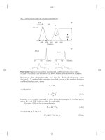
error-free. An example of an external calibration plot is presented in Figure 11.8
using the data from Table 11.1. The slope of the calibration plot, S
,isthenusedto
calculate the concentration of unknown samples:
ng/mL analyte =
area analyte
S
(11.5)
In the case of Figure 11.8 (with a linear-regression forced through the origin; i.e.,
x = 0, y = 0), S
= 200.8 (area units)/(ng/mL analyte). So an analyte peak of 862
area counts would have a concentration of (862/200.8) = 4.3 ng/mL. Equation
(11.5) assumes that the trend line intercepts the x-axis at y = 0, which may or
522 QUALITATIVE AND QUANTITATIVE ANALYSIS
0
0.5
1
1.5
2
2.5
0 200 400 600 800 1000
Concentration
(
n
g
/mL
)
Peak Area (x10
−5
)
(data forced through x = 0, y = 0)
y = 200.8x
r
2
= 0.9999
Figure 11.8 Calibration curve based on external standardization data of Table 11.1 (curve
forced through zero).
Table 11.1
Calibration Curve Data for Figures 11.8 to 11.10 for Same Analyte and Separation Conditions
Response
Concentration (ng/mL) External Standard
a
Internal Standard
b
Standard Addition
c
0 487
1 215 0.0408 729
2 416 0.0789 911
5 976 0.185 1,435
10 2,056 0.390 2,529
20 4,060 0.770
50 9,921 1.88
100 20,140 3.82
200 40,163 7.62
500 99,796 18.9
1000 201,123 38.2
a
Area units (Fig. 11.8).
b
Analyte/IS ratio (Fig. 11.9).
c
Area with standard added at concentration in column 1 (Fig. 11.10).
may not be the case (Section 11.2.4.5). Alternatively, carry out the linear-regression
without forcing the fit through zero (0, 0 point); this is the usual approach taken
by data processing software. In the present case, the data of Table 11.1 yield a
regression equation (without forcing zero) of y = 201x − 38. Solving for x and
inserting y = 862 gives x = 4.5 ng/mL, which adjusts for the (slight) nonzero
intercept. The calculated value represents the concentration of analyte in the injected
sample; any weighing, dilution, or other sample processing corrections need to be
applied to this value before the final sample concentration is reported.
11.4 QUANTITATIVE ANALYSIS 523
Because the external standard method assumes that the area of the analyte peak
accurately represents the concentration of analyte in the original sample, external
standardization is best used with methods that involve minimal sample manipulation
between the initial sampling process and injection. Therefore solid or liquid samples
that undergo weighing, pipetting, dilution, dissolution, and/or filtration processes are
good candidates for external standardization. Pharmaceutical dissolution analysis
involves placing one or more drug tablets in a dissolution bath of known volume,
taking samples at specific time points, filtering the samples, and injecting them. An
environmental water sample might be aliquotted by volume, shaken in a measured
volume of solvent, filtered, and injected. In both of these cases, it is easy to track the
concentration of the injected sample relative to the initial untreated sample, so they
would be good candidates for external standardization.
A variation of the external standardization method is single-point calibration
(Section 11.2.4.5). In this technique experiments during method development and
validation are performed to show that analyte response is proportional to its
concentration over the method range. Then a single standard is injected, and the
analyte concentration in an unknown sample is determined by the ratio of the areas
of the standard and the unknown (equivalent to use of Eq. 11.5). Usually the range
of the method is narrow, such as ±20% of the target dose of a drug in tablet form.
For example, dissolution testing of drug products designed for immediate release
can be tested with single-point calibration if supporting data have been gathered to
show the validity of this technique [11].
11.4.1.2 Internal Standardization
Internal standardization is superior to external standardization whenever there
are sample preparation steps (Chapter 16) in which sample loss can take place.
For example, the determination of drugs in plasma often involves solid-phase or
liquid–liquid extraction with variable volume recovery, evaporation to dryness, and
reconstitution in the injection solvent. At each of these steps the initial and final
sample volume seldom are the same for every sample, but the internal standard (IS)
tracks such changes, making it possible to obtain precise and accurate results.
The primary difference between internal standardization and external stan-
dardization is that an IS is added to samples and calibrators prior to sample
pretreatment; calculations of analyte concentration are based on the ratio of areas
for the analyte and IS. Calibrators are prepared by weighing and serial dilution, just
as for the external standard method (Section 11.4.1.1). Aliquots of the calibration
standards (e.g., 200 μL of spiked plasma) are then mixed with an IS solution (e.g.,
10 μL), as are all samples—standards and samples are then processed in the same
way. The IS solution is prepared in water or buffer at a concentration such that a
small volume (e.g., ≤5% of the sample volume) will generate a peak of sufficient
size (e.g., S/N
>
100) for measurement with suitable precision and accuracy. As
in the case of external standardization, standards are injected in a low-to-high
concentration sequence.
The ratio of the area of analyte to area of IS in each of the calibration samples is
calculated (e.g., Table 11.1) and a plot of this ratio against the analyte concentration
is made (Fig. 11.9). The linear regression for these data gives y = 0.0381x − 0.0073,
with a standard error of the y-intercept of 0.0172. Since the y-intercept (absolute
524 QUALITATIVE AND QUANTITATIVE ANALYSIS
0
10
20
30
40
50
0 200 400 600 800 1000
Concentration
(
n
g
/mL
)
Area Ratio (analyte/IS)
(data forced through x = 0, y = 0)
(data of Table 11.1)
y = 0.0381x
r
2
= 0.9999
Figure 11.9 Calibration curve based on internal standardization data of Table 11.1.
value) is <SE, it is appropriate to force the curve through the origin (Section 11.2.4.5);
that is, y = 0.0381x. For this example, S
= (0.0381ratio units)/(ng/mL analyte).
Calculations of unknown samples are carried out in the same manner as for external
standardization, except that the analyte-to-IS ratio is used instead of the absolute
analyte area:
ng/mL analyte =
analyte/IS area-ratio
S
(11.6)
Thus a sample for which the analyte area is 15,345 and the IS = 4725, the ratio
= 3.25. From Equation (11.6), (3.25 ratio units)/0.0381 = 85.2 ng/mL analyte.
For an IS to properly perform its functions, it must have certain properties,
several of which are summarized in Table 11.2. The IS or a sample component with
the same retention time must never be present in the sample, or an invalid (low)
assay result will be obtained. It is desirable to have the IS peak elute near the analyte
so that it experiences a similar chromatographic history. If possible, elution just after
the analyte is preferable, because if the IS has the correct retention time and area
Table 11.2
Internal Standard Properties
1. Never found in sample
2. Similar k to analyte, preferably eluted after analyte
3. Equivalent sample preparation properties to analyte (pK
a
,logP
o/w
,etc.)
4. Well-resolved from analyte (or stable-label for MS)
5. Stable
6. Pure or known purity
7. Compatible detector response (IS peak should have S/N
>
100)
8. Structurally similar to analyte (desirable, but not essential if property 3 is applicable)
11.4 QUANTITATIVE ANALYSIS 525
response, it is known that all peaks eluted earlier also were eluted under the proper
chromatographic conditions. Peaks eluted after the IS may be subject to bubbles or
equipment errors that would not be apparent from an examination of the IS peak.
One of the main roles of the IS is to correct for variations in analyte recovery
during sample pretreatment; the IS should therefore possess chemical properties
(extraction coefficients, pK
a
values for partly ionized analytes, etc.) that are similar
to those of the analyte. When several analytes are present in a single sample, it
may be desirable to have more than one IS, but this usually is not essential. The
IS needs to be well-resolved from the analyte so that peak measurements are not
compromised. The exception is for LC-MS applications where a stable, isotopically
labeled compound often is used as the IS. Stable-label standards do not require
chromatographic resolution if the MS resolution is adequate to distinguish clearly
between the analyte and IS (usually the case). Isotopically labeled standards that
co-elute with the analyte (typical for
13
C standards) will not be subject to changes
in separation conditions (e.g., a bubble) or detector conditions (e.g., spray plume
irregularity in the MS interface). Thus co-eluted standards more closely mimic
the analyte than standards that do not co-elute (often the case for deuterated
standards).
The IS needs to be sufficiently stable that it will not change during sample
preparation and chromatographic separation. Although it is not necessary for the
IS to be highly pure, as long as it is stable, it is important that no IS impurities
interfere with the analyte response. The IS also requires an acceptable detector
response. Some lists of IS requirements suggest that the IS should be structurally
similar to the analyte. There is no basis for this requirement—as long as the IS
has the other required properties. However, an IS that is structurally similar to the
analyte is more likely to be suitable, so most users choose a structurally similar IS.
Good IS candidates are structural analogues of the analyte or related compounds
that are not likely to be present in the sample. In some cases it is convenient to
make a ‘‘flip-flop’’ method, where two related compounds are used as the IS for
each other. For example, in method 1, compound X is used as the IS for analyte Y,
and in method 2, compound Y is used as the IS for analyte X; the limitation of this
technique, of course, is that both X and Y can never be present in the same sample.
This technique would work with two related drugs that were never co-dosed but
had closely related extraction and chromatographic properties
11.4.1.3 Area Normalization
A standard feature of data systems is to report percent peak area. This is obtained
by adding the areas of all peaks in a chromatogram and reporting each peak
as a percentage of the total. This report format is convenient for screening a
chemical reaction for completion and approximate product purity as well as other
applications. A related report is area normalization, in which one peak is chosen as
a reference peak and all other peaks are reported as a percentage of the reference
peak. Area normalization is common for methods used to test drug stability or
assay impurities in drug products. In the latter case, any peak ≥0.1% of the
active pharmaceutical ingredient (API) must be reported and identified, whereas
peaks ≥0.05% must be reported but not necessarily identified [10]. Both area-%
and area normalization are convenient because they do not require standards for