Introduction to Modern Liquid Chromatography, Third Edition part 58 potx
Bạn đang xem bản rút gọn của tài liệu. Xem và tải ngay bản đầy đủ của tài liệu tại đây (133.84 KB, 10 trang )
526 QUALITATIVE AND QUANTITATIVE ANALYSIS
each peak. However, both procedures rely on the assumption that the detector
response for nonstandardized (e.g., unknown) peaks is the same as for peaks for
which standards are available; this assumption may or may not be appropriate,
depending on the sample composition and choice of detector. UV detection is
notorious for order-of-magnitude differences in sensitivity for different compounds.
11.4.1.4 Standard Addition
The method of standard addition (‘‘spiking’’) can be useful when a sample blank
cannot be obtained, and the sample matrix can affect analyte recovery and/or
response. For example, when measuring insulin levels in plasma, it is impossible to
obtain plasma without insulin, so standard addition can be used.
Standard addition can be based on a single-point or multiple-point calibration.
For single-point calibration, the sample is split into two fractions. One fraction is
spiked with a known concentration of the standard, and both fractions are analyzed.
The calibration factor is obtained as
S
=
area
s
− area
ns
conc
s
(11.7)
where area
s
and area
ns
are the areas of the spiked and nonspiked samples, respec-
tively, and conc
s
is the concentration of standard added to the spiked sample.
Using the data of Table 11.1, we see that for conc
s
= 2ng/mL,area
s
= 911 and
area
ns
= 487. S
= (911 − 487)/(2 ng/mL) = 212 area units/(ng/mL). Now the con-
centration of the non-spiked sample (conc
ns
; shown as 0 ng/mL in Table 11.1) can
be determined as
conc
ns
=
area
ns
S
(11.8)
or conc
ns
= 487/212 = 2.3ng/mL.
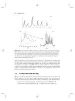
For multiple-point calibration using standard addition, the sample is split into
n + 1 fractions, where n is the number of standards to be used. Then n samples are
spiked, each with a different concentration of standard and all samples are analyzed.
A calibration curve is plotted, as shown in Figure 11.10 for the data of Table 11.1.
Note that the regression line is extended to the left until it intersects the x-axis (arrow
in Fig. 11.10). The value of the intercept x corresponds to −conc
ns
. Linear regression
of the data of Table 11.1 gives y = 201x + 496. Solving for x and inserting y = 0
gives x =−2.5, so conc
ns
= 2.5 ng/mL. The result (<1 SD difference) is the same as
that obtained above with Equations (11.7) and (11.8).
It should be stressed that the method of standard addition does not correct for
baseline variation or other sample interferences. These problems must be handled
in the usual way, before the standard addition procedure is applied. In effect, this
approach is a form of in situ calibration, and it can be very useful when the more
traditional techniques of external or internal standardization cannot be used. As
noted in Section 11.3.1, the method of standard addition also can be useful in
confirming peak identity, although its value for this purpose is no greater than the
use of a retention time.
11.4 QUANTITATIVE ANALYSIS 527
0
5
10
15
20
25
30
−50510
y = 201x + 496
r
2
= 0.9976
Peak Area (x10
−2
)
Concentration Added (ng/mL)
Figure 11.10 Use of the method of standard addition to determine analyte concentration at
x-axis intercept (arrow); data of Table 11.1.
11.4.1.5 Evaluating Calibration Curves
Plots for wide concentration-range calibration curves, such as those of Figures 11.8
and 11.9 can be hard to interpret when a linear x-axis is used, because the low
concentration points are crowded together. An alternate way to examine the data is
to make a plot of %-error against log concentration (a ‘‘%-error plot’’). %-Error
is determined by using the regression equation to calculate the theoretical y-value
for each concentration; error is calculated as (experimental value − theoretical
value)/theoretical value, and expressed as %-error.
The benefit of the %-error plot is shown in Figure 11.11 for a data from a
hypothetical method-validation study. Calibrators were injected at 10 concentra-
tions: 1, 2, 5, 10, 20, 50, 100, 200, 500, and 1000 ng/mL. The plot of %-error versus
(linear) concentration of Figure 11.11a shows that there is much more relative error
at lower concentrations, but this figure is hard to interpret because of the crowding
of data points at low concentrations. The %-error versus log concentration plot of
Figure 11.11b solves the crowding problem, and now the error at each concentration
can be easily examined. The data fall into two sets—those above 20 ng/mL with a
relatively constant error of ≈±1% (1 SD)—while remaining concentrations show
increasing relative error as concentration is reduced (dashed lines of Fig. 11.11b).
This pattern is expected, as S/N makes a larger contribution to relative error
(Eq. 11.1) at low concentrations. Other than the (normal) increase in error at
low concentrations, the data of Figure 11.11 have acceptable regression statistics
(r
2
= 0.9999, y-intercept <SE), so a multiple-point calibration curve with forced
zero (y = mx; Sections 11.4.1.1, 11.4.1.2) is appropriate.
The %-error plot can highlight problems with calibration curves. Data similar
to that of Figure 11.11a, b are shown in the example of Figure 11.11c,which
emphasizes the importance of picking the proper y-intercept. If a multiple-point
calibration is chosen, with the curve forced through zero, the %-error plot of
Figure 11.11c results. Lower concentrations show increasing error, with an average
error ≈50% for 1 ng/mL. For a bioanalytical method, where maximum error
allowed at the LLOQ =±20%, the lowest concentration with an average error
of ≤20% is 5 ng/mL (≈9% error); this limits the application of the method to a
528 QUALITATIVE AND QUANTITATIVE ANALYSIS
–15
–10
–5
0
0 200 400 600 800 1000
5
10
15
% – Error
Concentration (ng/mL)
–15
–10
–5
0
1 10 100 1000
5
10
15
% – Error% – Error
Concentration (ng/mL)
Concentration (n
g
/mL)
±1 RSD
(c)
(b)
(a)
70
60
50
40
30
20
10
0
mean
values
1000100101
Figure 11.11 Use of %-error plots to examine calibration-curve data. (a) %-Error versus (lin-
ear) concentration; (b)dataof(a) plotted as %-error versus log concentration (y-intercept
<SE; curve forced through zero); (c) %-error versus log concentration (y-intercept
>
SE; curve
improperly forced through zero).
11.5 SUMMARY 529
range of 5 to 1000 ng/mL. For this data set the y-intercept
>
SE, so the proper
curve fit is y = mx + b (with b = 0). The resulting %-error plot (not shown) closely
resembles Figure 11.11b, with an average error of <6% throughout the curve. Now
the calibration curve allows a bioanalytical method to be applied over a range of
1 to 1000 ng/mL. It is interesting to note that for both curve fits (with b = 0or
b = 0), r
2
>
0.9999, so r
2
alone is not sufficient to ensure good performance of a
multiple-point calibration.
Low r-orr
2
-values can indicate that there are problems with a calibration
curve, but the converse is not necessarily true—large r
2
-values do not guarantee
a well-behaved curve. A plot of %-error against log concentration (%-error plot)
is a useful way to make a visual examination of data. We recommend examining
all calibration-curve data with a %-error plot as a means of highlighting potential
problems with the method.
11.4.2 Trace Analysis
HPLC is used for the analysis of samples of widely varying concentration. The term
trace analysis often is used to describe small sample concentrations. One way to
define trace analysis is to describe samples for which the precision of measurement
is affected by the concentration, often with a transition point to trace analysis when
S/N < ≈100 (Section 4.3). Other than the problems associated with dealing with
low concentrations and small signals, trace analysis is little different from the analysis
of more concentrated samples. With trace analysis, peak height measurements may
be preferred over peak area. We recommend evaluating both peak height and
peak area; choose the final measurement technique based on the one that gives
the best precision and accuracy. For additional information, see Sections 2.6.3.2,
4.2.4, and 11.2.5; for discussion related to specific detectors, consult the appropriate
detector discussion in Sections 4.4 through 4.16.
11.5 SUMMARY
Use of the HPLC as a qualitative or quantitative analytical tool requires that the sys-
tem be operating properly and that the data system be set up to accurately determine
peak retention times and peak areas or heights. Consideration has to be taken relative
to resolution requirements for the peaks of interest, including the influence of relative
peak size and shape. Although the HPLC system is not as useful a tool for qualitative
analysis as some dedicated instruments (e.g., FTIR, NMR, or high-resolution MS),
with the help of the appropriate detector(s) it can provide valuable qualitative infor-
mation for many applications. Liquid chromatography shows its strongest assets
with quantitative analysis. HPLC can be used for trace analysis of pollutants in river
water, drugs and their metabolites in biological systems, or impurities in reagents.
It is also useful for determining content uniformity of pharmaceutical products with
high precision and accuracy. Quantitative analysis relies on selection of appropriate
reference standards and proper calibration so that the results are of high quality and
can withstand the scrutiny of review by regulatory agencies.
530 QUALITATIVE AND QUANTITATIVE ANALYSIS
REFERENCES
1. N. Dyson, Chromatographic Integration Methods, 2nd ed., Royal Society of Chemistry,
Letchworth, UK, 1998.
2. L. R. Snyder and J. J. Kirkland, Introduction to Modern Liquid Chromatography, 2nd
ed., Wiley, New York, 1974.
3. V. R. Meyer, J. Chromatogr. Sci., 33 (1995) 26.
4. R. Q. Thompson, J. Chem. Ed., 62 (1985) 866.
5. Guidance for Industry: Part 11, Electronic Records; Electronic Signatures—Scope
and Application, USFDA-CDER, Aug. 2003, />ref/
part11.
6. Guidance for Industry: Bioanalytical Method Validation, USFDA-CDER, May 2001,
/>7. Reviewer Guidance: Validation of Chromatographic Methods, USFDA-CDER, Nov.
1994, />8. L. R. Snyder, J. J. Kirkland, and J. L. Glajch, Practical HPLC Method Development,
2nd ed., Wiley-Interscience, New York, 1997, p. 71.
9. Validation of Analytical Procedures: Text and Methodology Q2(R1), International Con-
ference on Harmonization, Nov. 2005, />10. Impurities in New Drug Substances Q3A(R2), International Conference on Harmoniza-
tion, Oct. 2006, />11. Specifications: Test Procedures and Acceptance Criteria for New Drug Substances
and New Drug Products: Chemical Substances Q6A, International Conference on
Harmonization, Oct. 1999, />12. J. C. Miller and J. N. Miller, Statistics for Analytical Chemistry, Halsted Press-Wiley,
New York, 1984, secs. 4.9–4.10.
CHAPTER TWELVE
METHOD VALIDATION
with Michael Swartz
12.1 INTRODUCTION, 532
12.2 TERMS AND DEFINITIONS, 534
12.2.1 Accuracy, 535
12.2.2 Precision, 536
12.2.3 Specificity, 539
12.2.4 Limit of Detection and Limit of Quantification, 539
12.2.5 Linearity and Range, 540
12.2.6 Robustness, 540
12.3 SYSTEM SUITABILITY, 542
12.4 DOCUMENTATION, 543
12.4.1 Validation Protocol, 544
12.4.2 Test Method, 544
12.4.3 Validation Report, 545
12.5 VALIDATION FOR DIFFERENT PHARMACEUTICAL-METHOD
TYPES, 546
12.5.1 Category 1 Methods, 546
12.5.2 Category 2 Methods, 547
12.5.3 Category 3 Methods, 547
12.5.4 Category 4 Methods, 548
12.6 BIOANALYTICAL METHODS, 548
12.6.1 Reference Standard Preparation, 549
12.6.2 Bioanalytical Method Development and Validation, 549
12.6.3 Routine Application of the Bioanalytical Method, 552
12.6.4 Bioanalytical Method Documentation, 553
12.7 ANALYTICAL METHOD TRANSFER (AMT), 554
12.7.1 Analytical Method-Transfer Options, 555
12.7.2 Essentials of AMT, 556
12.7.3 Potential AMT Pitfalls, 558
12.8 METHOD ADJUSTMENT OR METHOD MODIFICATION, 561
12.8.1 pH Adjustments, 563
Introduction to Modern Liquid Chromatography, Third Edition, by Lloyd R. Snyder,
Joseph J. Kirkland, and John W. Dolan
Copyright © 2010 John Wiley & Sons, Inc.
531
532 METHOD VALIDATION
12.8.2 Concentration of Buffer Salts, 563
12.8.3 Ratio of Components in the Mobile Phase, 563
12.8.4 Wavelength of the UV-Visible Detector, 564
12.8.5 Temperature Adjustments, 564
12.8.6 Column Length, Diameter, and Particle-Size Adjustments, 564
12.9 QUALITY CONTROL AND QUALITY ASSURANCE, 564
12.9.1 Quality Control, 565
12.9.2 Quality Assurance, 565
12.10 SUMMARY, 565
12.1 INTRODUCTION
Quality is a commonly used word in the world of analytical chemistry. Quality
encompasses many aspects of the laboratory; in this chapter it refers to the develop-
ment and application of HPLC methods. Our primary focus will be the validation of
HPLC methods, a process that underlies the quality of the method and laboratory
results. Other aspects of quality in the HPLC laboratory are also discussed in this
chapter, especially quality control and quality assurance (Section 12.9).
Method validation establishes, by means of laboratory studies, that the per-
formance characteristics of the test method meet the requirements of the intended
analytical application. Method validation provides an assurance of reliability during
normal use, and this process is sometimes referred to as providing documented
evidence that the method does what it is intended to do. Regulated laboratories must
carry out method validation in order to be in compliance with governmental or other
regulatory agencies. A well-defined and documented method-validation process not
only satisfies regulatory compliance requirements but also provides evidence that the
system and method are suitable for their intended use, and aids in method transfer.
In 1987, the US Food and Drug Association (FDA) first designated the specifications
listed in the current edition of the United States Pharmacopeia (USP) as those legally
recognized to determine compliance with the Federal Food, Drug, and Cosmetic Act
[1–2]. More recently, new information has been published that updates the previous
guidelines and provides more detail and harmonization with International Confer-
ence on Harmonization (ICH) guidelines [3–4]. The inclusion and/or definition of
some terms differs for the FDA, USP, and ICH, but harmonization on a global basis
has provided much more detail than was available in the past. So it may be useful to
downplay any differences between global regulatory requirements.
An HPLC method may be referred to as an ‘‘analytical procedure,’’ ‘‘analyt-
ical method,’’ ‘‘assay procedure,’’ ‘‘test method,’’ or just ‘‘method.’’ In the present
discussion, we generally will use ‘‘test method’’ or (less often) ‘‘HPLC method’’ for
12.1 INTRODUCTION 533
any of these methods. The largest number of HPLC methods in use today is carried
out in the pharmaceutical industry; for this reason we will describe method valida-
tion for pharmaceutical applications. Other regulated industries have well-defined
processes in place for method validation as well. For example, environmental moni-
toring laboratories are under the oversight of the Environmental Protection Agency
(EPA) [5], whereas some other organizations rely on directives of the International
Organization for Standardization (ISO) [6].
Validation and other laboratory practices are regulated by the FDA, USP, ICH,
EPA, and related organizations. Because HPLC methods developed by an industrial
analytical laboratory often are used in a manufacturing department, the analytical
laboratory may be constrained by manufacturing practices and regulations. Two of
the most common references to these practices are cGMP (current Good Manufac-
turing Practice, e.g., [7–8]) and the ISO 9000 Global Management Standards [9]
and related ISO (International Organization on Standardization) documents. These
aspects of laboratory regulation are not discussed further in this chapter.
In nonregulated industries and academic laboratories, there also is a need
for high-quality test methods that provide reliable data. The use of good scientific
practices is often assumed but is not always the case, so method validation is strongly
recommended even where it is not required by regulation. The reader should be able
to take the information presented here for pharmaceutical applications and use it as
a basis for other areas of application.
Method validation can be regarded as just one part of an overall validation
process that encompasses at least four distinct steps: (1) software validation,
(2) analytical instrument qualification or validation (AIQ; Section 3.10.1),
(3) method validation, and (4) system suitability. The overall validation process
begins with validated software and a qualified instrument; then a test method
is developed and subsequently validated using the qualified system. Finally, the
performance of the test method on a given day can be confirmed by means of a
system suitability test. Each of these four steps is critical to method performance.
Two guidelines are important for any method validation process: USP Chapter
1225, Validation of Compendial Methods [2], and the International Conference
on Harmonization (ICH) Guideline, Validation of Analytical Procedures: Text and
Methodology Q2 (R1) [4]. Although the subject of the current discussion is HPLC,
both the USP and ICH guidelines apply to any analytical procedure, technique, or
technology used in a regulated laboratory. It should be noted that the USP publishes
official test methods, often called compendial methods, that are accepted by the USP
as already validated. The USP also publishes guidelines that should be applied to
the validation of test methods not developed by the USP (it is assumed that the USP
published methods were subject to the same guidelines). This chapter concentrates
on the application of the USP (and other regulatory agency) guidelines to HPLC
methods developed by independent laboratories (i.e., not by the agencies themselves).
Even though the USP is the sole legal document in the eyes of the FDA, this
chapter draws from both USP and ICH guidelines, as appropriate, for definitions
and methodology. For the most part the FDA, USP, and ICH guidelines agree. Where
the guidelines disagree, it is up to the user to decide on an appropriate interpretation
of the guidelines. Often this is the responsibility of the user’s quality assurance unit
(Section 12.9), and may be aided by review of the latest regulatory actions (e.g.,
FDA-issued Form 483 Inspectional Observations). For the present discussion, the
534 METHOD VALIDATION
regulatory publications will be referred to generically as ‘‘guidelines.’’ In addition to
these guidelines, sometimes a regulatory body publishes other information that can
be useful for interpretation of the guidelines. One of these is a ‘‘reviewer guidance’’
[10] published by the FDA. This document is intended to help FDA auditors
determine what comprises a good test method, so many users try to adhere to the
suggestions of this document to ensure that their test methods will pass regulatory
scrutiny. In addition to the general process of method validation, we will discuss
terms, definitions, and related topics, and—where possible—provide examples to
illustrate how these general guidelines apply to HPLC.
A major difference between this chapter and other chapters in this book is
the regulatory oversight of validated methods in the pharmaceutical, environmental,
and certain other industries. Rules pertaining to method validation are described in
official documents that originate at different times, are written by different people
(with different writing skills), and are released by different agencies (with varying
internal policies). This bureaucratic process inevitably results in documents that can
be ambiguous, inconsistent, and difficult to interpret. In this chapter we try to impart
some unity to the requirements and guidelines contained in these various regulatory
pronouncements. We also try to make the discussion practical for the average
user. Nevertheless, the bureaucratic language of regulatory recommendations and
requirements could not entirely be masked. Consider this limitation as good practice
for dealing with the official documents.
Method validation may seem to have a vocabulary of its own. This chapter
therefore begins with a discussion of important terms and definitions (Section 12.2).
A procedure that ensures that a test method can provide valid data on a given day
is the system suitability test, described in Section 12.3. Without documentation,
there is no proof of method validity; some aspects of method documentation are
described in Section 12.4. Validation of test methods for drug substance (pure
drug) and drug product (formulated drug) have requirements (Section 12.5) that
are distinctly different from bioanalytical methods that measure drugs in biological
matrices (Section 12.6). Once a test method has been validated, it often must be
transferred to another laboratory for routine application; some of the principles
of analytical method transfer (AMT) are discussed in Section 12.7. Many times
when test methods are transferred, they do not work exactly as they did in the
original laboratory, and over time, most methods require some adjustment to meet
system suitability requirements. Methods can be adjusted to meet system suitability,
but if they require more substantial changes (are modified), they must undergo
as least some re-validation; the topic of adjustment vs. modification is covered in
Section 12.8. Finally, a good test method and its application require strong quality
control and quality assurance programs, as described in Section 12.9.
12.2 TERMS AND DEFINITIONS
Several analytical performance characteristics may be investigated during any method
validation protocol:
• accuracy
• precision/ruggedness
12.2 TERMS AND DEFINITIONS 535
• specificity
• limit of detection
• limit of quantitation
• linearity
• range
• robustness
Although most of these terms are familiar and are used daily in any regulated
HPLC laboratory, they sometimes mean different things to different people.
For example, ruggedness, which forms a part of any well-designed precision
study, is often confused with robustness. The following standard definitions
(Sections 12.2.1–12.2.6), of applications in the pharmaceutical industry should
clarify any confusion. In this context, drug substance refers to the pure chemical
drug, also called the active pharmaceutical ingredient (API). The drug product refers
to the product that is sold to the consumer; it usually contains one or more drug
substances plus excipients—(other chemicals, fillers, colors, etc.).
Several types of test methods are used to measure the API and/or impurities,
related substances, excipients, and so forth. The major method types discussed in this
chapter are assay, impurity (also related substances), dissolution, and bioanalytical
methods. A test method used for assay is one that measures the active ingredient
concentration in a drug product or substance. A content uniformity method is
similar to an assay method, but it targets the measurement of the variability in drug
concentration within a batch of samples. An impurity test measures the (generally
unintentional) minor components present in the substance or product that originate
from raw material manufacturing, product manufacturing, or degradation during
storage or processing. A stability-indicating method is used to quantify the presence
of impurities (degradants) generated through a forced degradation of the API;
it is assumed that this test will enable measurement of any impurities generated
during normal or accelerated shelf-life testing of a drug substance or product. Any
degradants found in this way may be included in the impurity test. A dissolution
assay measures the concentration of API in a solution designed to simulate release
of the drug from a formulation under the conditions of administration of the drug
(e.g., in simulated stomach fluids). Whereas the preceding test methods are for drug
product or drug substance, a bioanalytical method (Section 12.6) is used to determine
the concentration of a drug in a biological system, most commonly plasma.
12.2.1 Accuracy
Accuracy is the measure of exactness of an analytical method, or the closeness of
agreement between an accepted reference value and the value found in a sample.
Established across the range of the method, accuracy is measured as the percentage
of analyte recovered by the assay. For the drug substance, accuracy measurements
are obtained by comparison of the results to the analysis of a standard reference
material, or by comparison to results from a second, well-characterized method.
For the assay of the drug product, accuracy is evaluated by the analysis of synthetic
mixtures spiked with known quantities of the analytes. For the quantification of
impurities, accuracy is determined by the analysis of samples (drug substance or drug