Introduction to Modern Liquid Chromatography, Third Edition part 62 ppt
Bạn đang xem bản rút gọn của tài liệu. Xem và tải ngay bản đầy đủ của tài liệu tại đây (180.54 KB, 10 trang )
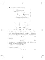
566 METHOD VALIDATION
scrutiny of any reviewer. The contents of this chapter concentrate on pharmaceutical
methods, but the same principles can be applied to any HPLC method so as to ensure
that it is suitable for its intended use
REFERENCES
1. Guideline for Submitting Samples and Analytical Data for Methods Validation,
USFDA-CDER (February 1987), />2. United States Pharmacopeia No. 31-NF 26, (2008), ch. 1225.
3. Analytical Procedures and Methods Validation, USFDA-CDER (Aug. 2000),
/>4. Harmonized Tripartite Guideline, Validation of Analytical Procedures, Text and
Methodology, Q2 (R1), International Conference on Harmonization, (Nov. 2005),
/>5. Guidance for Methods Development and Methods Validation for the Resource Con-
servation and Recovery Act (RCRA) Program, US EPA, (1995), />epawaste/hazard/testmethods/pdfs/methdev.pdf.
6. ISO/IEC 17025, General Requirements for the Competence of Testing and
Calibration Laboratories, (2005), />catalogue/catalogue tc/
catalogue
detail.htm?csnumber=39883.
7. Current Good Manufacturing Practice in Manufacturing, Processing, Packing, or Holding
Of Drugs, 21 CFR Part 210, />8. Current Good Manufacturing Practice for Finished Pharmaceuticals, 21 CFR Part 211,
/>9. International Organization for Standardization, />10. Reviewer Guidance, Validation of Chromatographic Methods, USFDA (November 1994)
/>11. L. R. Snyder, J. J. Kirkland, and J. L. Glajch, Practical HPLC Method Development, 2nd
ed., Wiley-Interscience, New York, 1997.
12. United States Pharmacopeia No. 31-NF 26, (2008), ch. 621.
13. V. P. Shah, K. K. Midha, and S. V. Dighe, Pharm. Res., 9 (1992) 588.
14. V. P. Shah, K. K. Midha, J. W. A. Findlay, H. M. Hill, J. D. Hulse, I. J. McGilvaray,
G. McKay, K. J. Miller, R. N. Patnaik, M. L. Powell, A. Tonelli, C. T. Viswanathan, and
A. Yacobi, Pharm. Res., 17 (2000) 1551.
15. C. T. Viswanathan, S. Bansal, B. Booth, A. J. DeStafano, M. J. Rose, J. Sailstad,
V. P. Shah, J. P. Skelly, P. G. Swann, and R. Weiner, AAPS J., 9(1), (2007) E30. See also:
www.aapsj.org.
16. Guidance for Industry, Bioanalytical Method Validation, USFDA-CDER (May 2001),
/>17. ISPE Good Practice Guide: Technology Transfer, ISPE, Tampa, FL (Mar. 2003),
/>good practice guides section/ispe good practice guides.
18. PhRMA Analytical Research and Development Workshop, Wilmington DE, 20 Sept.
2000.
19. S. Scypinski, D. Roberts, M. Oates, and J. Etse, Pharm. Tech., (Mar. 2004) 84.
20. J. C. Miller and J. N. Miller, Statistics for Analytical Chemistry, Ellis Horwood,
Chichester, UK, 1986.
REFERENCES 567
21. NIST/SEMATECH e-Handbook of Statistical Methods, />handbook.
22. P. C. Meier and R. E. Zund, Statistical Methods in Analytical Chemistry, Wiley,
New York, 1993.
23. M. Swartz and R. Plumb, unpublished results.
24. H. Pappa and M. Marques, presentation at USP Annual Scientific Meeting, Denver, 28
September 2006. See also: />25. M. E. Swartz and I. S. Krull, LCGC, 23 (2005) 1100.
26. Pharmacopeial Forum, 31(2) (Mar.–Apr. 2005) 555.
27. FDA ORA Laboratory Procedure, ORA-LAB.5.4.5, USFDA (09/09/ 2005). See also:
/>ref/lm/vol2/section/5 04 05.pdf.
28. W. B. Furman, J. G. Dorsey, and L. R. Snyder, Pharm. Technol., 22(6) (1998) 58.
29. Pharmacopeial Forum, 31(3) (May–Jun. 2005) 825.
30. Pharmacopeial Forum, 31(6) (Nov.–Dec. 2005) 1681.
31. M. E. Swartz and I. S. Krull, LCGC, 23 (2005) 46.
32. M. E. Swartz, unpublished data on the analysis of tricyclic amines at pH-7.2.
33. M. E. Swartz and I. S. Krull, LCGC, 24 (2006) 770.
CHAPTER THIR TEEN
BIOCHEMICAL AND
SYNTHETIC POLYMER
SEPARATIONS
with Timothy Wehr, Carl Scandella, and Peter Schoenmakers
13.1 BIOMACROMOLECULES, 570
13.2 MOLECULAR STRUCTURE AND CONFORMATION, 571
13.2.1 Peptides and Proteins (Polypeptides), 571
13.2.2 Nucleic Acids, 574
13.2.3 Carbohydrates, 576
13.2.4 Viruses, 578
13.3 SPECIAL CONSIDERATIONS FOR BIOMOLECULE HPLC, 579
13.3.1 Column Characteristics, 579
13.3.2 Role of Protein Structure in Chromatographic Behavior, 583
13.4 SEPARATION OF PEPTIDES AND PROTEINS, 584
13.4.1 Reversed-Phase Chromatography (RPC), 584
13.4.2 Ion-Exchange Chromatography (IEC) and Related
Techniques, 597
13.4.3 Hydrophobic Interaction Chromatography (HIC), 608
13.4.4 Hydrophilic Interaction Chromatography (HILIC), 613
13.4.5 Multidimensional Liquid Chromatography (MDLC) in
Proteomics, 616
13.5 SEPARATION OF NUCLEIC ACIDS, 618
13.5.1 Anion-Exchange Chromatography, 619
13.5.2 Reversed-Phase Chromatography, 620
13.5.3 Hydrophobic Interaction Chromatography, 624
13.6 SEPARATION OF CARBOHYDRATES, 625
13.6.1 Hydrophilic Interaction Chromatography, 625
13.6.2 Ion-Moderated Partition Chromatography, 626
13.6.3 High-Performance Anion-Exchange Chromatography, 628
Introduction to Modern Liquid Chromatography, Third Edition, by Lloyd R. Snyder,
Joseph J. Kirkland, and John W. Dolan
Copyright © 2010 John Wiley & Sons, Inc.
569
570 BIOCHEMICAL AND SYNTHETIC POLYMER SEPARATIONS
13.7 SEPARATION OF VIRUSES, 630
13.8 SIZE-EXCLUSION CHROMATOGRAPHY (SEC), 631
13.8.1 SEC Retention Process, 632
13.8.2 Columns for Gel Filtration, 633
13.8.3 Mobile Phases for Gel Filtration, 636
13.8.4 Operational Considerations, 637
13.8.5 Advantages and Limitations of SEC, 638
13.8.6 Applications of SEC, 639
13.9 LARGE-SCALE PURIFICATION OF LARGE BIOMOLECULES, 641
13.9.1 Background, 641
13.9.2 Production-Scale Purification of rh-Insulin, 642
13.9.3 General Requirements for Prep-LC Separations of Proteins, 648
13.10 SYNTHETIC POLYMERS, 648
13.10.1 Background, 648
13.10.2 Techniques for Polymer Analysis, 651
13.10.3 Liquid-Chromatography Modes for Polymer Analysis, 653
13.10.4 Polymer Separations by Two-Dimensional Chromatography, 657
13.1 BIOMACROMOLECULES
Since liquid chromatography was first developed, it has been an important tool for
the isolation and characterization of biomolecules. However, the extension of HPLC
to the successful separation of biopolymers such as polypeptides, nucleic acids, and
carbohydrates required the development of column packings that were tailored for
these molecules. This chapter will concentrate on the HPLC separation of these three
most important classes of biomacromolecules, with an emphasis on analytical and
semipreparative applications. We can assume that the general principles of HPLC
separation for ‘‘small’’ molecules apply equally to the separation of biopolymers.
However, the size and structure of a biomolecule lead to some important differences
that will be examined in this chapter. As an introduction to the present chapter,
the reader is encouraged to first review relevant earlier chapters, especially Chapter
2 on basic concepts and the control of separation, and Chapter 9 on gradient
elution.
The primary chromatographic modes for the low-pressure separation of
biomacromolecules have been ion exchange, size exclusion, hydrophobic inter-
action, metal chelate, and affinity chromatography; the HPLC versions of the first
four techniques will be discussed here. For a detailed discussion of affinity chro-
matography, see [1]. In addition reversed-phase HPLC (RPC) has been hugely
13.2 MOLECULAR STRUCTURE AND CONFORMATION 571
successful in the separation and characterization of peptides, and it serves as one of
the major analytical tools for the development and characterization of protein-based
biopharmaceuticals. The RPC separation of peptides and proteins will therefore
be a major topic in this chapter. For more general guidelines for the preparative
separation of all samples, see Chapter 15.
13.2 MOLECULAR STRUCTURE AND CONFORMATION
Macromolecules found in living cells are polymers consisting of subunits of similar
chemical properties, such as amino acids, nucleotides, and sugars. The amino-acid
sequence of proteins and the nucleotide sequences of RNA and DNA are precisely
specified by the genetic code. In contrast, the carbohydrate sequences in glycoprotein
side chains are determined by the specificity of the biosynthetic enzyme systems and
the availability of substrates, so they may be more variable with respect to structure
and sites of attachment on the polypeptide backbone. The properties of the assembled
polymer depend on the properties of the individual subunits, as well as how they are
positioned within the molecule. These two aspects of biopolymer organization (sub-
unit properties and three-dimensional structure) influence both biological function
and chromatographic behavior. Although it was earlier thought that the chromatog-
raphy of biopolymers depends on different principles than for small molecules, it has
been shown that biopolymers interact chromatographically in the same manner as
small molecules, albeit with complexities introduced by polymer size, folding state,
and three-dimensional structure [2, 3]. These macromolecules, proteins in particular,
show complex behavior in solution with respect to their structure, stability, and
aggregation state. This behavior restricts the choice of chromatographic conditions.
13.2.1 Peptides and Proteins (Polypeptides)
The fundamental subunits of polypeptides are amino acids, each of which consists
of a carboxylic acid group, an amino group, and a side chain (Fig. 13.1). Amino
acids differ in their side chains, which can be neutral and hydrophilic (e.g., serine,
threonine), neutral and hydrophobic (e.g., leucine, phenylalanine), acidic (aspartic
acid, glutamic acid), or basic (lysine, arginine, histidine). In polypeptide biosynthesis
the carboxyl group of one amino acid (or residue) is linked to the amino group of the
next amino acid with loss of water to form an amide or peptide bond (–CONH–).
Of special interest is the amino acid cysteine, whose side-chain –SH group can be
linked to that of another cysteine to form a disulfide bond (–SS–). Also noteworthy
is the imidazole group of histidine, which can form coordination complexes with
metal cations. The structures of the 20 common protein amino acids are shown
in Figure 13.1, with their single- and three-letter codes, and the pKa values of the
ionogenic side chains.
13.2.1.1 Primary Sequence
This comprises the sequence of amino acids in the molecule (Fig. 13.2a). Peptides
consist of 40 amino acids or less, with a mass of no more than about 5000 Da.
Proteins are larger polypeptide chains that contain up to several hundred amino
acids, with masses from 5000 to 250,000 Da or greater. Peptides with fewer than 15
572 BIOCHEMICAL AND SYNTHETIC POLYMER SEPARATIONS
Acidic
The Common Amino Acids
3.9
Aspartic acid (Asp, D)
Lysine (Lys, K)
Arginine (Arg, R)
Glycine (Gly, G)
Alanine (Ala, A)
Valine (Val, V)
Leucine (Leu, L)
Asparagine (Asp, N)
Glutamine (Gln, Q)
Isoleucine (Ile, I)
Histidine (His, H)
Phenylalanine (Phe, F)
Tryptophan (Trp, W)
Tyrosine (Tyr, Y)
Glutamic acid (Glu, E)
Proline (Pro, P)
Serine (Ser, S)
Threonine (Thr, T)
Cysteine (Cys, C)
Methionine (Met, M)
4.3
pK
a
=
pK
a
= 1.8-2.6
pK
a
= 8.8-10.8
10.8
12.5
6.0
pK
a
=
Basic
Aliphatic
Imine
Aliphatic alcohol
Sulfur containing
Amides
Aromatic
HO NH
2
O
OH
O
NH
2
OH
O
HO
NH
2
OH
O
H
2
N
H
2
N
NH
2
OH
O
NH
HN
NH
NH
2
OH
O
N
NH
2
OH
O
NH
2
OH
O
NH
NH
2
OH
O
HO
NH
2
R1
OH
O
α
NH
2
OH
O
NH
2
OH
O
NH
2
OH
O
NH
2
OH
O
NH
2
OH
O
NH
OH
O
HO
NH
2
OH
O
NH
2
OH
OHO
HS
NH
2
OH
O
NH
2
OH
O
S
NH
2
NH
2
O
OH
O
NH
2
OH
O
NH
2
O
Figure 13.1 Structures of the amino acids commonly found in proteins. The amino acids are
divided into groups according to the chemical properties of the side chains. The pK
a
values for
the ionogenic side chains are shown for acidic and basic amino acids. Adapted from [7].
13.2 MOLECULAR STRUCTURE AND CONFORMATION 573
Protein Structural Heirarchies
(a) Primary Structure (b) Secondary Structure
H
2
N-Asp-Glu-Phe-Arg-Asp-Ser
Gly-Tyr-Glu-Val-His-Gln-Lys-Leu-COOH
(c) Tertiary Structure (d) Quaternary Structure
Figure 13.2 Polypeptide structures. (a) Linear arrangement of amino acids in a polypeptide
determines the primary structure. (b) Arrangement of amino acids of a 14-residue alanine
homo-oligomer as an α-helical secondary structure, showing representation as a stick figure,
and with only the backbone shown, overlain with a ribbon representation of the helix. (c)Rib-
bon diagram of the backbone of the hemoglobin β-subunit. (d ) Schematic representation of the
multi-sub-unit enzyme catalase. Adapted from [7, 8].
amino acid residues exist in solution as random coils, and they behave substantially
like small organic molecules in chromatography. As peptide length begins to exceed
15 residues, molecular folding introduces increasing structure, as described below.
13.2.1.2 Secondary Structure
The spontaneous intramolecular interactions of a polypeptide during biosynthesis
results in a secondary structure in which the three-dimensional shape of the final
molecule is determined. Examples of the secondary structure (Fig. 13.2b) include the
α-helix, which is stabilized by hydrogen bonds between residues located at intervals
of about four amino acids along the primary sequence, and the β-sheet, which forms
by hydrogen bonding between adjacent linear segments of primary sequence.
574 BIOCHEMICAL AND SYNTHETIC POLYMER SEPARATIONS
13.2.1.3 Tertiary and Quaternary Structure
The final folded structure of a single polypeptide chain is the tertiary structure,
which may consist of combinations of helices, β-sheets, turns, and random coil
sections (Fig. 13.2c). Combinations of secondary-structure elements may exist as
domains, the fundamental units of tertiary structure; each domain contains an
individual hydrophobic core built from secondary structural units. The tertiary
structure is stabilized by the summation of a great number of weak interactions,
including hydrogen bonding, ionic bonds, and hydrophobic forces. In addition the
tertiary structure may depend on disulfide bonds between cysteine residues, which
can covalently join remote segments of the primary sequence.
Quaternary structure represents the association of two or more folded protein
chains to form a complex (13.2d) and depends on the same interactions involved in
tertiary structure. The association of protein subunits (and conformational changes
within the subunits) often plays a functional role in the regulation of protein
activity. Similarly protein aggregation can be altered by the binding of substrates
and small-molecule effectors.
Denaturation refers to both a functional and a physical change in the state
of the native (bioactive) protein. Functionally, denaturation results in a loss of
biological activity. Physically, denaturation occurs when the folding state of protein
is altered or abolished, resulting in loss of secondary and higher order structures.
Denatured proteins in a random-coil state often form aggregates that precipitate
from solution. The environment of the protein molecule (either dissolved in the
mobile phase or bound to the stationary phase) is a common cause of denaturation.
Denaturation with loss of secondary, tertiary, and quaternary structure commonly
occurs during RPC, but is less likely in ion-exchange, hydrophobic interaction, or
size-exclusion chromatography.
13.2.1.4 Post-translational Modifications
A protein’s primary sequence, which is a direct reflection of the nucleotide sequence
in its associated gene, largely determines folding. However, many proteins are
modified after translation (the initial creation of the protein) by the addition of one
or more groups, and these post-translational modifications (PTMs) are not inferable
from the gene sequence. The same gene sequence may direct the synthesis of proteins
with different PTMs when expressed in different cells. A huge variety of PTMs have
been described, but the most frequent are addition of sugar groups to the side chains
of serine, threonine, or asparagine residues (glycosylation) and phosphorylation of
serine, threonine, or tyrosine groups. Some PTMs are important biologically because
they are involved in the regulation of protein function, in signal transduction, and
in receptor-ligand interactions, while others result from mistreatment of the protein
during isolation and handling. From a separation standpoint, the presence of PTMs
may alter the interaction of a protein with a chromatographic surface and its
retention.
13.2.2 Nucleic Acids
13.2.2.1 Single-Stranded Nucleic Acids
Single-stranded nucleic acids consist of a linear chain of nucleotides (Fig. 13.3),
with each nucleotide consisting of a purine (adenine or guanine) or pyrimidine base
13.2 MOLECULAR STRUCTURE AND CONFORMATION 575
RNA
DNA
(a)(b)Oligonucleotide
composition
B2
B2
B2
B1
B1
B1
O
O
O
O
O
SS
S
P
P
O
O
O
O
O
O
O
O
O
O
O
O
P
H
3
C
Common nucleobases
Methylphosphonate Phosphorothioate Phosphorodithioate
(c) Backbone-modified oligonucleotides
B1
B2
O
OH
P
P
O
O
O
O
O
O
O
O
O
NH
NH
2
NH
2
NH
2
N
N
N
N
N
N
NH
NH
NH
NH
NH
NH
NH
O
O
O
O
O
O
Adenine Guanine
Thymine
Cytosine
Uracil
Figure 13.3 Structure of nucleic acids. (a) Schematic composition of a single-stranded
oligonucleotide; in RNA the 2
ribose position is hydroxylated (circled), whereas it is not in
DNA. B1 and B2 represent the nucleobases, shown in (b). Adapted from [7].
(cytosine or thymine for DNA, cytosine or uracil for RNA) (Fig. 13.3b) linked to the
C-1 carbon of ribose (RNA) or deoxyribose (DNA) (Fig. 13.3a). Nucleotide residues
are linked through phosphodiester bonds between the 3
hydroxyl of one nucleotide
and the 5
hydroxyl of the successive nucleotide. Oligonucleotides are short (usually
single-stranded) nucleic acids, typically 13 to 25 bases in length, although lengths
of 100 bases are sometimes referred to as oligonucleotides. Backbone-modified
oligonucleotides (Fig. 13.3c) are synthetic derivatives used in ‘‘antisense’’ therapy,
where the modified compound is able to combine with and deactivate the messenger
RNA associated with a pathogen—because of the complementarity of the two
molecular entities (as in following Section 13.2.2.2).
13.2.2.2 Double-Stranded Nucleic Acids
These consist of two complementary polynucleotide chains in a helical structure,
with both chains coiled around a common axis, and with the two chains oriented
in opposite directions (Fig. 13.4). Bases attached to the external sugar-phosphate
backbone are situated inside the helix and participate in specific, interchain hydro-
gen bonds, with adenine (A) pairing with thymine (T) or uracil (U), and guanine
(G) pairing with cytosine (C). As with native proteins, the molecular structure