Introduction to Modern Liquid Chromatography, Third Edition part 65 pdf
Bạn đang xem bản rút gọn của tài liệu. Xem và tải ngay bản đầy đủ của tài liệu tại đây (151.94 KB, 10 trang )
596 BIOCHEMICAL AND SYNTHETIC POLYMER SEPARATIONS
Table 13.3
Initial Conditions for RPC Method Development
Values for Different Samples
Condition Peptides Proteins
Sample 1<M < 5kDa 5< M < 20 kDa M
>
20 kDa
Sample treatment
prior to injection
None Add 8 M urea, store
for 30 min
Add 8M urea, store
for 30 min
Column
a
150 × 4.6-mm, type-B
C
18
(8–12 nm
pore-diameter),
3-μm particles
150 × 4.6-mm, type-B
C
18
(12–30 nm
pore-diameter),
3-μm particles
50 × 4.6-mm, type-B
C
4
(≥ 30 nm
pore-diameter),
3-μm particles
Solvent A 0.1% TFA—water 0.1% TFA—water 0.1% TFA—water
Solvent B 0.10% TFA—ACN 0.10% TFA—ACN 0.10% TFA—ACN
Gradient range 0–60% B 5–100% B 5–100% B
Temperature 30–35
◦
C30–35
◦
C
b
30–35
◦
C
b
Flow rate (mL/min) 2.0 1.0 0.5
Gradient time
(min)
25 50 50
k
∗
211
%B/min 2.4 1.2 1.2
Value of S assumed 25 40 70
a
Columns should be stable at low pH and temperatures ≤ 60
◦
C; other column lengths, diameters and par-
ticle sizes can be used, in which case gradient time and flow rate should be adjusted to maintain similar
values of k
∗
with acceptable pressure drop. The choice of ligand length (C
8
,C
18
)islesscritical.
b
Higher temperatures (e.g., 60–80
◦
C) can be desirable for some protein samples, especially those
with M
>
20 kDa; column stability for these conditions should be verified before the use
>
50
◦
Cand
pH < 2.5.
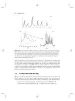
Figure 13.10, the initial four runs a–d can be used to predict the best combination of
temperature and gradient time for optimal resolution (Fig. 13.10e). Once acceptable
peak spacing is achieved, the gradient range can be trimmed to shorten overall
separation time. For example, the gradient can be initiated at a %B-value just prior
to elution of the first peak, and terminated at the %B-value just after elution of the
last peak (Fig. 13.10f ).
If no combination of gradient time and temperature yields acceptable reso-
lution, the next step could be a change in the column or the composition of the
A- or B-solvent; for example, an increase in TFA concentration, a change in pH,
or the substitution of isopropanol for acetonitrile as B-solvent. After one or more
of the latter changes in conditions, the four-run change in both gradient time and
temperature (as in Fig. 13.10a–d) should be repeated, using the new conditions for
other variables.
Finally, segmented gradients can be used to address particular separation
problems. In the case of strongly adsorbed contaminants that must be removed from
the column prior to the next sample injection, a final, steep gradient to 100% B can
be used to clean the column. In the case of complex samples with clusters of poorly
13.4 SEPARATION OF PEPTIDES AND PROTEINS 597
resolved components, a segment with a shallow gradient ramp can be inserted to
improve their separation. This strategy is of limited value for small molecules; it is
more likely to be successful for peptides, and especially for proteins [36].
13.4.2 Ion-Exchange Chromatography (IEC) and Related Techniques
Ion-exchange chromatography (IEC) can be used for analytical separations of
peptides and proteins, but it is more frequently employed for the isolation and
purification of proteins from laboratory to process scale [37]. The most important
advantages of IEC for protein isolation include (1) the tendency of proteins to
maintain their native conformation and biological activity during separation, (2)
the relatively high binding capacity of IEC packings, and (3) high mass recov-
eries. Features (1) and (2) are favored by the use of mobile phases of moderate
ionic strength and near-physiological pH. The most important feature of IEC
for analytical applications is its unique selectivity relative to other modes of col-
umn chromatography. Three other chromatographic techniques (chromatofocusing,
hydroxyapaptite chromatography, and immobilized-metal affinity chromatography;
Sections 13.4.2.3–13.4.2.5) are related to IEC in that they also rely on ionic
interactions between the column and sample.
Ion exchange is based on the reversible electrostatic interaction of charged
groups on the packing with oppositely charged groups on the polypeptide (Section
7.4.1). The retention of a peptide or protein molecule P occurs as a result of the
displacement of mobile-phase counterions X
+
by P
+z
(or X
−
by P
−z
).
(cation exchange) P
+z
(m) + z(R
−
)X
+
(s) ⇔ (R
−
)
z
P
+z
(s) + zX
+
(m) (13.4)
(anion exchange) P
−z
(m) + z(R
+
)X
−
(s) ⇔ (R
+
)
z
P
−z
(s) + zX
−
(m) (13.5)
Here R
−
or R
+
refers to a charged group (ligand) in the stationary phase, z is the
charge on the protein molecule P
+z
or P
−z
,and(m)or(s) refers to a molecule
in the mobile or stationary phase, respectively. A monovalent counter-ion X
+
or
X
−
is assumed in Equations (13.4) and (13.5). In cation-exchange chromatogra-
phy, an anionic ligand (R
−
) associates with cationic sites on the polypeptide. In
anion-exchange chromatography, a positively charge ligand (R
+
) binds to anionic
groups on the polypeptide. Sample retention can be varied by altering the charge
on the solute or—in some cases—the column ligand (Section 7.5.4) via a change
in mobile-phase pH. A more common elution strategy is to vary the concentration
of X
+
or X
−
in the mobile phase, as discussed in Section 7.4.1, or to use gradient
elution where the concentration of X
+
or X
−
increases during the gradient (salt
gradient). For reasons discussed below, the apparent charge ±z on the protein in
Equations (13.4) and (13.5) can differ from the net charge.
Charged groups at the protein amino and carboxyl termini (as well as on
amino-acid side-chains) strongly affect IEC retention. These groups have pK
a
values
between 2 and 13 (Table 13.4 and Fig. 13.1), so retention will be strongly dependent
on mobile-phase pH. Note that the local environment of a charged amino-acid
residue in a protein (i.e., surrounding mobile phase, and adjacent amino-acid groups
within the molecule) can shift its apparent pK
a
from the nominal value for the free
amino acid. Charged post-translational modifications such as sialic acid, phosphate,
and sulfate groups can also contribute to ionic retention.
598 BIOCHEMICAL AND SYNTHETIC POLYMER SEPARATIONS
Table 13.4
pK
a
Values for Charged Amino Acids
Residue pK
a
in Amino Acid pK
a
in Protein
Terminal amino 8.8–10.8 6.8–7.9
Arginyl 12.5 ≥12
Histidyl 6.0 6.4–7.4
Lysyl 10.8 5.9–10.4
Terminal carboxyl 1.8–2.6 3.5–4.3
Aspartyl 3.9 4.0–7.3
Glutamyl 4.3 4.0–7.3
Source: Reprinted from [37] with permission from Validated Biosystems.
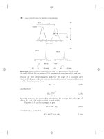
The net charge ±z on a protein will depend on mobile-phase pH. At the pH
where the sum of positive and negative charges are equal (the isoelectric point, or
pI), no net IEC retention is expected. At pH values below its pI, a protein will have a
net positive charge and should bind to a cation exchanger. At pH values above its pI,
the protein will possess a net negative charge and should bind to an anion exchanger
(Fig. 13.15a). This simple model can serve as a guide for selecting a column and
mobile-phase pH, but in practice, a protein may exhibit anomalous binding behavior
at or near its isoelectric point (Fig. 13.15b). The reason is that the charge on a protein
may not be homogeneously distributed across its surface but instead clustered into
different regions (contact areas) on the molecule (Section 13.3.2). As a result regions
of excess charge can appear at different parts of the molecule, and these regions
can interact with the column more or less independently of each other. Anomalous
binding behavior can include binding at the isoelectric point, binding to an anion
exchanger below the protein pI, or binding to a cation exchanger above the pI.
Similarly a protein may fail to bind to an anion exchanger above its pI or to a cation
exchanger below its pI. For example, β glucosidase (pI = 7.3) binds at pH-7.3 on an
anion exchanger but fails to bind to a cation exchanger until the mobile-phase pH
is two units below its pI (Fig. 13.15b). Chymotrypsin, with a pI of 9, binds at pH-9
on both an anion and a cation exchanger more than (Fig. 13.15c).
As a guideline, anion-exchange separations are often carried out at 1 to 1.5 pH
units above a protein’s pI, and cation-exchange separations at 1 to 1.5 pH units
below the pI. Solubility and stability properties of the protein(s) of interest can limit
the allowable ionic conditions for the separation. Virtually all protein purification
schemes used in the biopharmaceutical industry contain one or multiple anion-
and/or cation-exchange steps.
Since only a limited number of charged residues on the protein surface may
interact with the stationary phase, small differences in the nature and positions of
these charged residues can profoundly affect selectivity in ion-exchange chromatog-
raphy [14]. In addition amino-acid substitutions within the interior of the protein
may alter its conformation and affect ion-exchange selectivity indirectly by changing
the positions of charged groups on the protein surface.
13.4 SEPARATION OF PEPTIDES AND PROTEINS 599
(a)
(b)
(c)
20
10
0
2468 4 6 8 1010
2
pI pI
t
R
(min)
pH
cation exchange
anion exchange
+z −z
t
R
t
R
pI
pH
Cation exchange Anion exchange
Protein
charge z,
retention
time t
R
β−glucosidase chymotrypsinogen
Figure 13.15 Protein retention on ion exchangers as a function of pH. Ideal behavior (a);
actual behavior of β-glucosidase (b) and chymotrypsinogen (c). Adapted from [38].
13.4.2.1 Column Selection
Column-selection criteria include:
• particle size and pore diameter
• support composition
• ligand type
• ligand density
Particle size and pore diameter considerations are the same as described in
Sections 13.3.1.1 and 13.3.1.2 for RPC.
Support Composition. The first supports for high-performance IEC were silica
based, for the same reasons that silica was chosen for other modes of HPLC.
However, early silica packings were unstable under preferred ion-exchange condi-
tions (physiological pH, moderate salt concentration) and were gradually replaced
by polymeric packings based on polystyrene-divinyl benzene or polymethacrylate.
Although modern silica-based packings exhibit improved stability at neutral to
600 BIOCHEMICAL AND SYNTHETIC POLYMER SEPARATIONS
alkaline pH, many labs continue to use polymer-based columns. For process chro-
matography, large-particle supports composed of semi-rigid gels such as cross-linked
dextran, agarose, or polyacrylamide are preferred for their lower cost, and because
they can withstand highly alkaline cleaning steps for the removal of endotoxins and
other biological contaminants.
Ligand Type and Density. Within the respective categories of cation and anion
exchange, IEC packings can be further divided into ‘‘strong’’ or ‘‘weak’’—depending
on the pK
a
of the stationary-phase ionic ligand. Consequently the charge on the
column and its binding capacity can vary with mobile-phase pH (Fig. 13.16).
Strong ion-exchangers have pK
a
values outside the normal pH-operating range of
the column, and are therefore fully ionized—regardless of mobile-phase pH; see
Table 13.5 for some common examples of IEC column ligands. Ionic groups in
strong ion-exchangers include –SO
3
−
for cation exchange and –N(CH
3
)
3
+
for anion
exchange. Weak ion-exchangers have pK
a
values within the operating range of the
column, so their ion-exchange capacity varies with mobile-phase pH. Examples of
0 2468101214
02468101214
Strong CEX
Strong AEX
Exchange Capacity
Weak CEX
Weak AEX
Exchange Capacity
(a)
(b)
pH
p
H
Figure 13.16 Capacities of ion-exchange groups. (a) Strong ion exchangers; (b)weakion
exchangers. Adapted from [39].
13.4 SEPARATION OF PEPTIDES AND PROTEINS 601
Table
13.5
Strong and Weak Ion-Exchange Ligands
Anion Exchange (AEX) Cation Exchange (CEX)
Weak Weak
DEAE (diethylaminoethyl) –O–CH
2
–CH
2
–N
+
H(CH
2
CH
3
)
2
CM (Carboxymethyl) –O–CH
2
–COO
−
PEI (polyethyleneimine)
(–NHCH
2
CH
2
)
n
–N(CH
2
CH
2
–)
n
.
|
CH
2
CH
2
NH
2
Strong Strong
Q (quaternary ammonium) –CHOH–CH
2
–N
+
(CH
3
)
3
S (sulfonate) –CH
2
–CH
2
–CH
2
–SO
−
3
weak IEC groups include –N(C
2
H
5
)
2
H
+
for weak anion exchange and –COO
−
for
weak cation exchange. Weak anion-exchange columns of polyethyleneimine consist
of a dense polymeric coating onto a silica support, yielding a column with high
capacity and good stability under alkaline conditions. Strong ion-exchangers are
often preferred, as their exchange capacity is independent of mobile-phase pH and
their behavior is more predictable. The binding capacity of ion-exchangers depends
on the surface area of the support and its charge density (μmoles/m
2
). Typical
ion-exchange capacities (i.e., for maximum uptake of sample by the column) for
large-pore silica or polymer-based columns are in the range of 30 to 120 mg protein
per milliliter of packing.
The linker group that joins the ion-exchange group to the support can con-
tribute to the chromatographic properties of the column. For example, hydrophobic
groups in the linker may participate in hydrophobic (reversed-phase) interactions
with the solute. Such interactions can account for differences in column selectivity
among different vendors who use the same ion-exchange functionality. Tentacle IEC
stationary phases have a flexible hydrophilic linker (the ‘‘tentacle’’) that connects
the charged group to the support [40]. These columns improve access of the protein
to the charged group of the packing, thus enhancing binding capacity. In addition
tentacle columns may exhibit reduced nonspecific interaction, improved binding
kinetics, and reduced protein denaturation.
13.4.2.2 Mobile-Phase Selection
As noted above, control of retention (solvent strength) is usually achieved by varying
the concentration of a displacing salt (counter-ion), rather than by changes in
mobile-phase pH. Conditions that affect selectivity include:
• column (Section 13.4.2.1)
• mobile-phase buffer
• counter-ion salt type (as in Fig. 13.17)
602 BIOCHEMICAL AND SYNTHETIC POLYMER SEPARATIONS
01020
30
010
1
2
3
+
4
5
1
2
3
4
5
(a)(b)
NaCl Na
2
SO
4
(
min
)(
min
)
Figure 13.17 Effect of salt type on anion exchange separation of five proteins. Conditions:
50 × 4-mm Shim-pack WAX-2 column (Shimadzu); 0–0.5M of indicated salt in 20 min; pH-8
phosphate buffer; 1 mL/min. Adapted from [41].
• gradient steepness
• organic B-solvent (if used)
• other mobile-phase additives (especially surfactants)
• temperature
See also the discussion of Section 7.5.
Mobile-Phase Buffer. Achieving the desired retention and selectivity requires a
careful selection and control of the mobile-phase pH. For a cation-exchange column,
a mobile-phase pH near 6 is a good starting point, while a mobile-phase pH of 8
is appropriate for an anion exchanger. For good buffering capacity, the buffering
agent should have a pK
a
value within roughly 1.0 units of the target pH (Section
7.2.1.1), and a concentration of 0.02 to 0.1 M. Common buffers used for IEC are
listed in Table 7.1. Note that some of these buffers absorb strongly at shorter UV
wavelengths, especially if higher concentrations are used.
Counter-Ion. The most common elution strategy in IEC is the use of a gradient
of increasing concentration of the counter-ion. The relative strength of different
counter-ions follows their ranking in the Hofmeister series [37, 42]; see Table 13.6
or a similar series in Section 7.5.2. However, gradients that involve an increase in
NaCl are most often used for both anion and cation exchange. Note that chloride is
corrosive for stainless steel at low pH (<5) and should be removed from the HPLC
system after use. However, special-purpose HPLC systems have been designed that
enable the use of chloride under acidic conditions.
Organic Solvents and Surfactants. Organic solvents (e.g 1–10% methanol,
propanol, or acetonitrile) can be added to the mobile phase to suppress hydrophobic
13.4 SEPARATION OF PEPTIDES AND PROTEINS 603
Table
13.6
Hofmeister Series of Lyotropic and Chaotropic Ions [36]
Increasing lyotropic (salting out) effect
SCN
−
(least) < ClO
4
−
< NO
3
−
< Br
−
< Cl
−
< COO
−
< SO
4
2−
< PO
4
3−
(most)
Increasing chaotropic (salting in) effect
Ba
2+
(most)
>
Ca
2+
>
Mg
2+
>
Li
+
>
Cs
+
>
Na
+
>
K
+
>
Rb
+
>
NH
4
+
(least)
Source: Data from [36].
interactions with the support or linker groups, and to decrease peak broadening or
tailing (addition of as much as 50% organic solvent may be required in some cases,
as in the example of Fig. 11.15 of [43]). Nonionic surfactants can also be used
for the same reasons. Either of these mobile-phase additives can also maintain the
solubility of very hydrophobic solutes such as membrane proteins. Ionic surfactants
can not be used in ion-exchange chromatography.
13.4.2.3 Chromatofocusing
Chromatofocusing is a specialized form of IEC in which proteins are eluted from
the column with a pH gradient [44–49]. Chromatofocusing is unique in that the
pH gradient is formed within the column, by means of a single mobile phase that
is a complex mixture of different buffering species. Although chromatofocusing can
be performed with cation- or anion-exchangers, commercially available products
are limited to anion exchange [48]. At the start of separation, proteins are retained
by the anion exchanger, which has been pre-equilibrated at high pH for maximum
retention of the sample. Then a low-pH buffer mixture is used as mobile phase,
which, upon moving through the column, progressively titrates the charge on the
column so that pH increases along the column, from inlet to outlet. Proteins migrate
down the column in response to the changing pH and elute at or near their isoelectric
points—a pH at which they can no longer bind to the exchanger. Elution is in order
of descending protein pI values. Chromatofocusing is characterized by very high
capacity, so it is useful for preparative separations. The technique is also capable
of very high resolution, by virtue of focusing effects that generate sharp peaks 0.04
to 0.05 pH units in width. As is the case for conventional IEC (Figs. 13.15b,c),
a protein can elute from a chromatofocusing column at a pH that is significantly
different from its pI.
Successful and reproducible chromatofocusing separations depend on the use
of buffers that contain multiple species, whose pK
a
values span the range of the
pH gradient, and that can achieve effective buffering across this range. Commercial
chromatofocusing buffers are composed of a mixture of ampholytes (substances
that may act as either an acid or a base). Alternatively, a combination of biological
buffers such as Good’s buffers [50] can be used. The ionic strength of the elution
buffer must be kept low, in order to minimize salt-mediated elution (displacement of
proteins by counter-ions). The improved resolution (or faster separation) of proteins
whose pI values fall within a narrow range of values can be achieved by narrowing
the pH range of the ampholyte or buffer blend (similar to a decrease in φ in
604 BIOCHEMICAL AND SYNTHETIC POLYMER SEPARATIONS
gradient elution). Strong ion-exchange columns are preferred for chromatofocusing,
since they are fully ionized—regardless of pH.
One shortcoming of chromatofocusing is the reduced solubility of proteins at
their isoelectric point, a limitation which is exacerbated by the low ionic strength
of the elution buffer. Protein solubility can be enhanced by an increase in salt
concentration, but this will increase mobile-phase strength and compromise the
separation. A preferred strategy for dealing with protein precipitation is the addition
of zwitterions to the elution buffer. Additives such as taurine, glycine, and betaine
promote protein solubility and can be used in concentrations up to 2M without
affecting the ionic strength of the buffer. The addition of urea at concentrations of
1 to 2M also helps solubilize proteins; nonionic and zwitterionic surfactants may
be used as well. Note, however, the tendency of urea to decompose to carbamates,
which can covalently modify a protein.
Chromatofocusing is able to resolve isoforms of proteins that have different
charge states, for example, post-translationally modified proteins that differ in the
number of sialic acids or phosphate groups. The resolution of isoforms can be a
limitation, if the goal is protein purification. The target protein is then resolved
into multiple peaks, which dilutes the target protein and increases the risk of
co-elution with sample contaminants. On the other hand, this characteristic of
chromatofocusing can be an advantage, if only the characterization of isoforms is
desired.
13.4.2.4 Hydroxyapatite Chromatography
This technique is frequently used in process chromatography for protein purification
and the removal of contaminants [37]. Hydroxyapatite (HA) is a crystalline material
composed of Ca
10
(PO
4
)
6
(OH)
2
that serves both as the support and the stationary
phase [51]. The multifunctional surface consists of positively charged pairs of
calcium ions (C-sites) and clusters of six anionic oxygen atoms associated with
triplets of phosphate ions (P-sites). The C- and P-sites and hydroxyls are distributed
in a fixed pattern on the crystal surface [51–53], as illustrated in Figure 13.18.
Early preparations of HA were unstable, but modern HA materials are sintered
at high temperature to form ceramic hydroxyapatite (CHT), which is stable under
chromatographic conditions. Columns packed with either 5- or 10-μm CHT particles
are available for both analytical and preparative applications.
Protein interactions with CHT are complex (Fig. 13.18). Electrostatic inter-
actions include attraction of protonated amino groups by P-sites and repulsion by
C-sites (Fig. 13.18a). Similarly ionized carboxyl groups are attracted by C-sites and
repelled by P-sites (Fig. 13.18b). Although the initial attraction of carboxyls to C-sites
is electrostatic, the actual binding involves formation of much stronger coordination
complexes between C-sites and clusters of protein carboxyl-groups [37]. Protein
phosphate-groups bind C-sites even more strongly than protein carboxyl-groups.
The selectivity of CHT for basic proteins is distinct from that of conventional
cation exchange, due to the repulsion of amines by C-sites. Binding of weakly basic
proteins can be enhanced by the addition of a low concentration of phosphate,
which suppresses C-site repulsion of amines but does not block their interaction
with P-sites [54]. Basic proteins can be eluted by gradients of sodium chloride or
phosphate; a final salt concentration as high as 0.5M may be required. Although the
13.4 SEPARATION OF PEPTIDES AND PROTEINS 605
OH
OH
OH
Ca
+))
Ca
+))
PO
4
=
PO
4
=
PO
4
=
((+
H
2
N
((+
H
2
N
+
H
2
N
+
H
2
N
(a)
(b)
COO
−
COO
−
COO
−))
COO
−))
((
PO
4
=
PO
4
=
((
PO
4
=
+
Ca
+
Ca
HO
HO
HO
Protein
Protein
CHT
CHT
C-sites
P-sites
Figure 13.18 Binding to ceramic hydroxyapatite (CHT) of a basic protein (a) and an acidic
protein (b). Double parenthesis indicate repulsion, dotted lines indicate ionic bonds, and trian-
gular linkages indicate coordination bonds. Adapted from [37].
binding of basic proteins increases at lower pH, CHT is unstable below pH 5. Acidic
proteins cannot be eluted with sodium chloride—even at concentrations
>
0.3M;
their elution requires the use of phosphate, citrate, or fluoride. This characteristic of
CHT permits separation of basic proteins with an initial NaCl gradient, followed by
elution of acidic proteins with a phosphate gradient.
CHT typically provides excellent recovery of protein mass and biological
activity; it is used for protein purification from laboratory to process scale. The
unique selectivity of CHT can enable the resolution of closely related species such as
protein variants and glycoforms. It is used in the biopharmaceutical industry for the
purification of antibodies and removal of contaminants such as endotoxins, nucleic
acids, and viruses. The stability of CHT toward concentrated base, organic solvents,
and chaotropes enables aggressive cleaning regimes to be applied after use.
13.4.2.5 Immobilized-Metal Affinity Chromatography (IMAC)
This separation mode, also known as metal-interaction chromatography (MIC), is
based on the differential interaction of proteins with a metal ion [55–57]. The metal
ion is immobilized by chelating groups that are attached to the support via a linker;
see the example of Figure 13.19, which includes the various steps in its use. Several