Introduction to Modern Liquid Chromatography, Third Edition part 71 docx
Bạn đang xem bản rút gọn của tài liệu. Xem và tải ngay bản đầy đủ của tài liệu tại đây (134.16 KB, 10 trang )
656 BIOCHEMICAL AND SYNTHETIC POLYMER SEPARATIONS
13.10.3.5 Chemical Composition as a Function of Molecular Size
A copolymer typically exhibits both molecular-weight and chemical-composition
distributions. Depending on polymerization conditions, the chemical composition
may or may not vary with polymer molecular weight. To investigate the presence
of such chemical heterogeneity, we can couple SEC with a spectroscopic technique
that yields chemical-composition information. Such a combined technique provides
the average composition at each point in the SEC chromatogram, that is, for
each molecular size. If only one of two monomers can be detected by UV, the
combination of a UV detector and another concentration-sensitive detector (e.g.,
refractive index, RI) can in principle be used to follow the concentration of each
monomer. Additional information can be obtained from combining SEC with either
FTIR or NMR spectroscopy.
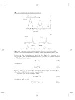
Although information about chemical composition as a function of molecular
size can be very valuable, even the smallest SEC fractions can contain a variety of
molecules that vary in both chemical composition and molecular weight. That is,
differences in chemical composition can result in molecules with different molecular
weights having the same molecular ‘‘size’’ in solution, as illustrated in Figure 13.49.
A fraction obtained from a high-resolution SEC separation (rectangular box in
Fig. 13.49) will contain molecules with the same molecular size (gyration radius
R
g
) in solution, but with different molecular weights. It is often important to know
the chemical-composition distribution, rather than just the average chemical com-
position. Likewise the functionality-type distribution (FTD) may be more important
than the average number of functional groups per molecule. This will be especially
true if the chemical composition or the number of functional groups per molecule is
known (or suspected) to vary. An example is reactive (pre-)polymers that are used in
many formulations for sealants, adhesives, and coatings. Molecules without reactive
(functional) groups will not react, molecules with one functional group will locally
terminate the polymerization process, molecules with two functional groups will
0.025
0.020
0.015
0.010
0.005
0.000
Radius R
g
(μm)
0 50 100 150 200 250 300
Molecular wei
g
ht
(
x10
−3
)
homopolymer A
homopolymer B
co-polymers of A and B
fraction
Figure 13.49 Schematic illustration of the relationship between molecular size and molecu-
lar weight for (co-)polymers of different composition. Lines represent (from top to bottom)
homopolymer A, copolymer AB (75:25), AB (50:50), AB (25:75), and homopolymer B.
13.10 SYNTHETIC POLYMERS 657
sustain the polymerization, and molecules with more than two functional groups
promote the formation of resinous polymeric networks. Knowledge of only the
average number of functional groups per molecule would be insufficient in this case.
13.10.4 Polymer Separations by Two-Dimensional Chromatography
In comprehensive two-dimensional liquid chromatography (LC × LC; Sections
9.3.10, 13.4.5), the entire sample is subjected to two different successive separations,
while the separation obtained in the first dimension is preserved. To simultaneously
determine two mutually dependent distributions, such as the combination of MWD
and CCD (MWD × CCD), a technique that separates according to molecular weight
(e.g., SEC) must be combined with one that separates (largely) according to com-
position, such as i-LC. Combination of the two separations (i-LC × SEC) then
yields a two-dimensional chromatogram that represents an analysis of the sample
according to both molecular weight and chemical composition; an example is shown
in Figure 13.48. Corresponding one-dimensional separations are shown for SEC
at the side, and for i-LC at the top of Figure 13.48. While neither of the latter
one-dimensional separations provides an adequate separation of the total sample,
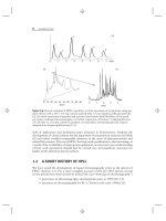
the corresponding two-dimensional separation does. Another i-LC × SEC separa-
tion is shown in Figure 13.50, for a more complex sample: chain-end-functionalized
poly(methyl methacrylates). The horizontal time-axis for the i-LC separation is
indicative of the chemical composition of the copolymer (note labels at top of figure
for the number of functional groups in the molecule); while the vertical time-axis
for the SEC separation is related to its molecular weight.
Two-dimensional chromatograms such as those in Figures 13.48 and 13.50
can provide a useful qualitative picture of the composition of a copolymer. Different
samples can be compared in great detail, and the results of such a comparison
groups per molecule
1.2
1.1
1.0
0.9
0.8
0.7
SEC (min)
i-LC (hr)
012
0.5 0.6 0.7 0.8 0.9 1.0 1.1 1.2 1.3 1.4
Figure 13.50 Two-dimensional separation of chain-end-functionalized poly(methyl
methacrylates). The dashed lines indicate areas in the 2D-chromatogram that correspond
to molecules with zero, one or two functional groups, as indicated at the top of the figure.
Adapted from [172].
658 BIOCHEMICAL AND SYNTHETIC POLYMER SEPARATIONS
can be used to better understand the properties of polymeric materials or related
polymerization processes [173]. Unfortunately, it is much more difficult to obtain
quantitative information from such figures, as a number of complications arise. First,
the relationship between SEC retention time and molecular weight depends also on
polymer chemical composition and topology (e.g., degree of branching). Second,
detector response also depends on these polymer properties.
To solve the first problem (retention not completely defined by molecular
weight), we must know retention in SEC as a function of solute molecular weight
and chemical composition; this can be accomplished by the use of appropriate
copolymer standards. The second problem (varying response factor) is more of
a challenge. When homopolymers are studied, the response factor may be nearly
constant (i.e., independent of molecular weight) for UV detection. However, many
polymers lack chromophors, which necessitates the use of refractive-index (RI)
detection. Here the response factor (usually referred to as the refractive-index
increment or dn/dc) tends to be nonconstant in the oligomeric region.
REFERENCES
1. G. T. Hermanson, Bioconjugate Techniques, Academic Press, San Diego, CA, 1996.
2. L. R. Snyder and M. A. Stadalius, High-Performance Liquid Chromatography:
Advances and Perspectives,Vol.4,C.Horv
´
ath, ed. Academic Press, San Diego,
1986, p. 195.
3. L. R. Snyder and J. W. Dolan, High-Performance Gradient Elution, Wiley-Interscience,
Hoboken, NJ, 2007.
4. J. O. Konz, R. C. Livingood, A. J. Bett, A. R. Goerke, M. E. Laska, and S. L. Sagar,
Hum. Gene Ther., 16 (2005) 1346.
5. E. I. Trilisky and A. M. Lenhoff, J.Chromatogr., 1142 (2007) 2.
6. M. A. Stadalius, B. F. D. Ghrist, and L. R. Snyder, J. Chromatogr., 387 (1987) 21.
7. J. S. Richardson, Adv. Protein Chem., 34 (1981) 167.
8. W. R. Melick-Adayan, V. V. Barynin, A. A. Vagin, V. V. Borisov, B. K. Vainshtein, B.
K.Fita,M.R.N.Murthy,andM.G.Rossman,J. Mol. Biol., 188 (1986) 63.
9. R. L. Cunico, K. M. Gooding, and T. Wehr, Basic HPLC and CE of Biomolecules,Bay
Bioanalytical Laboratory, Richmond, CA, 1998.
10. M. T. W. Hearn and B. Grego, J. Chromatogr., 282, (1983) 541.
11. W. Doerfler, in Medical Microbiology, 4th ed., S. Baron (ed.), Univ. TX Medical
Branch, Galveston, 1996.
12. N. B. Afeyan, N. F. Gordon, I. Mazsaroff, L. Varady, S. P. Fulton, Y. B. Yang, and F.
E. Regnier, J. Chromatogr., 519, (1990) 1.
13. F. B. Rudolph, D. P. Wiesenborn, J. Greenhut, and M. L. Harrison, in HPLC of
Biological Macromolecules, K. M. Gooding and F. E. Regnier, eds., Dekker, New
York, 1990, p. 333.
14. F. E. Regnier, Science, 238 (1987).
15. M. A. Stadalius, H. S. Gold, and L. R. Snyder, J. Chromatogr., 296 (1984) 31.
16. M. Kawakatsu, H. Kotaniguchi, H. Freiser, and K. M. Gooding, J. Liq. Chromatogr.,
18 (1995) 633.
17. A. Apfel, S. Fischer, G. Goldberg, P. C. Goodley, and F. E. Kuhlmann, J. Chromatogr.
A, 712 (1995) 177.
REFERENCES 659
18. D. V. McCalley, LCGC, 23 (2005) 162.
19. D. V. McCalley, J. Chromatogr. A, 1075 (2005) 57.
20. D. Guo, C. T. Mant, and R. S. Hodges, J. Chromatogr., 386 (1987) 205.
21. M. T. W. Hearn, in HPLC of Biological Macromolecules, 2nd ed., K. M. Gooding and
F. E. Regnier, eds., Dekker, New York, 2002, pp. 195–312.
22. J. E. Rivier, J. Liq. Chromatogr., 1 (1978) 343.
23. D. Guo, C. T. Mant, A. K. Taneja, J. M. R. Parker, and R. S. Hodges, J. Chromatogr.,
359 (1986) 499.
24. W. Hancock, R. C. Chloupek, J. J. Kirkland, and L. R. Snyder, J. Chromatogr. A, 686
(1994) 31.
25. S. Terabe, S. Nishi, and T. Ando, J. Chromatogr., 212 (1981) 295.
26. J.L.Glajch,M.A.Quarry,J.F.Vasta,andL.R.Snyder,Anal. Chem., 58 (1986) 280.
27. C. T. Wehr and L. Correia, LC at Work, LC-121, Varian, Walnut Creek, CA 1980.
28. D. Guo, C. T. Mant, A. K. Taneja, and R. S. Hodges, J. Chromatogr., 359 (1986) 519.
29. C. T. Mant, T. W. L. Burke, J. A. Black, and R. S. Hodges, J. Chromatogr., 458 (1988)
193.
30. M. T. W. Hearn and B. Grego, J. Chromatogr., 296 (1984) 61.
31. W. R. Melander, J. Jacobson, and C. Horv
´
ath, J. Chromatogr., 234 (1982) 269.
32. S. Cohen, K. Benedek, Y. Tapuhi, J. C. Ford, and B. L Karger, Anal. Biochem., 144
(1985) 275.
33. W. G. Burton, K. D. Nugent, T. K. Slattery, B. F. Johnson, and L. R. Snyder,
J.Chromatogr., 443 (1988) 363.
34. K. D. Nugent, W. G. Burton, T. K. Slattery, B. F. Johnson, and L. R. Snyder,
J.Chromatogr., 443 (1988) 381.
35. L.J.Licklider,C.C.Thoreen,J.Peng,andS.P.Gygi,Anal. Chem., 74 (2002) 3076.
36. B. F. D. Ghrist and L. R. Snyder, J. Chromatogr., 459 (1989) 43.
37. P. Gagnon, Purification Tools for Monoclonal Antibodies, Validated Biosystems,
Tuscon, AZ, 1996.
38. W. Kopaciewicz, M. A. Rounds, J. Fausnaugh, and F. E. Regnier, J. Chromatogr., 266
(1983) 3.
39. C. D. Scott in Modern Practice of Liquid Chromatography, J. J. Kirkland, ed.,
Wiley-Interscience, New York, 1971.
40. W. Muller, J. Chromatogr., 510 (1990) 133.
41. M. T. Ueda and Y. Ishida, J. Chromatogr., 386 (1987) 273.
42. R. Chicz and F. Regnier, Met. Enzymol., 182 (1990) 392.
43. L. R. Snyder, J. J. Kirkland, and J. L. Glajch, Practical HPLC Method Development,
2nd ed., Wiley-Interscience, New York, 1997, p. 515.
44. L. Sluyterman and O. Elermsa, J. Chromatogr., 150 (1978) 17.
45. L. Sluyterman and J. Wijdenes, J. Chromatogr., 150 (1978) 31.
46. L. Sluyterman and J. Wijdenes, J. Chromatogr., 206 (1981) 429.
47. L. Sluyterman and J. Wijdenes,
J. Chromatogr.,
206 (1981) 441.
48. Chromatofocusing with Polybuffer and PBE Handbook, ed. AB, Publication
18-1009-07, Amersham Pharmacia Biotech, Uppsala, Sweden.
49. P. Gagnon, Quarterly Resource Guide to Downstream Processing, Validated Biosys-
tems, Tuscon, AZ, 1999.
660 BIOCHEMICAL AND SYNTHETIC POLYMER SEPARATIONS
50. N. G. Good, G. D. Winget, W. Winter, T. N. Connally, S. Izawa, and R. M. M. Singh,
Biochemistry, 5 (1966) 467.
51. T. Kawasaki and S. Takahashi, Eur. J. Biochem., 152 (1985) 361.
52. T. Kawasaki, J. Chromatogr., 151 (1978) 95.
53. T. Kawasaki, J. Chromatogr., 157 (1978) 7.
54. M. J. Gorbunoff, Anal. Biochem., 136 (1984) 425.
55. K. M. Gooding, Z. El Rassi, and C. Horv
´
ath, in HPLC of Biologicial Macromolecules,
2nd ed., K. M. Gooding and F. E. Regnier, eds., Dekker, New York, 2002, pp.
247–280.
56. L. Kagedal, in Protein Purification, J. C. Janson and L. Ryden, eds., VCH, New York,
1989, pp. 227–251.
57. F. H. Arnold, Biotechnology, 151 (1991) 9.
58. J. Porath and B. Olin, Biochemistry, 22 (1983) 162.
59. E. Hochuli, W. Bannwarth, H. Dobeli, R. Gentz, and D. Stuber, Biotechnology,6
(1988) 1321.
60. B. Bodenmiller, L. N. Mueller, M. Mueller, B. Domon, and R. Aebersold, Nature
Methods, 4 (2007) 231.
61. J. Porath, J. Chromatogr., 443 (1988) 3.
62. Z. El Rassi and C. Horv
´
ath, J. Chromatogr., 359 (1986) 241.
63. A. Tiselius, Ark. Kem. Min. Geol., 26B (1948).
64. J. Porath, Biochem. Biophys. Acta, 39 (1960) 193.
65. B. Gelotte, J. Chromatogr., 3 (1960) 330.
66. Y. Kato, T. Kitamura, and T. Hashimoto, J. Chromatogr., 266 (1983) 49.
67. Y. Kato, T. Kitamura, and T. Hashimoto, J. Chromatogr., 292 (1984) 418.
68. R. E. Shansky, S L. Wu, A. Figueroa, and B. L. Karger, in HPLC of Biological
Macromolecules, K. M. Gooding and F. E. Regnier, eds., Dekker, New York, 1990, p.
95.
69. H. S. Frank and M. J. Evans, J. Chem. Phys., 13 (1945) 507.
70. S. Shaltiel, Z. Er-el, Proc. Natl. Acad. Sci. USA, 52 (1973) 430.
71. D. L. Gooding, M. N. Schmuck, M. P. Nowlan, and K. M. Gooding, J. Chromatogr.,
359 (1986) 331.
72. J. L. Fausnaugh and F. E. Regnier, J. Chromatogr., 359 (1986) 131.
73. D. B. Wetlaufer and M. R. Koenigbauer, J. Chromatogr., 359 (1986) 55.
74. S. L. Wu, K. Benedek, and B. L. Karger, J. Chromatogr., 359 (1986) 3.
75. A. J. Alpert, J. Chromatogr., 499 (1990) 177.
76. M. Lafosse, B. Herbreteau, M. Dreux, and L. Morinallorym, J. Chromatogr., 472
(1989) 209.
77. W. Naidong, J. Chromatogr. B, 796 (2003) 209.
78. B. A. Olsen,
J. Chromatogr. A,
913 (2001) 113.
79. T. Yoshida, Anal. Chem., 68 (1997) 3038.
80. H. Tanaka, X. Zhou, and O. Masayoshi, J. Chromatogr. A, 987 (2003) 119.
81. T. K. Chambers and J. S. Fritz, J. Chromatogr. A, 797 (1998) 139.
82. M. Wuhrer, C. A. M. Koeleman, A. M. Deelder, and C. N. Hokke, Anal. Chem., 76
(2004) 833.
83. M. Wuhrer, C. A. M. Koeleman, C. H. Hokke, and A. M. Deelder, Anal. Chem., 77
(2005) 886.
REFERENCES 661
84. A. J. Ytterberg, R. R. Ogorzalek-Loo, P. Boontheung, J. Wohlschlegel, and J. A. Loo,
abstract WP 523, 55th ASMS Conference on Mass Spectrometry and Allied Topics,
Indianapolis, 2007.
85. C.T.Mant,J.R.Litowski,andR.S.Hodges,J. Chromatogr. A, 816 (1998) 65.
86. C. A. Mizzen, A. J. Alpert, L. Levesque. T. P. A. Kruck, and D. R. McLachlan, J.
Chromatogr. B, 744 (2000) 33.
87. A. Jungbauer, C. Machold, and R. Hahn, J. Chromatogr. A, 1079 (2005) 221.
88. H. Lindner, B. Sarg, C. Meraner, and W. Helliger, J. Chromatogr. A, 743 (1996) 137.
89. H. Lindner, B. Sarg, C. Meraner, and W. Helliger, J. Chromatogr. A, 782 (1997) 55.
90. B. Sarg, W. Helliger, H. Talasz, E. Kooutzamani, and H. Lindner, J. Biol. Chem., 279
(2004) 53–58.
91. A. J. Alpert, Anal. Chem., 80 (2008) 62.
92. A. J. Alpert, G. Mitulovic, and M. Mechtler, poster P2412-W, 32nd Annual Symposium
on High Performance Liquid Phase Separations and Related Techniques, Baltimore,
2008.
93. U. Lewandrowski, K. Lohrig, R. P. Zahedi, D. Wolters, and A. Sickmann, Clin.
Proteom., 4 (2008) 25.
94. P. H. O’Farrell, J. Biol. Chem., 250 (1975) 4007.
95. M. Gilar, P. Olivova, A. E. Daly, and J. C. Gebler, Anal. Chem., 77 (2005) 6426.
96. S. P. Gygi, B. Rist, S. A. Gerber, F. Turecek, M. H. Gelb, and R. Aebersold, Nat.
Biotechnol., 17 (1999) 994.
97. A. J. Link, J. Eng, D. M. Schieltz, E. Carmac, G. J. Mize, D. R. Morris, B. M. Garvik,
andJ.R.Yates,Nat. Biotechnol., 17 (1999) 676.
98. D. A. Wolter, M. P. Washburn, and J. R. Yates, Anal. Chem., 73 (2001) 5683.
99. M. P. Washburn, D. Wolters, and J. R. Yates, Nat. Biotechnol., 19 (2001) 5683.
100. M. T. Davis, J. Beierle, E. T. Bures, M. D. McGinley, J. Mort, J. H. Robinson, C. S.
Spahr, W. Yu, R. Luethy, and S. D. Patterson, J. Chromatogr. B, 752 (2001) 281.
101. G. J. Opiteck and J. W. Jorgenson, Anal. Chem., 69 (1997) 2283.
102. G. J. Opiteck, S. M. Ramirez, J. W. Jorgenson, and M. A. Moseley Anal. Biochem.,
258 (1998) 349.
103. K. Wagner, T. Miliotis, G. Marko-Varga, R. Bischoff, and K. K. Unger, Anal.Chem.,
74 (2002) 809.
104. R. Bischoff and L. W. McLaughlin, in HPLC of Biologicial Macromolecules,K.M.
Gooding and F. E. Regnier, eds., Dekker, New York, 1990, pp. 641–667.
105. R. Hecker, M. Colpan, and D. Riesner, J. Chromatogr., 326 (1985) 251.
106. S. Nakatani, T. Tsuda, Y. Yamasaki, M. Moriyama, H. Watanabe, and Y. Kato,
Technical Report 78, TosoHaas, Tokyo, 1995.
107. R. R. Drager and F. E. Regnier, Anal. Biochem., 145 (1985) 47.
108. G. Zon, in Characterization of Proteins: New Methods in Peptide Mapping,W.S.
Hancock, ed., CRC Press, Boca Raton, 1995, p. 301.
109. W. Xiao and P. J. Oefner, Human Mutation, 17 (2001) 439.
110. A. Premstaller and P. J. Oefner, in Methods in Molecular Biology,. 211, P Y. Kwok,
ed., Humana Press, Totowa, NJ, 2002, p. 15.
111. R. L. Pearson, J. F. Weiss, and A. D. Kelmers, Biochim. Biophys. Acta, 228 (1971)
770.
112. R. Bischoff and L. W. McLaughlin, Anal. Biochem., 151 (1985) 526.
113. J. D. Pearson, M. Mitchell, and F. E. Regnier,
J. Liq. Chromatogr.,
6 (1983) 1441.
662 BIOCHEMICAL AND SYNTHETIC POLYMER SEPARATIONS
114. R. Bischoff and L. W. McLaughlin, J. Chromatogr., 296 (1984) 329.
115. Z. el Rassi and C. Horv
´
ath, J. Chromatogr., 326 (1985) 79.
116. Z. el Rassi and C. Horv
´
ath, Chromatographia, 19 (1984) 9.
117. S. C. Churms, CRC Handbook of Chromatography: Carbohydrates,Vol.2,CRCPress,
Boca Raton, 1991.
118. S. C. Churms, J. Chromatogr. A, 720 (1996) 75.
119. K. Koizumi, T. Utamura, Y. Kubota, and S. Hizukuri, J. Chromatogr., 409 (1987) 396.
120. C. Brons and C. Olieman, J. Chromatogr., 159 (1983) 79.
121. D. W. Armstrong and H. L. Jin, J. Chromatogr., 462 (1989) 219.
122. S. Honda and S. Suzuki, Anal. Biochem., 142 (1984).
123. A. S. Feste and I. Khan, J. Chromtogr., 607 (1992) 7.
124. Guide to Aminex
®
HPLC Columns, Bulletin 1928, Bio-Rad Laboratories.
125. T. Jupille, Amer. Lab., 13 (1981) 80.
126. R. W. Goulding, J. Chromatogr., 103 (1975) 229.
127. Analysis of Carbohydrates by High Performance Anion Exchange Chromatography
with Pulsed Amperometric Detection (HPAE-PAD), Dionex Technical Note 20 (2000).
128. Glycoprotein Oligosaccharide Analysis Using High-Performance Anion-Exchange
Chromatography, Dionex Technical Note 42 (1997).
129. Optimal Settings for Pulsed Amperometric Detection of Carbohydrates Using the
Dionex ED40 Electrochemical Detector, Dionex Technical Note 21 (1998).
130. B. G. Huyghe, X. Liu, S. Sutjipto, B. J. Sugarman, M. T. Horn, H. M. Shepard, C. J.
Scandella, and P. Shabram, Hum. Gene Ther., 6 (1995) 1403.
131. W. W. Yau, J. J. Kirkland, and D. D. Bly, Modern Size-Exclusion Liquid Chromatog-
raphy, Wiley-Interscience, New York, 1979.
132. J. Porath and P. Flodin, Nature (London), 183, (1959) 1657.
133. S. Hjerten and R. Mosbach, Anal. Biochem., 3, (1962) 109.
134. S. Hjerten, Arch. Biochem. Biophys., 99, (1962) 466.
135. E. L. Johnson and R. L. Stevenson, in Basic Liquid Chromatography, Varian, Walnut
Creek, CA, 1978, p. 150.
136. L. Hagel and J. C. Janson, in Chromatography, 5th ed., E. Heftmann, ed., Elsevier,
Amsterdam, 1992, A267.
137. K. M. Gooding and F. E. Regnier, in HPLC of Biological Macromolecules, 2nd ed., K.
M Gooding and F. E. Regnier, eds., Dekker, New York, 2002, p. 59.
138. B. F. D. Ghrist, M. A. Stadalius, and L. R. Snyder, J. Chromatogr., 387 (1987) 1.
139. R. L. Cunico, K. M. Gooding, and T. Wehr, in Basic HPLC and CE of Biomolecules,
Bay Bioanalytical Laboratory, Richmond, CA, 1999, p. 135.
140. E. Pfannkoch, K. C. Lu, F. E. Regnier, and H. G. Barth, J. Chromatogr. Sci., 18, (1980)
430.
141. E. Folta-Stogniew and K. R. Williams,
J. Biomol. Techniques, 10 (1999) 51.
142. V. N. Uversky, Biochem.,
32 (1993) 13288.
143. L. Hagel, J. Chromatogr., 648, (1993) 19.
144. B. Sebille and N. Thuaud, In Handbook of HPLC for the Separation of Amino Acids,
Peptides, and Proteins, Vol. 2, W. S. Hancock, ed., CRC Press, Boca Raton, 1984, pp.
379–391.
145. J. P. Hummel and W. J. Dreyer, Biochim. Biophys. Acta, 63 (1962) 530.
146. J. Curling., Biopharm International, (Feb. 2007) 10.
REFERENCES 663
147. G. Walsh., Appl. Microbiol Biotechnol., 67 (2005) 151.
148. L. Hagel, G. Jagschies, and G. K. Sofer, Handbook of Process Chromatography: Devel-
opment, Manufacturing, Validation and Economics, 2nd ed., Elsevier, Amsterdam,
2007.
149. H. Chase, Trends Biotechnol., 12 (1994) 296.
150. A. Jungbauer and E. Boschetti., J. Chromatogr. B, 662 (1994) 143.
151. A. Jungbauer, J. Chromatogr. A, 1065 (2005) 3.
152. P. Lu, C. D. Carr, P. Chadwick, M. Li, and K. Harrison, BioPharm., (Sep. 2001) 19.
153. J. Rivier and R. McClintock, J. Chromatogr., 268 (1983) 112.
154. J. Rivier, R. McClintock, R. Galyean, and H. Anderson. J. Chromatogr., 288 (1983)
303.
155. E. I. Grimm and E. E. Logsdon, US Patent 4,612,367 (1986).
156. P. H. Lai and T. W. Strickland, US Patent 4,667,016 (1987).
157. R. Bischoff, D. Clesse, O. Whitechurch, P. Lepage, and C. Roitsch, J. Chromatogr. A,
476 (1989) 245.
158. D. I. Urdal, D. Mochizuki, P. J. Conlon, C. J. March, M. L. Remerowski, J. Eisenman,
C. Ramthun, and S. Gillis, J. Chromatogr. A, 296 (1984) 171.
159. S. Hershenson, Z. Shaked, and J. Thomson, US Patent 4,961,969 (1990).
160. C. V. Olsen, D. H. Reifsnyder, E. Canova-Davis, V. T. Ling, and S. E. Builder, J.
Chromatogr. A 675 (1994) 101.
161. V. Price, D. Mochizuki, C. J. March, D. Cosman, M. C. Deeley, R. Klinke, W.
Clevenger, S. Gillis, P. Baker, and D. Urdal, Gene, 55 (1987) 28.
162. E. P. Kroeff, R. A. Owens, E. L. Campbell, R. D. Johnson, and H. I. Marks, J.
Chromatogr., 461 (1989) 45.
163. J. Brange, The Physico-chemical and Pharmaceutical Aspects of Insulin,Springer,
Berlin, 1987.
164. E. P. Kroeff and R. E. Chance, Proceedings of the FDA-USP Workshop on Drug and
Reference Standards for Insulins, Somatotrophins and Thyroid-Axis Hormones,United
States Pharmacopeia Convention, Rockville, MD, 1982, pp. 148–162.
165. Current Good Manufacturing Practice in Manufacturing, Processing, Packing, or
Holding Of Drugs, 21 CFR Part 210, />166. Current Good Manufacturing Practice for Finished Pharmaceuticals, 21 CFR Part 211,
/>167. International Organization for Standardization, />168. M. T. W. Hearn, Reversed-Phase High Performance Liquid Chromatography,Aca-
demic Press, New York, 1984.
169. A. M Striegel, W. W Yau, J. J Kirkland, and D. D Bly, Modern Size-Exclusion Liquid
Chromatography, 2nd ed., Wiley-Interscience, New York, 2009.
170. T. H. Mourey, Int. J. Polym. Anal. Charact., 9 (2004) 97.
171. A. M Striegel, Anal. Chem., 77 (2005) 104A.
172. W. F Reed, in Multiple Detection in Size-Exclusion Chromatography, A. M. Striegel,
ed., ACS, New York, 2005, ch. 2.
173. W. M. C. Decrop et al., submitted for publication.
174. X. Jiang, P. J. Schoenmakers, X. Lou, V. Lima, J. L. J. van Dongen, and J. Brokken-Zijp,
J. Chromatogr., 1055 (2004) 123.
175. X. Jiang, A. van der Horst, V. Lima, and P. J. Schoenmakers, J. Chromatogr, 1076
(2005) 51.
CHAPTER FOUR TEEN
ENANTIOMER
SEPARATIONS
with Michael L
¨
ammerhofer, Norbert M.Maier, and Wolfgang Lindner
14.1 INTRODUCTION, 666
14.2 BACKGROUND AND DEFINITIONS, 666
14.2.1 Isomerism and Chirality, 667
14.2.2 Chiral Recognition and Enantiomer Separation, 669
14.3 INDIRECT METHOD, 670
14.4 DIRECT METHOD, 675
14.4.1 Chiral Mobile-Phase-Additive Mode (CMPA), 675
14.4.2 Chiral Stationary-Phase Mode (CSP), 677
14.4.3 Principles of Chiral Recognition, 679
14.5 PEAK DISPERSION AND TAILING, 681
14.6 CHIRAL STATIONARY PHASES
AND THEIR CHARACTERISTICS, 681
14.6.1 Polysaccharide-Based CSPs, 682
14.6.2 Synthetic-Polymer CSPs, 689
14.6.3 Protein Phases, 691
14.6.4 Cyclodextrin-Based CSPs, 697
14.6.5 Macrocyclic Antibiotic CSPs, 699
14.6.6 Chiral Crown-Ether CSPs, 706
14.6.7 Donor-Acceptor Phases, 707
14.6.8 Chiral Ion-Exchangers, 711
14.6.9 Chiral Ligand-Exchange CSPs (CLEC), 713
14.7 THERMODYNAMIC CONSIDERATIONS, 715
14.7.1 Thermodynamics of Solute-Selector Association, 715
Introduction to Modern Liquid Chromatography, Third Edition, by Lloyd R. Snyder,
Joseph J. Kirkland, and John W. Dolan
Copyright © 2010 John Wiley & Sons, Inc.
665