Chapter 015. Headache (Part 6) doc
Bạn đang xem bản rút gọn của tài liệu. Xem và tải ngay bản đầy đủ của tài liệu tại đây (52.06 KB, 5 trang )
Chapter 015. Headache
(Part 6)
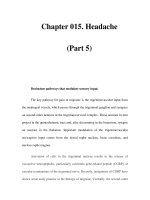
Positron emission tomography (PET) activation in migraine.
In spontaneous attacks of episodic migraine (A) there is activation of the
region of the dorsolateral pons (intersection of dark blue lines); an identical pattern
is found in chronic migraine (not shown). This area, which includes the
noradrenergic locus coeruleus, is fundamental to the expression of migraine.
Moreover, lateralization of changes in this region of the brainstem correlates with
lateralization of the head pain in hemicranial migraine; the scans shown in panels
B and C are of patients with acute migraine headache on the right and left side,
respectively. (From S Afridi et al: Arch Neurol 62:1270, 2005; Brain 128:932,
2005.)
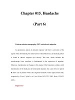
Figure 15-3
Posterior hypothalamic gray matter activationon positron emission
tomography (PET) in a patient with acute cluster headache. Posterior
hypothalamic gray matter activation on positron emission tomography (PET) in a
patient with acute cluster headache (A). (From A May et al: Lancet 352:275,
1998.) High-resolution T1 weighted MRI obtained using voxel-based
morphometry demonstrates increased gray matter activity, lateralized to the side of
pain in a patient with cluster headache (B). (From A May et al: Nat Med 5:836,
1999.)
Diagnosis and Clinical Features
Diagnostic criteria for migraine headache are listed in Table 15-4. A high
index of suspicion is required to diagnose migraine: the migraine aura, consisting
of visual disturbances with flashing lights or zigzag lines moving across the visual
field or of other neurologic symptoms, is reported in only 20–25% of patients. A
headache diary can often be helpful in making the diagnosis; this is also helpful in
assessing disability and the frequency of treatment for acute attacks. Patients with
episodes of migraine that occur daily or near-daily are considered to have chronic
migraine (see "Chronic Daily Headache," below). Migraine must be differentiated
from tension-type headache (discussed below), the most common primary
headache syndrome seen in clinical practice. Migraine at its most basic level is
headache with associated features, and tension-type headache is headache that is
featureless. Most patients with disabling headache probably have migraine.
Table 15-4 Simplified Diagnostic Criteria for Migraine
Repeated attacks of headache lasting 4–72 h in patie
nts with a normal
physical examination, no other reasonable cause for the headache, and:
At least 2 of the following
features:
Plus at least 1 of the following
features:
Unilateral pain Nausea/vomiting
Throbbing pain Photophobia and phonophobia
Aggravation by movement
Moderate or severe intensity
Source: Adapted from the International Headache Society Classification
(Headache Classification Committee of the International Headache Society, 2004).
Patients with acephalgic migraine experience recurrent neurologic
symptoms, often with nausea or vomiting, but with little or no headache. Vertigo
can be prominent; it has been estimated that one-third of patients referred for
vertigo or dizziness have a primary diagnosis of migraine.