Chapter 016. Back and Neck Pain (Part 7) ppsx
Bạn đang xem bản rút gọn của tài liệu. Xem và tải ngay bản đầy đủ của tài liệu tại đây (14.56 KB, 5 trang )
Chapter 016. Back and Neck Pain
(Part 7)
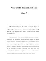
MRI of lumbar herniated disk; left S1 radiculopathy. Sagittal T1-
weighted image on the left with arrows outlining disk margins. Sagittal T2 image
on the right reveals a protruding disk at the L5-S1 level (arrows), which displaces
the central thecal sac.
The mechanism by which intervertebral disk injury causes back pain is
controversial. The inner annulus fibrosus and nucleus pulposus are normally
devoid of innervation. Inflammation and production of proinflammatory cytokines
within the protruding or ruptured disk may trigger or perpetuate back pain.
Ingrowth of nociceptive (pain) nerve fibers into inner portions of a diseased disk
may be responsible for chronic "diskogenic" pain. Nerve root injury
(radiculopathy) from disk herniation may be due to compression, inflammation, or
both; pathologically, demyelination and axonal loss are usually present.
Symptoms of a ruptured disk include back pain, abnormal posture,
limitation of spine motion (particularly flexion), or radicular pain. A dermatomal
pattern of sensory loss or a reduced or absent deep tendon reflex is more
suggestive of a specific root lesion than is the pattern of pain. Motor findings
(focal weakness, muscle atrophy, or fasciculations) occur less frequently than
focal sensory or reflex changes. Symptoms and signs are usually unilateral, but
bilateral involvement does occur with large central disk herniations that compress
multiple descending nerve roots within the spinal canal. Clinical manifestations of
specific nerve root lesions are summarized in Table 16-2. There is suggestive
evidence that lumbar disk herniation with a nonprogressive nerve root deficit can
be managed nonsurgically. The size of the disk protrusion may naturally decrease
over time.
The differential diagnosis covers a variety of serious and treatable
conditions, including epidural abscess, hematoma, or tumor. Fever, constant pain
uninfluenced by position, sphincter abnormalities, or signs of spinal cord disease
suggest an etiology other than lumbar disk disease. Bilateral absence of ankle
reflexes can be a normal finding in old age or a sign of bilateral S1 radiculopathy.
An absent deep tendon reflex or focal sensory loss may indicate injury to a nerve
root, but other sites of injury along the nerve must also be considered. For
example, an absent knee reflex may be due to a femoral neuropathy or an L4 nerve
root injury. A loss of sensation over the foot and lateral lower calf may result from
a peroneal or lateral sciatic neuropathy or an L5 nerve root injury. Focal muscle
atrophy may reflect a nerve root or peripheral nerve injury, an anterior horn cell
disease, or disuse.
An MRI scan or CT-myelogram is necessary to establish the location and
type of pathology. Spinal MRI yields exquisite views of intraspinal and adjacent
soft tissue anatomy. Bony lesions of the lateral recess or intervertebral foramen are
optimally visualized by CT-myelography. The correlation of neuroradiologic
findings to symptoms, particularly pain, is not simple. Contrast-enhancing tears in
the annulus fibrosus or disk protrusions are widely accepted as common sources of
back pain; however, many studies have found that most asymptomatic adults have
similar findings. Asymptomatic disk protrusions are also common and may
enhance with contrast. Furthermore, in patients with known disk herniation treated
either medically or surgically, persistence of the herniation 10 years later had no
relationship to the clinical outcome. In summary, MRI findings of disk protrusion,
tears in the annulus fibrosus, or contrast enhancement are common incidental
findings that, by themselves, should not dictate management decisions for patients
with back pain.
There are four indications for intervertebral disk surgery: (1) progressive
motor weakness from nerve root injury demonstrated on clinical examination or
EMG, (2) bowel or bladder disturbance or other signs of spinal cord compression,
(3) incapacitating nerve root pain despite conservative treatment for 4 weeks at a
minimum, and (4) recurrent incapacitating pain despite conservative treatment.
The latter two criteria are more subjective and less well established than the
others. Surgical treatment should also be considered if steady pain and/or
neurologic findings do not substantially improve over 4–12 weeks.
The usual surgical procedure is a partial hemilaminectomy with excision of
the prolapsed disk. Fusion of the involved lumbar segments should be considered
only if significant spinal instability is present (i.e., degenerative spondylolisthesis
or isthmic spondylolysis). Over a recent 5-year period, the number of lumbar
fusion procedures performed in the United States more than doubled, for uncertain
reasons. There are no large prospective, randomized trials comparing fusion to
other types of surgical intervention. In one study, patients with persistent low back
pain despite an initial diskectomy fared no better with spine fusion than with a
conservative regimen of cognitive intervention and exercise.
Cauda equina syndrome (CES) signifies an injury of multiple lumbosacral
nerve roots within the spinal canal. Low back pain, weakness and areflexia in the
legs, saddle anesthesia, and loss of bladder function may occur. The problem must
be distinguished from disorders of the lower spinal cord (conus medullaris
syndrome), acute transverse myelitis (Chap. 372), and Guillain-Barré syndrome
(Chap. 380). Combined involvement of the conus medullaris and cauda equina can
occur. CES is commonly due to a ruptured lumbosacral intervertebral disk,
lumbosacral spine fracture, hematoma within the spinal canal (e.g., following
lumbar puncture in patients with coagulopathy), compressive tumor, or other mass
lesion. Treatment options include surgical decompression, sometimes urgently in
an attempt to restore or preserve motor or sphincter function, or radiotherapy for
metastatic tumors (Chap. 374).