Chapter 023. Weakness and Paralysis (Part 3) ppt
Bạn đang xem bản rút gọn của tài liệu. Xem và tải ngay bản đầy đủ của tài liệu tại đây (745.08 KB, 5 trang )
Chapter 023. Weakness and Paralysis
(Part 3)
Lower Motor Neuron Weakness
This pattern results from disorders of cell bodies of lower motor neurons in
the brainstem motor nuclei and the anterior horn of the spinal cord, or from
dysfunction of the axons of these neurons as they pass to skeletal muscle (Fig. 23-
2). Weakness is due to a decrease in the number of muscle fibers that can be
activated, through a loss of α motor neurons or disruption of their connections to
muscle. Loss of γmotor neurons does not cause weakness but decreases tension on
the muscle spindles, which decreases muscle tone and attenuates the stretch
reflexes elicited on examination. An absent stretch reflex suggests involvement of
spindle afferent fibers.
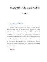
Figure 23-2
Lower motor neurons are divided into α and γ types.
The larger α motor neurons are more numerous and innervate the extrafusal
muscle fibers of the motor unit. Loss of α motor neurons or disruption of their
axons produces lower motor neuron weakness. The smaller, less numerous γ
motor neurons innervate the intrafusal muscle fibers of the muscle spindle and
contribute to normal tone and stretch reflexes. The α motor neuron receives direct
excitatory input from corticomotoneurons and primary muscle spindle afferents.
The α and γ motor neurons also receive excitatory input from other descending
upper motor neuron pathways, segmental sensory inputs, and interneurons. The α
motor neurons receive direct inhibition from Renshaw cell interneurons, and other
interneurons indirectly inhibit the α and γ motor neurons.
A tendon reflex requires the function of all illustrated structures. A tap on a
tendon stretches muscle spindles (which are tonically activated by γ motor
neurons) and activates the primary spindle afferent neurons. These stimulate the α
motor neurons in the spinal cord, producing a brief muscle contraction, which is
the familiar tendon reflex.When a motor unit becomes diseased, especially in
anterior horn cell diseases, it may spontaneously discharge, producing
fasciculations that may be seen or felt clinically or recorded by electromyography
(EMG). When α motor neurons or their axons degenerate, the denervated muscle
fibers may also discharge spontaneously. These single muscle fiber discharges, or
fibrillation potentials, cannot be seen or felt but can be recorded with EMG. If
lower motor neuron weakness is present, recruitment of motor units is delayed or
reduced, with fewer than normal activated at a given discharge frequency. This
contrasts with weakness of upper motor neuron type, in which a normal number of
motor units is activated at a given frequency but with a diminished maximal
discharge frequency.
Myopathic Weakness
Myopathic weakness is produced by disorders of the muscle fibers.
Disorders of the neuromuscular junctions also produce weakness, but this is
variable in degree and distribution and is influenced by preceding activity of the
affected muscle. At a muscle fiber, if the nerve terminal releases a normal number
of acetylcholine molecules presynaptically and a sufficient number of postsynaptic
acetylcholine receptors are opened, the end plate reaches threshold and thereby
generates an action potential that spreads across the muscle fiber membrane and
into the transverse tubular system. This electrical excitation activates intracellular
events that produce an energy-dependent contraction of the muscle fiber
(excitation-contraction coupling).
Myopathic weakness is produced by a decrease in the number or contractile
force of muscle fibers activated within motor units. With muscular dystrophies,
inflammatory myopathies, or myopathies with muscle fiber necrosis, the number
of muscle fibers is reduced within many motor units. On EMG, the size of each
motor unit action potential is decreased, and motor units must be recruited more
rapidly than normal to produce the desired power. Some myopathies produce
weakness through loss of contractile force of muscle fibers or through relatively
selective involvement of the type II (fast) fibers. These may not affect the size of
individual motor unit action potentials and are detected by a discrepancy between
the electrical activity and force of a muscle.
Diseases of the neuromuscular junction, such as myasthenia gravis, produce
weakness in a similar manner, but the loss of muscle fibers is functional (due to
inability to activate them) rather than related to muscle fiber loss. The number of
muscle fibers that are activated varies over time, depending on the state of rest of
the neuromuscular junctions. Thus, fatigable weakness is suggestive of myasthenia
gravis or other disorders of the neuromuscular junction.