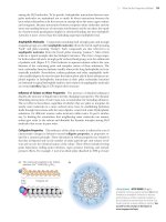
Biochemistry, 4th Edition P7 pot
Bạn đang xem bản rút gọn của tài liệu. Xem và tải ngay bản đầy đủ của tài liệu tại đây (815.76 KB, 10 trang )
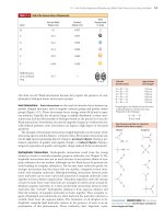
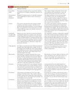
1.6 What Are Viruses? 23
Structure Molecular Composition Function
Extracellular
matrix
Cell membrane
(plasma
membrane)
Nucleus
Endoplasmic
reticulum (ER)
and ribosomes
Golgi apparatus
Mitochondria
Lysosomes
Peroxisomes
Cytoskeleton
TABLE 1.8
Major Features of a Typical Animal Cell
The surfaces of animal cells are covered with a flexible
and sticky layer of complex carbohydrates, proteins, and
lipids.
Roughly 50Ϻ50 lipidϺprotein as a 5-nm-thick continuous
sheet of lipid bilayer in which a variety of proteins are
embedded.
The nucleus is separated from the cytosol by a double
membrane, the nuclear envelope. The DNA is com-
plexed with basic proteins (histones) to form chromatin
fibers, the material from which chromosomes are made.
A distinct RNA-rich region, the nucleolus, is the site of
ribosome assembly.
Flattened sacs, tubes, and sheets of internal membrane
extending throughout the cytoplasm of the cell and en-
closing a large interconnecting series of volumes called
cisternae. The ER membrane is continuous with the outer
membrane of the nuclear envelope. Portions of the sheet-
like areas of the ER are studded with ribosomes, giving
rise to rough ER. Eukaryotic ribosomes are larger than
prokaryotic ribosomes.
The Golgi is an asymmetrical system of flattened membrane-
bounded vesicles often stacked into a complex. The face of
the complex nearest the ER is the cis face; that most distant
from the ER is the trans face. Numerous small vesicles found
peripheral to the trans face of the Golgi contain secretory
material packaged by the Golgi.
Mitochondria are organelles surrounded by two mem-
branes that differ markedly in their protein and lipid com-
position. The inner membrane and its interior volume—
the matrix—contain many important enzymes of energy
metabolism. Mitochondria are about the size of bacteria,
Ϸ1 m. Cells contain hundreds of mitochondria, which
collectively occupy about one-fifth of the cell volume.
Lysosomes are vesicles 0.2–0.5 m in diameter, bounded
by a single membrane. They contain hydrolytic enzymes
such as proteases and nucleases that act to degrade cell
constituents targeted for destruction. They are formed as
membrane vesicles budding from the Golgi apparatus.
Like lysosomes, peroxisomes are 0.2–0.5 m, single-
membrane–bounded vesicles. They contain a variety of
oxidative enzymes that use molecular oxygen and gener-
ate peroxides. They are also formed from membrane vesi-
cles budding from the smooth ER.
The cytoskeleton is composed of a network of protein
filaments: actin filaments (or microfilaments), 7 nm in
diameter; intermediate filaments, 8–10 nm; and micro-
tubules, 25 nm. These filaments interact in establishing
the structure and functions of the cytoskeleton. This in-
teracting network of protein filaments gives structure and
organization to the cytoplasm.
This complex coating is cell specific, serves in cell–
cell recognition and communication, creates cell
adhesion, and provides a protective outer layer.
The plasma membrane is a selectively permeable
outer boundary of the cell, containing specific
systems—pumps, channels, transporters, receptors—
for the exchange of materials with the environment
and the reception of extracellular information. Impor-
tant enzymes are also located here.
The nucleus is the repository of genetic information
encoded in DNA and organized into chromosomes.
During mitosis, the chromosomes are replicated and
transmitted to the daughter cells. The genetic informa-
tion of DNA is transcribed into RNA in the nucleus and
passes into the cytosol, where it is translated into pro-
tein by ribosomes.
The endoplasmic reticulum is a labyrinthine organelle
where both membrane proteins and lipids are synthe-
sized. Proteins made by the ribosomes of the rough
ER pass through the ER membrane into the cisternae
and can be transported via the Golgi to the periphery
of the cell. Other ribosomes unassociated with the ER
carry on protein synthesis in the cytosol. The nuclear
membrane, ER, Golgi, and additional vesicles are all
part of a continuous endomembrane system.
Involved in the packaging and processing of macro-
molecules for secretion and for delivery to other
cellular compartments.
Mitochondria are the power plants of eukaryotic cells
where carbohydrates, fats, and amino acids are oxi-
dized to CO
2
and H
2
O. The energy released is
trapped as high-energy phosphate bonds in ATP.
Lysosomes function in intracellular digestion of
materials entering the cell via phagocytosis or pino-
cytosis. They also function in the controlled degrada-
tion of cellular components. Their internal pH is
about 5, and the hydrolytic enzymes they contain work
best at this pH.
Peroxisomes act to oxidize certain nutrients, such as
amino acids. In doing so, they form potentially toxic
hydrogen peroxide, H
2
O
2
, and then decompose it to
H
2
O and O
2
by way of the peroxide-cleaving enzyme
catalase.
The cytoskeleton determines the shape of the cell and
gives it its ability to move. It also mediates the internal
movements that occur in the cytoplasm, such as the
migration of organelles and mitotic movements of
chromosomes. The propulsion instruments of cells—
cilia and flagella—are constructed of microtubules.
(c)
(a)
(b)
24 Chapter 1 The Facts of Life: Chemistry Is the Logic of Biological Phenomena
Structure Molecular Composition Function
Cell wall
Cell membrane
Nucleus
Endoplasmic reticulum,
Golgi apparatus,
ribosomes, lysosomes,
peroxisomes, and
cytoskeleton
Chloroplasts
Mitochondria
Vacuole
TABLE 1.9
Major Features of a Higher Plant Cell:A Photosynthetic Leaf Cell
Cellulose fibers embedded in a polysaccharide/
protein matrix; it is thick (Ͼ0.1 m), rigid, and
porous to small molecules.
Plant cell membranes are similar in overall struc-
ture and organization to animal cell membranes
but differ in lipid and protein composition.
The nucleus, nucleolus, and nuclear envelope of
plant cells are like those of animal cells.
Plant cells also contain all of these characteristic
eukaryotic organelles, essentially in the form
described for animal cells.
Chloroplasts have a double-membrane envelope, an
inner volume called the stroma, and an internal
membrane system rich in thylakoid membranes,
which enclose a third compartment, the thylakoid
lumen. Chloroplasts are significantly larger than
mitochondria. Other plastids are found in special-
ized structures such as fruits, flower petals, and roots
and have specialized roles.
Plant cell mitochondria resemble the mitochondria
of other eukaryotes in form and function.
The vacuole is usually the most obvious compart-
ment in plant cells. It is a very large vesicle en-
closed by a single membrane called the tonoplast.
Vacuoles tend to be smaller in young cells, but in
mature cells, they may occupy more than 50% of
the cell’s volume. Vacuoles occupy the center of the
cell, with the cytoplasm being located peripherally
around it. They resemble the lysosomes of animal
cells.
Protection against osmotic or mechanical rupture.
The walls of neighboring cells interact in cement-
ing the cells together to form the plant. Channels
for fluid circulation and for cell–cell communica-
tion pass through the walls. The structural material
confers form and strength on plant tissue.
The plasma membrane of plant cells is selectively
permeable, containing transport systems for the
uptake of essential nutrients and inorganic ions. A
number of important enzymes are localized here.
Chromosomal organization, DNA replication, tran-
scription, ribosome synthesis, and mitosis in plant
cells are generally similar to the analogous features
in animals.
These organelles serve the same purposes in plant
cells that they do in animal cells.
Chloroplasts are the site of photosynthesis, the
reactions by which light energy is converted to
metabolically useful chemical energy in the form of
ATP. These reactions occur on the thylakoid mem-
branes. The formation of carbohydrate from CO
2
takes place in the stroma. Oxygen is evolved during
photosynthesis. Chloroplasts are the primary
source of energy in the light.
Plant mitochondria are the main source of energy
generation in photosynthetic cells in the dark and
in nonphotosynthetic cells under all conditions.
Vacuoles function in transport and storage of
nutrients and cellular waste products. By accumu-
lating water, the vacuole allows the plant cell to
grow dramatically in size with no increase in cyto-
plasmic volume.
© Science Source/Photo Researchers, Inc.
BSIP/Photo Researchers, Inc.
Eye of Science/Photo Researchers, Inc.
FIGURE 1.23 Viruses are genetic elements enclosed in a protein coat. Viruses are not free-living organisms and
can reproduce only within cells.Viruses show an almost absolute specificity for their particular host cells, infect-
ing and multiplying only within those cells.Viruses are known for virtually every kind of cell. Shown here are
examples of (a) an animal virus, adenovirus; (b) bacteriophage T
4
on E. coli; and (c) a plant virus, tobacco
mosaic virus.
Summary 25
Protein
coat
Genetic
material
(DNA or RNA)
Entry of virus
genome into cell
Transcription
RNA
Translation
Assembly
Replication
Host
cell
Coat
proteins
Release
from cell
ACTIVE FIGURE 1.24 The virus life cycle.Viruses are mobile bits of genetic information
encapsulated in a protein coat.The genetic material can be either DNA or RNA. Once this genetic material
gains entry to its host cell, it takes over the host machinery for macromolecular synthesis and subverts it to the
synthesis of viral-specific nucleic acids and proteins.These virus components are then assembled into mature
virus particles that are released from the cell. Often, this parasitic cycle of virus infection leads to cell death and
disease. Test yourself on the concepts in this figure at www.cengage.com/login
SUMMARY
1.1 What Are the Distinctive Properties of Living Systems? Living sys-
tems display an astounding array of activities that collectively constitute
growth, metabolism, response to stimuli, and replication. In accord with
their functional diversity, living organisms are complicated and highly or-
ganized entities composed of many cells. In turn, cells possess subcellular
structures known as organelles, which are complex assemblies of very
large polymeric molecules, or macromolecules. The monomeric units of
macromolecules are common organic molecules (metabolites). Biologi-
cal structures play a role in the organism’s existence. From parts of or-
ganisms, such as limbs and organs, down to the chemical agents of me-
tabolism, such as enzymes and metabolic intermediates, a biological
purpose can be given for each component. Maintenance of the highly
organized structure and activity of living systems requires energy that
must be obtained from the environment. Energy is required to create and
maintain structures and to carry out cellular functions. In terms of the ca-
pacity of organisms to self-replicate, the fidelity of self-replication resides
ultimately in the chemical nature of DNA, the genetic material.
1.2 What Kinds of Molecules Are Biomolecules? C, H, N, and O are
among the lightest elements capable of forming covalent bonds
through electron-pair sharing. Because the strength of covalent bonds
is inversely proportional to atomic weight, H, C, N, and O form the
strongest covalent bonds. Two properties of carbon covalent bonds
merit attention: the ability of carbon to form covalent bonds with itself
and the tetrahedral nature of the four covalent bonds when carbon
atoms form only single bonds. Together these properties hold the po-
tential for an incredible variety of structural forms, whose diversity is
multiplied further by including N, O, and H atoms.
1.3
What Is the Structural Organization of Complex Biomolecules?
Biomolecules are built according to a structural hierarchy: Simple mol-
ecules are the units for building complex structures. H
2
O, CO
2
, NH
4
ϩ
,
NO
3
Ϫ
, and N
2
are the inorganic precursors for the formation of simple
organic compounds from which metabolites are made. These metabo-
lites serve as intermediates in cellular energy transformation and as
building blocks (amino acids, sugars, nucleotides, fatty acids, and glyc-
erol) for lipids and for macromolecular synthesis (synthesis of proteins,
polysaccharides, DNA, and RNA). The next higher level of structural or-
ganization is created when macromolecules come together through
noncovalent interactions to form supramolecular complexes, such as
multifunctional enzyme complexes, ribosomes, chromosomes, and cyto-
skeletal elements.
The next higher rung in the hierarchical ladder is occupied by the
organelles. Organelles are membrane-bounded cellular inclusions ded-
icated to important cellular tasks, such as the nucleus, mitochondria,
chloroplasts, endoplasmic reticulum, Golgi apparatus, and vacuoles, as
well as other relatively small cellular inclusions. At the apex of the bio-
molecular hierarchy is the cell, the unit of life, the smallest entity dis-
playing those attributes associated uniquely with the living state—
growth, metabolism, stimulus response, and replication.
1.4 How Do the Properties of Biomolecules Reflect Their Fitness to the
Living Condition? Some biomolecules carry the information of life;
others translate this information so that the organized structures essen-
tial to life are formed. Interactions between such structures are the
processes of life. Properties of biomolecules that endow them with the
potential for creating the living state include the following: Biological
macromolecules and their building blocks have directionality, and thus
biological macromolecules are informational; in addition, biomolecules
have characteristic three-dimensional architectures, providing the
means for molecular recognition through structural complementarity.
Weak forces (H bonds, van der Waals interactions, ionic attractions, and
hydrophobic interactions) mediate the interactions between biological
molecules and, as a consequence, restrict organisms to the narrow
range of environmental conditions where these forces operate.
1.5 What Is the Organization and Structure of Cells? All cells share a
common ancestor and fall into one of two broad categories—prokaryotic
and eukaryotic—depending
on whether the cell has a nucleus. Prokary-
otes are typically single-celled organisms and have a rather simple cellu-
lar organization. In contrast, eukaryotic cells are structurally more com-
plex, having organelles and various subcellular compartments defined
by membranes. Other than the Protists, eukaryotes are multicellular.
1.6 What Are Viruses? Viruses are supramolecular complexes of nu-
cleic acid encapsulated in a protein coat and, in some instances, sur-
rounded by a membrane envelope. Viruses are not alive; they are not
even cellular. Instead, they are packaged bits of genetic material that
26 Chapter 1 The Facts of Life: Chemistry Is the Logic of Biological Phenomena
can parasitize cells in order to reproduce. Often, they cause disintegra-
tion, or lysis, of the cells they’ve infected. It is these cytolytic properties
that are the basis of viral disease. In certain circumstances, the viral nu-
cleic acid may integrate into the host chromosome and become quies-
cent, creating a state known as lysogeny. If the host cell is damaged, the
replicative capacities of the quiescent viral nucleic acid may be acti-
vated, leading to viral propagation and release.
PROBLEMS
Preparing for an exam? Create you own study path for this
chapter at www.cengage.com/login
1. The nutritional requirements of Escherichia coli cells are far simpler
than those of humans, yet the macromolecules found in bacteria
are about as complex as those of animals. Because bacteria can
make all their essential biomolecules while subsisting on a simpler
diet, do you think bacteria may have more biosynthetic capacity and
hence more metabolic complexity than animals? Organize your
thoughts on this question, pro and con, into a rational argument.
2. Without consulting the figures in this chapter, sketch the charac-
teristic prokaryotic and eukaryotic cell types and label their perti-
nent organelle and membrane systems.
3. Escherichia coli cells are about 2 m (microns) long and 0.8 m in
diameter.
a. How many E. coli cells laid end to end would fit across the diame-
ter of a pinhead? (Assume a pinhead diameter of 0.5 mm.)
b. What is the volume of an E. coli cell? (Assume it is a cylinder, with
the volume of a cylinder given by V ϭ r
2
h, where ϭ 3.14.)
c. What is the surface area of an E. coli cell? What is the surface-to-
volume ratio of an E. coli cell?
d. Glucose, a major energy-yielding nutrient, is present in bacterial
cells at a concentration of about 1 mM. What is the concentration
of glucose, expressed as mg/mL? How many glucose molecules
are contained in a typical E. coli cell? (Recall that Avogadro’s
number ϭ 6.023 ϫ 10
23
.)
e. A number of regulatory proteins are present in E. coli at only one
or two molecules per cell. If we assume that an E. coli cell contains
just one molecule of a particular protein, what is the molar con-
centration of this protein in the cell? If the molecular weight of
this protein is 40 kD, what is its concentration, expressed as
mg/mL?
f. An E. coli cell contains about 15,000 ribosomes, which carry out
protein synthesis. Assuming ribosomes are spherical and have a di-
ameter of 20 nm (nanometers), what fraction of the E. coli cell vol-
ume is occupied by ribosomes?
g. The E. coli chromosome is a single DNA molecule whose mass
is about 3 ϫ 10
9
daltons. This macromolecule is actually a linear
array of nucleotide pairs. The average molecular weight of a
nucleotide pair is 660, and each pair imparts 0.34 nm to the
length of the DNA molecule. What is the total length of the
E. coli chromosome? How does this length compare with the over-
all dimensions of an E. coli cell? How many nucleotide pairs does
this DNA contain? The average E. coli protein is a linear chain of
360 amino acids. If three nucleotide pairs in a gene encode one
amino acid in a protein, how many different proteins can the
E. coli chromosome encode? (The answer to this question is a rea-
sonable approximation of the maximum number of different
kinds of proteins that can be expected in bacteria.)
4. Assume that mitochondria are cylinders 1.5 m in length and
0.6 m in diameter.
a. What is the volume of a single mitochondrion?
b. Oxaloacetate is an intermediate in the citric acid cycle, an im-
portant metabolic pathway localized in the mitochondria of
eukaryotic cells. The concentration of oxaloacetate in mito-
chondria is about 0.03 M. How many molecules of oxaloacetate
are in a single mitochondrion?
5. Assume that liver cells are cuboidal in shape, 20 m on a side.
a. How many liver cells laid end to end would fit across the diameter
of a pinhead? (Assume a pinhead diameter of 0.5 mm.)
b. What is the volume of a liver cell? (Assume it is a cube.)
c. What is the surface area of a liver cell? What is the surface-
to-volume ratio of a liver cell? How does this compare to the
surface-to-volume ratio of an E. coli cell (compare this answer with
that of problem 3c)? What problems must cells with low surface-
to-volume ratios confront that do not occur in cells with high
surface-to-volume ratios?
d. A human liver cell contains two sets of 23 chromosomes, each
set being roughly equivalent in information content. The total
mass of DNA contained in these 46 enormous DNA molecules is
4 ϫ 10
12
daltons. Because each nucleotide pair contributes
660 daltons to the mass of DNA and 0.34 nm to the length of
DNA, what is the total number of nucleotide pairs and the com-
plete length of the DNA in a liver cell? How does this length
compare with the overall dimensions of a liver cell? The maximal
information in each set of liver cell chromosomes should be re-
lated to the number of nucleotide pairs in the chromosome set’s
DNA. This number can be obtained by dividing the total number
of nucleotide pairs just calculated by 2. What is this value? If this
information is expressed in proteins that average 400 amino
acids in length and three nucleotide pairs encode one amino
acid in a protein, how many different kinds of proteins might a
liver cell be able to produce? (In reality, liver cell DNA encodes
approximately 20,000 different proteins. Thus, a large discrep-
ancy exists between the theoretical information content of DNA
in liver cells and the amount of information actually expressed.)
6. Biomolecules interact with one another through molecular surfaces
that are structurally complementary. How can various proteins in-
teract with molecules as different as simple ions, hydrophobic
lipids, polar but uncharged carbohydrates, and even nucleic acids?
7. What structural features allow biological polymers to be informa-
tional macromolecules? Is it possible for polysaccharides to be in-
formational macromolecules?
8. Why is it important that weak forces, not strong forces, mediate bio-
molecular recognition?
9. What is the distance between the centers of two carbon atoms (their
limit of approach) that are interacting through van der Waals forces?
What is the distance between the centers of two carbon atoms
joined in a covalent bond? (See Table 1.4.)
10. Why does the central role of weak forces in biomolecular interac-
tions restrict living systems to a narrow range of environmental
conditions?
11. Describe what is meant by the phrase “cells are steady-state systems.”
12. The genome of the Mycoplasma genitalium consists of 523 genes, en-
coding 484 proteins, in just 580,074 base pairs (Table 1.6). What
fraction of the M. genitalium genes encode proteins? What do you
think the other genes encode? If the fraction of base pairs devoted
to protein-coding genes is the same as the fraction of the total
genes that they represent, what is the avera
ge number of base pairs
per protein-coding gene? If it takes 3 base pairs to specify an amino
acid in a protein, how many amino acids are found in the average
M. genitalium protein? If each amino acid contributes on average
Further Reading 27
120 Daltons to the mass of a protein, what is the mass of an average
M. genitalium protein?
13. Studies of existing cells to determine the minimum number of
genes for a living cell have suggested that 206 genes are sufficient.
If the ratio of protein-coding genes to non–protein-coding genes is
the same in this minimal organism as the genes of Mycoplasma geni-
talium, how many proteins are represented in these 206 genes? How
many base pairs would be required to form the genome of this min-
imal organism if the genes are the same size as M. genitalium genes?
14. Virus genomes range in size from approximately 3500 nucleotides
to approximately 280,000 base pairs. If viral genes are about the
same size as M. genitalium genes, what is the minimum and maxi-
mum number of genes in viruses?
15. The endoplasmic reticulum (ER) is a site of protein synthesis. Pro-
teins made by ribosomes associated with the ER may pass into the
ER membrane or enter the lumen of the ER. Devise a pathway by
which:
a. a plasma membrane protein may reach the plasma membrane.
b. a secreted protein may be deposited outside the cell.
Preparing for the MCAT Exam
16. Biological molecules often interact via weak forces (H bonds, van
der Waals interactions, etc.). What would be the effect of an in-
crease in kinetic energy on such interactions?
17. Proteins and nucleic acids are informational macromolecules. What
are the two minimal criteria for a linear informational polymer?
FURTHER READING
General Biology Textbooks
Campbell, N. A., and Reece, J. B., 2005, Biology, 7th ed. San Francisco:
Benjamin/Cummings.
Solomon, E. P., Berg, L. R., and Martin, D. W., 2004. Biology, 7th ed.
Pacific Grove, CA: Brooks/Cole.
Cell and Molecular Biology Textbooks
Alberts, B., Johnson, A., Lewis, J., Raff, M., et al., 2007. Molecular Biology
of the Cell, 5th ed. New York: Garland Press.
Lewin, B., Cassimeris, L., Lingappa, V. R., and Plopper, G., 2007. Cells.
Boston, MA: Jones and Bartlett.
Lodish, H., Berk, A., Kaiser, C. A., Kreiger, M., et al., 2007. Molecular Cell
Biology, 5th ed. New York: W. H. Freeman.
Snyder, L., and Champness, W., 2002. Molecular Genetics of Bacteria, 2nd
ed. Herndon, VA: ASM Press.
Watson, J. D., Baker, T. A., Bell, S. T., Gann, A., et al., 2007. Molecular
Biology of the Gene, 6th ed. Menlo Park, CA: Benjamin/Cummings.
Papers on Cell Structure
Gil, R., Silva, F. J., Pereto, J., and Moya, A., 2004. Determination of the
core of a minimal bacterial gene set. Microbiology and Molecular Biol-
ogy Reviews 68:518–537.
Goodsell, D. S., 1991. Inside a living cell. Trends in Biochemical Sciences
16:203–206.
Lewis, P. J., 2004. Bacterial subcellular architecture: Recent advances
and future prospects. Molecular Microbiology 54:1135–1150.
Lloyd, C., ed., 1986. Cell organization. Trends in Biochemical Sciences
11:437–485.
Papers on Genomes
Cho, M. K., et al., 1999. Ethical considerations in synthesizing a minimal
genome. Science 286:2087–2090.
Kobayashi, K., Ehrlich, S. D., Albertini, A., Amati, G., et al., 2003. Es-
sential Bacillus subtilis genes. Proceedings of the National Academy of Sci-
ence, U.S.A. 100:4678–4683.
Lartigue, C., Glass, J. I., Alperovich, N., Pieper, R., et al., 2007. Genome
transplantation in bacteria: changing one species to another.
Science 317:632–638.
Szathmary, E., 2005. In search of the simplest cell. Nature 433:469–470.
Papers on Early Cell Evolution
Margulis, L., 1996. Archaeal-eubacterial mer
gers in the origin of Eu-
karya: Phylogenetic classification of life. Proceedings of the National
Academy of Science, U.S.A. 93:1071–1076.
Pace, N. R., 2006. Time for a change. Nature 441:289.
Service, R. F., 1997. Microbiologists explore life’s rich, hidden king-
doms. Science 275:1740–1742.
Wald, G., 1964. The origins of life. Proceedings of the National Academy of
Science, U.S.A. 52:595–611.
Whitfield, J., 2004. Born in a watery commune. Nature 427:674–676.
Woese, C. R., 2002. On the creation of cells. Proceedings of the National
Academy of Science, U.S.A. 99:8742–8747.
A Brief History of Life
De Duve, C., 2002.
Life-Evolving: Molecules, Mind, and Meaning. New Y
ork:
Oxford University Press.
Morowitz, H., and Smith, E., 2007. Energy flow and the organization of
life. Complexity 13:51–59.
© Paul Steel/CORBIS
2
Water: The Medium of Life
Water is a major chemical component of the earth’s surface. It is indispensable to
life. Indeed, it is the only liquid that most organisms ever encounter. We are
prone to take it for granted because of its ubiquity and bland nature, yet we mar-
vel at its many unusual and fascinating properties. At the center of this fascination
is the role of water as the medium of life. Life originated, evolved, and thrives in
the seas. Organisms invaded and occupied terrestrial and aerial niches, but none
gained true independence from water. Typically, organisms are 70% to 90% water.
Indeed, normal metabolic activity can occur only when cells are at least 65% H
2
O.
This dependency of life on water is not a simple matter, but it can be grasped by
considering the unusual chemical and physical properties of H
2
O. Subsequent
chapters establish that water and its ionization products, hydrogen ions and hy-
droxide ions, are critical determinants of the structure and function of many bio-
molecules, including amino acids and proteins, nucleotides and nucleic acids,
and even phospholipids and membranes. In yet another essential role, water is an
indirect participant—a difference in the concentration of hydrogen ions on op-
posite sides of a membrane represents an energized condition essential to bio-
logical mechanisms of energy transformation. First, let’s review the remarkable
properties of water.
2.1 What Are the Properties of Water?
Water Has Unusual Properties
Compared with chemical compounds of similar atomic organization and molecular
size, water displays unexpected properties. For example, compare water, the hy-
dride of oxygen, with hydrides of oxygen’s nearest neighbors in the periodic table,
namely, ammonia (NH
3
) and hydrogen fluoride (HF), or with the hydride of its
nearest congener, sulfur (H
2
S). Water has a substantially higher boiling point, melt-
ing point, heat of vaporization, and surface tension. Indeed, all of these physical
properties are anomalously high for a substance of this molecular weight that is nei-
ther metallic nor ionic. These properties suggest that intermolecular forces of at-
traction between H
2
O molecules are high. Thus, the internal cohesion of this sub-
stance is high. Furthermore, water has an unusually high dielectric constant, its
maximum density is found in the liquid (not the solid) state, and it has a negative
volume of melting (that is, the solid form, ice, occupies more space than does the
liquid form, water). It is truly remarkable that so many eccentric properties occur
together in this single substance. As chemists, we expect to find an explanation for
these apparent eccentricities in the structure of water. The key to its intermolecular
attractions must lie in its atomic constitution. Indeed, the unrivaled ability to form hy-
drogen bonds is the crucial fact to understanding its properties.
Where there’s water, there’s life.
If there is magic on this planet, it is contained in
water.
Loren Eisley
(inscribed on the wall of the National Aquarium
in Baltimore, Maryland)
KEY QUESTIONS
2.1 What Are the Properties of Water?
2.2 What Is pH?
2.3 What Are Buffers, and What Do They Do?
2.4 What Properties of Water Give It a Unique
Role in the Environment?
ESSENTIAL QUESTION
Water provided conditions for the origin, evolution, and flourishing of life; water is
the medium of life.
What are the properties of water that render it so suited to its role as the
medium of life?
Create your own study path for
this chapter with tutorials, simulations, animations,
and Active Figures at www.cengage.com/ login
2.1 What Are the Properties of Water? 29
Hydrogen Bonding in Water Is Key to Its Properties
The two hydrogen atoms of water are linked covalently to oxygen, each sharing an
electron pair, to give a nonlinear arrangement (Figure 2.1). This “bent” structure of
the H
2
O molecule has enormous influence on its properties. If H
2
O were linear, it
would be a nonpolar substance. In the bent configuration, however, the electroneg-
ative O atom and the two H atoms form a dipole that renders the molecule distinctly
polar. Furthermore, this structure is ideally suited to H-bond formation. Water can
serve as both an H donor and an H acceptor in H-bond formation. The potential to
form four H bonds per water molecule is the source of the strong intermolecular at-
tractions that endow this substance with its anomalously high boiling point, melting
point, heat of vaporization, and surface tension. In ordinary ice, the common crys-
talline form of water, each H
2
O molecule has four nearest neighbors to which it is
hydrogen bonded: Each H atom donates an H bond to the O of a neighbor, and the
O atom serves as an H-bond acceptor from H atoms bound to two different water
molecules (Figure 2.2). A local tetrahedral symmetry results.
Hydrogen bonding in water is cooperative. That is, an H-bonded water mole-
cule serving as an acceptor is a better H-bond donor than an unbonded molecule
(and an H
2
O molecule serving as an H-bond donor becomes a better H-bond ac-
ceptor). Thus, participation in H bonding by H
2
O molecules is a phenomenon
of mutual reinforcement. The H bonds between neighboring molecules are weak
(23 kJ/mol each) relative to the HOO covalent bonds (420 kJ/mol). As a conse-
quence, the hydrogen atoms are situated asymmetrically between the two oxygen
atoms along the O-O axis. There is never any ambiguity about which O atom the
H atom is chemically bound to, nor to which O it is H bonded.
The Structure of Ice Is Based On H-Bond Formation
In ice, the hydrogen bonds form a space-filling, three-dimensional network. These
bonds are directional and straight; that is, the H atom lies on a direct line between
the two O atoms. This linearity and directionality mean that the H bonds in ice are
strong. In addition, the directional preference of the H bonds leads to an open lat-
tice structure. For example, if the water molecules are approximated as rigid
spheres centered at the positions of the O atoms in the lattice, then the observed
density of ice is actually only 57% of that expected for a tightly packed arrangement
of such spheres. The H bonds in ice hold the water molecules apart. Melting in-
volves breaking some of the H bonds that maintain the crystal structure of ice so
104.3°
Covalent bond
length = 0.095 nm
van der Waals radius
of oxygen = 0.14 nm
van der Waals radius
of hydrogen = 0.12 nm
Dipole moment
O
H
H
␦
+
␦
+
␦
–
ACTIVE FIGURE 2.1 The structure of
water.Two lobes of negative charge formed by the
lone-pair electrons of the oxygen atom lie above and
below the plane of the diagram.This electron density
contributes substantially to the large dipole moment of
the water molecule. Note that the HOOOH angle is
104.3°, not 109°, the angular value found in molecules
with tetrahedral symmetry, such as CH
4
. Many of the
important properties of water derive from this angular
value, such as the decreased density of its crystalline
state, ice. Test yourself on the concepts in this figure
at www.cengage.com/login
ANIMATED FIGURE 2.2 The structure of normal ice.The smallest number of H
2
O molecules in
any closed circuit of H-bonded molecules is six, so this structure bears the name hexagonal ice.See this figure
animated at www.cengage.com/login
30 Chapter 2 Water:The Medium of Life
that the molecules of water (now liquid) can actually pack closer together. Thus, the
density of ice is slightly less than that of water. Ice floats, a property of great impor-
tance to aquatic organisms in cold climates.
In liquid water, the rigidity of ice is replaced by fluidity and the crystalline pe-
riodicity of ice gives way to spatial homogeneity. The H
2
O molecules in liquid wa-
ter form a disordered H-bonded network, with each molecule having an average
of 4.4 close neighbors situated within a center-to-center distance of 0.284 nm
(2.84 Å). At least half of the hydrogen bonds have nonideal orientations (that is,
they are not perfectly straight); consequently, liquid H
2
O lacks the regular lat-
ticelike structure of ice. The space about an O atom is not defined by the pres-
ence of four hydrogens but can be occupied by other water molecules randomly
oriented so that the local environment, over time, is essentially uniform. Never-
theless, the heat of melting for ice is but a small fraction (13%) of the heat of sub-
limation for ice (the energy needed to go from the solid to the vapor state). This
fact indicates that the majority of H bonds between H
2
O molecules survive the
transition from solid to liquid. At 10°C, 2.3 H bonds per H
2
O molecule remain
and the tetrahedral bond order persists, even though substantial disorder is now
present.
Molecular Interactions in Liquid Water Are Based on H Bonds
The present interpretation of water structure is that water molecules are connected
by uninterrupted H-bond paths running in every direction, spanning the whole
sample. The participation of each water molecule in an average state of H bonding
to its neighbors means that each molecule is connected to every other in a fluid net-
work of H bonds. The average lifetime of an H-bonded connection between two
H
2
O molecules in water is 9.5 psec (picoseconds, where 1 psec ϭ 10
Ϫ12
sec). Thus,
about every 10 psec, the average H
2
O molecule moves, reorients, and interacts with
new neighbors, as illustrated in Figure 2.3.
In summary, pure liquid water consists of H
2
O molecules held in a disordered,
three-dimensional network that has a local preference for tetrahedral geometry, yet
contains a large number of strained or broken hydrogen bonds. The presence of
strain creates a kinetic situation in which H
2
O molecules can switch H-bond alle-
giances; fluidity ensues.
The Solvent Properties of Water Derive from Its Polar Nature
Because of its highly polar nature, water is an excellent solvent for ionic sub-
stances such as salts; nonionic but polar substances such as sugars, simple
alcohols, and amines; and carbonyl-containing molecules such as aldehydes and
ketones. Although the electrostatic attractions between the positive and negative
ions in the crystal lattice of a salt are very strong, water readily dissolves salts. For
example, sodium chloride is dissolved because dipolar water molecules partici-
pate in strong electrostatic interactions with the Na
ϩ
and Cl
Ϫ
ions, leading to the
formation of hydration shells surrounding these ions (Figure 2.4). Although hy-
dration shells are stable structures, they are also dynamic. Each water molecule in
the inner hydration shell around a Na
ϩ
ion is replaced on average every 2 to
4 nsec (nanoseconds, where 1 nsec ϭ 10
Ϫ9
sec) by another H
2
O. Consequently, a
water molecule is trapped only several hundred times longer by the electrostatic
force field of an ion than it is by the H-bonded network of water. (Recall that the
average lifetime of H bonds between water molecules is about 10 psec.)
Water Has a High Dielectric Constant The attractions between the water mole-
cules interacting with, or hydrating, ions are much greater than the tendency of op-
positely charged ions to attract one another. Water’s ability to surround ions in di-
pole interactions and diminish their attraction for each other is a measure of its
dielectric constant, D. Indeed, ionization in solution depends on the dielectric con-
stant of the solvent; otherwise, the strongly attracted positive and negative ions
psec
H bond
ACTIVE FIGURE 2.3 The fluid network
of H bonds linking water molecules in the liquid state. It
is revealing to note that, in 10 psec, a photon of light
(which travels at 3 ϫ 10
8
m/sec) would move a distance
of only 0.003 m. Test yourself on the concepts in this
figure at www.cengage.com/login
2.1 What Are the Properties of Water? 31
would unite to form neutral molecules. The strength of the dielectric constant is re-
lated to the force, F, experienced between two ions of opposite charge separated by
a distance, r, as given in the relationship
F ϭ e
1
e
2
/Dr
2
where e
1
and e
2
are the charges on the two ions. Table 2.1 lists the dielectric con-
stants of some common liquids. Note that the dielectric constant for water is more
than twice that of methanol and more than 40 times that of hexane.
Water Forms H Bonds with Polar Solutes In the case of nonionic but polar com-
pounds such as sugars, the excellent solvent properties of water stem from its abil-
ity to readily form hydrogen bonds with the polar functional groups on these com-
pounds, such as hydroxyls, amines, and carbonyls (see Figure 1.14). These polar
interactions between solvent and solute are stronger than the intermolecular at-
tractions between solute molecules caused by van der Waals forces and weaker hy-
drogen bonding. Thus, the solute molecules readily dissolve in water.
Hydrophobic Interactions The behavior of water toward nonpolar solutes is dif-
ferent from the interactions just discussed. Nonpolar solutes (or nonpolar func-
tional groups on biological macromolecules) do not readily H bond to H
2
O, and
as a result, such compounds tend to be only sparingly soluble in water. The
process of dissolving such substances is accompanied by significant reorganization
of the water surrounding the solute so that the response of the solvent water to
such solutes can be equated to “structure making.” Because nonpolar solutes must
occupy space, the random H-bonded network of water must reorganize to ac-
commodate them. At the same time, the water molecules participate in as many
H-bonded interactions with one another as the temperature permits. Conse-
quently, the H-bonded water network rearranges toward formation of a local cage-
like (clathrate) structure surrounding each solute molecule, as shown for a long-
chain fatty acid in Figure 2.5. This fixed orientation of water molecules around a
hydrophobic “solute” molecule results in a hydration shell. A major consequence
of this rearrangement is that the molecules of H
2
O participating in the cage layer
have markedly reduced options for orientation in three-dimensional space. Water
molecules tend to straddle the nonpolar solute such that two or three tetrahedral
directions (H-bonding vectors) are tangential to the space occupied by the inert
solute. “Straddling” allows the water molecules to retain their H-bonding possi-
bilities because no H-bond donor or acceptor of the H
2
O is directed toward the
caged solute. The water molecules forming these clathrates are involved in highly
ordered structures. That is, clathrate formation is accompanied by significant or-
dering of structure or negative entropy.
Cl
–
Cl
–
Cl
–
Cl
–
Cl
–
Cl
–
Cl
–
Cl
–
Cl
–
Cl
–
Cl
–
Cl
–
Cl
–
Na
+
Na
+
Na
+
Na
+
Na
+
Na
+
Na
+
Na
+
Na
+
Na
+
Na
+
Na
+
Na
+
–
+
+
–
–
–
–
–
–
–
–
–
–
–
–
–
–
–
–
–
–
–
–
–
–
–
–
–
–
–
–
–
–
–
–
–
+
+
+
+
+
+
+
+
+
+
+
+
+
+
+
+
+
+
+
+
+
+
+
+
+
+
+
+
+
+
+
+
+
+
+
+
+
+
+
+
+
+
+
+
+
+
+
+
+
+
+
+
+
+
+
+
+
+
+
+
+
+
+
+
ANIMATED FIGURE 2.4
Hydration shells surrounding ions in
solution.Water molecules orient so that
the electrical charge on the ion is
sequestered by the water dipole. See
this figure animated at www
.cengage.com/login
Solvent Dielectric Constant (D)
Formamide 109
Water 78.5
Methyl alcohol 32.6
Ethyl alcohol 24.3
Acetone 20.7
Acetic acid 6.2
Chloroform 5.0
Benzene 2.3
Hexane 1.9
*The dielectric constant is also referred to as relative permitiv-
ity by physical chemists.
TABLE 2.1
Dielectric Constants* of Some
Common Solvents at 25°C
32 Chapter 2 Water:The Medium of Life
Multiple nonpolar molecules tend to cluster together, because their joint solva-
tion cage involves less total surface area and thus fewer ordered water molecules
than in their separate cages. It is as if the nonpolar molecules had some net attrac-
tion for one another. This apparent affinity of nonpolar structures for one another
is called hydrophobic interactions (Figure 2.6). In actuality, the “attraction” be-
tween nonpolar solutes is an entropy-driven process due to a net decrease in order
C
HO
ANIMATED FIGURE 2.5 (left) Disordered network of H-bonded water
molecules. (right) Clathrate cage of ordered, H-bonded water molecules around a
nonpolar solute molecule. See this figure animated at www.cengage.com/
login
C
C
C
C
HO
C
HO
C
HO
C
HO
C
HO
FIGURE 2.6 Hydrophobic interactions between nonpolar molecules (or nonpolar regions of molecules) are due
to the increase in entropy of solvent water molecules.

