Biochemistry, 4th Edition P16 pptx
Bạn đang xem bản rút gọn của tài liệu. Xem và tải ngay bản đầy đủ của tài liệu tại đây (887.4 KB, 10 trang )
5.5 What Is the Nature of Amino Acid Sequences? 113
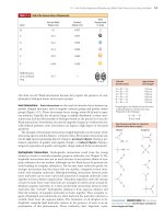
the same amino acid residues are always found (Figure 5.19). These invariant
residues serve roles crucial to the biological function of this protein, and thus
substitutions of other amino acids at these positions cannot be tolerated. The
number of amino acid differences between two cytochrome c sequences is propor-
tional to the phylogenetic difference between the species from which they are de-
rived. Cytochrome c in humans and in chimpanzees is identical; human and
another mammalian (sheep) cytochrome c differ at 10 residues. The human cyto-
chrome c sequence has 14 variant residues from a reptile sequence (rattlesnake),
18 from a fish (carp), 29 from a mollusc (snail), 31 from an insect (moth), and
more than 40 from yeast or higher plants (cauliflower).
The Phylogenetic Tree for Cytochrome c Figure 5.20 displays a phylogenetic tree
(a diagram illustrating the evolutionary relationships among a group of organisms)
constructed from the sequences of cytochrome c. The tips of the branches are occu-
pied by contemporary species whose sequences have been determined. The tree has
been deduced by computer analysis of these sequences to find the minimum num-
ber of mutational changes connecting the branches. Other computer methods can
be used to infer potential ancestral sequences represented by nodes, or branch
points, in the tree. Such analysis ultimately suggests a primordial cytochrome c se-
quence lying at the base of the tree. Evolutionary trees constructed in this manner,
that is, solely on the basis of amino acid differences occurring in the primary se-
quence of one selected protein, show remarkable agreement with phylogenetic re-
lationships derived from more classic approaches and have given rise to the field of
molecular evolution.
Related Proteins Share a Common Evolutionary Origin
Amino acid sequence analysis reveals that proteins with related functions often
show a high degree of sequence similarity. Such findings suggest a common
ancestry for these proteins.
Oxygen-Binding Heme Proteins Myoglobin and the ␣- and -globin chains of
hemoglobin constitute a set of paralogous proteins. Myoglobin, the oxygen-binding
heme protein of muscle, consists of a single polypeptide chain of 153 residues.
Hemoglobin, the oxygen transport protein of erythrocytes, is a tetramer composed
of two ␣-chains (141 residues each) and two -chains (146 residues each). These glo-
bin paralogs—myoglobin, ␣-globin, and -globin—share a strong degree of
sequence homology (Figure 5.21). Human myoglobin and the human ␣-globin
chain show 38 amino acid identities, whereas human ␣-globin and human -globin
have 64 residues in common. The relatedness suggests an evolutionary sequence of
events in which chance mutations led to amino acid substitutions and divergence in
primary structure. The ancestral myoglobin gene diverged first, after duplication of
a primordial globin gene had given rise to its progenitor and an ancestral hemo-
globin gene (Figure 5.22). Subsequently, the ancestral hemoglobin g
ene duplicated
to generate the progenitors of the present-day ␣-globin and -globin genes. The
ability to bind O
2
via a heme prosthetic group is retained by all three of these
polypeptides.
Serine Proteases Whereas the globins provide an example of gene duplication
giving rise to a set of proteins in which the biological function has been highly con-
served, other sets of proteins united by strong sequence homology show more di-
vergent biological functions. Trypsin, chymotrypsin (see Chapter 14), and elastase
are members of a class of proteolytic enzymes called serine proteases because of the
central role played by specific serine residues in their catalytic activity. Thrombin,
an essential enzyme in blood clotting, is also a serine protease. These enzymes show
sufficient sequence homology to conclude that they arose via duplication of a prog-
enitor serine protease gene, even though their substrate preferences are now quite
different.
Phe
Cys
His
Gly
Pro
Leu
Gly
Arg
Gly
Gly
Tyr
Trp
Leu
Asn
Pro
Lys
Lys
Pro
Thr
Lys
Met
Phe
Gly
Arg
17
18
29
30
32
34
38
41
45
48
59
68
70
71
76
78
79
80
82
84
91
10
72
73
Heme
Gly1
Gly6
Asn52
Tyr
74
100
FIGURE 5.19 The sequence of cyto-
chrome c from more than 40 different
species reveals that 28 residues are in-
variant.These invariant residues are scat-
tered irregularly along the polypeptide
chain, except for a cluster between
residues 70 and 80. All cytochrome c
polypeptide chains have a cysteine
residue at position 17, and all but one
have another Cys at position 14.These
Cys residues serve to link the heme
prosthetic group of cytochrome c to the
protein.
114 Chapter 5 Proteins:Their Primary Structure and Biological Functions
Neurospora
crassa
Candida
krusei
Debaryomyces
kloeckri
Baker's
yeast
Pig, bovine, sheep
Dog
Bullfrog
Gray whale
Pekin duck
Rabbit
Monkey
Gray
kangaroo
Pigeon
King penguin
Snapping turtle
Pacific lamprey
Chicken, turkey
Horse
Puget Sound dogfish
Silkworm
moth
Screwworm
fly
Fruit
fly
Human,
chimpanzee
Hornworm
moth
25
13
7.5
7.5
12
12.5
14.5
12
15
25
3
2
6
5
11
11
6
4
6.5
2.5
3
2
6
2
2.5
3
4
6
6
4
4
2
2
5
7.5
Tuna
Bonito
Carp
Wheat
Sesame
Castor
Mungbean
Sunflower
AlaThrAla
Asn GluThrAla
? Lys Gly Ala Lys Ile Phe Lys Thr ? Cys Ala Gln Cys His Thr Val Glu Gly ?LysAspGly ? Gly
Val Lys Gly Lys Lys Ile Phe Ile Met Lys Cys Ser Gln Cys His Thr Val Glu Gly LysGluAspGly Lys Gly
AlaPro
Ancestral
cytochrome
c
Human
cytochrome
c
110 20
Val Pro Asn Leu His Gly Leu Phe Gly Arg Lys ? Gly Gln Ala ? Gly Tyr Thr AspGlyLysHis Ser Tyr
Thr Pro Asn Leu His Gly Leu Phe Gly Arg Lys Thr Gly Gln Ala Pro Gly Tyr Thr AlaGlyLysHis Ser Tyr
504030
Lys Lys Gly ? ? Trp ? Glu Asn Thr Leu Phe Glu Tyr Leu Glu Asn Pro Tyr IleAsnAsnAla Lys Lys
Lys Lys Gly Ile Ile Trp Gly Glu Asp Thr Leu Met Gln Tyr Leu Glu Asn Pro Tyr ProAsnAsnAla Lys Lys
60 70
Thr Met ? Phe ? Gly Leu Lys Lys ? ? Asp Arg Ala Asp Leu Ile Ala Lys ?LysGlyPro Tyr Leu
Thr Met Ile Phe Val Gly Lys Lys Lys Glu Glu Arg Ala Asp Leu Ile Ala Lys LysLysGlyPro Tyr Leu
80 90 100
Ile
FIGURE 5.20 This phylogenetic tree depicts the evolutionary relationships among organisms as determined
by the similarity of their cytochrome c amino acid sequences.The numbers along the branches give the
amino acid changes between a species and a hypothetical progenitor.Note that extant species are located
only at the tips of branches. Below, the sequence of human cytochrome c is compared with an inferred an-
cestral sequence represented by the base of the tree. Uncertainties are denoted by question marks.
(Adapted
from Creighton,T. E., 1983. Proteins: Structure and Molecular Properties. San Francisco:W. H. Freeman.)
5.5 What Is the Nature of Amino Acid Sequences? 115
Gly Leu Ser Asp Gly Glu Trp Gln Leu Val Leu Asn Val Trp Gly Lys Val Glu Ala Asp Ile Pro Gly His Gly Gln Glu Val
Val Leu Ser Pro Ala Asp Lys Thr Asn Val Lys Ala Ala Trp Gly Lys Val Gly Ala His Ala Gly Gln Tyr Gly Ala Glu Ala
Val Leu Thr Pro Glu Glu Lys Ser Ala Val Thr Ala Leu Trp Gly Lys Val Asn Val Asp Glu Val Gly Gly Glu AlaHis
Arg Phe Lys Gly His Pro Glu Thr Leu Glu Lys Phe Asp Lys Phe Lys His Leu Glu Asp Glu Met Lys Ala Ser GluLeuIleLeu Lys Ser
Arg Phe Leu Ser Phe Pro Thr Thr Lys Thr Tyr Phe Pro His Phe Asp Leu Gly Ser AlaMetGluLeu Ser His
Arg Leu Val Val Tyr Pro Trp Thr Gln Arg Phe Phe Glu Ser Phe Gly Asp Leu Pro Asp Ala Val Met Gly Asn ProLeuGlyLeu Ser Thr
Lys His Gly Ala Thr Val Leu Thr Ala Leu Gly Gly Ile Leu Lys Lys Lys Gly Glu Ala Glu Ile Lys Pro Leu AlaLysLeuAsp His His
Lys His Gly Lys Lys Val Ala Asp Ala Leu Thr Asn Ala Val Ala His Val Asp Pro Asn Ala Leu Ser Ala Leu SerGlyValGln Asp Met
Lys His Gly Lys Lys Val Leu Gly Ala Phe Ser Asp Gly Leu Ala His Leu Asp Lys Gly Thr Phe Ala Thr Leu SerAlaValLys Asn Leu
His Thr Lys His Lys Ile Pro Val Lys Tyr Leu Glu Phe Ile Ser Glu Cys Ile Val Leu Gln Ser Lys His Pro GlyAlaSerGln Ile Gln
His His Lys Leu Arg Val Asp Pro Val Asn Phe Lys Leu Leu Ser His Cys Leu Thr Leu Ala Ala His Leu Pro AlaAlaLeuAsp Leu His
His Asp Lys Leu His Val Asp Pro Glu Asn Phe Arg Leu Leu Gly Asn Val Leu Val Leu Ala His His Phe Gly LysCysLeuGlu Val Asn
Gly Asp Ala Gln Gly Ala Met Asn Lys Ala Leu Glu Leu Phe Arg Lys Asp Met Asn Tyr Lys Glu Leu Gly Phe GlnAlaPheAsp Ala Ser
Thr Ala Val His Ala Ser Leu Asp Lys Phe Leu Ala Ser Val Ser Thr Val Leu Lys Tyr ArgProPheGlu Thr Ser
Thr Pro Val Gln Ala Ala Tyr Gln Lys Val Val Ala Gly Val Ala Asn Ala Leu Lys Tyr HisProPheGlu Ala His
Gly
Myoglobin
Hemoglobin
␣

110 20
30 40 50 60
70 80 90
100 110 120
130 140 150
␣-chain of horse methemoglobin -chain of horse methemoglobin S
p
erm whale myoglobin
FIGURE 5.21 The amino acid sequences of the globin chains of human hemoglobin and myoglobin show a
strong degree of homology.The ␣- and -globin chains share 64 residues of their approximately 140 residues
in common. Myoglobin and the ␣-globin chain have 38 amino acid sequence identities.This homology is fur-
ther reflected in these proteins’ tertiary structure.
Myoglobin
Ancestral
-globin
Ancestral
␣-globin
␣
Ancestral
hemoglobin
Ancestral globin
FIGURE 5.22 This evolutionary tree is inferred from
the homology between the amino acid sequences
of the ␣-globin, -globin, and myoglobin chains.
Duplication of an ancestral globin gene allowed
the divergence of the myoglobin and ancestral he-
moglobin genes. Another gene duplication event
subsequently gave rise to ancestral ␣ and  forms,
as indicated. Gene duplication is an important evo-
lutionary force in creating diversity.
116 Chapter 5 Proteins:Their Primary Structure and Biological Functions
Apparently Different Proteins May Share a Common Ancestry
A more remarkable example of evolutionary relatedness is inferred from sequence
homology between hen egg white lysozyme and human milk ␣-lactalbumin, pro-
teins of different biological activity and origin. Lysozyme (129 residues) and
␣-lactalbumin (123 residues) are identical at 48 positions. Lysozyme hydrolyzes the
polysaccharide wall of bacterial cells, whereas ␣-lactalbumin regulates milk sugar
(lactose) synthesis in the mammary gland. Although both proteins act in reactions
involving carbohydrates, their functions show little similarity otherwise. Neverthe-
less, their tertiary structures are strikingly similar (Figure 5.23). It is conceivable
that many proteins are related in this way, but time and the course of evolutionary
change erased most evidence of their common ancestry. In contrast to this case, the
proteins G-actin and hexokinase (Figure 5.24) share essentially no sequence homol-
C
129
N
Hen egg white lysozyme
123
N
C
Human milk
␣
-lactalbumin
FIGURE 5.23 The tertiary structures of hen egg white
lysozyme and human ␣-lactalbumin are very similar.
(a)
(b)
FIGURE 5.24 The tertiary structures of (a) hexokinase and (b) actin; ADP is bound to both proteins (purple).
5.6 Can Polypeptides Be Synthesized in the Laboratory? 117
ogy, yet they have strikingly similar three-dimensional structures, even though their
biological roles and physical properties are very different. Actin forms a filamentous
polymer that is a principal component of the contractile apparatus in muscle; hex-
okinase is a cytosolic enzyme that catalyzes the first reaction in glucose catabolism.
A Mutant Protein Is a Protein with a Slightly Different
Amino Acid Sequence
Given a large population of individuals, a considerable number of sequence variants
can be found for a protein. These variants are a consequence of mutations in a gene
(base substitutions in DNA) that have arisen naturally within the population. Gene mu-
tations lead to mutant forms of the protein in which the amino acid sequence is altered
at one or more positions. Many of these mutant forms are “neutral” in that the func-
tional properties of the protein are unaffected by the amino acid substitution. Others
may be nonfunctional (if loss of function is not lethal to the individual), and still oth-
ers may display a range of aberrations between these two extremes. The severity of the
effects on function depends on the nature of the amino acid substitution and its role
in the protein. These conclusions are exemplified by the hundreds of human hemo-
globin variants that have been discovered to date. Some of these are listed in Table 5.4.
A variety of effects on the hemoglobin molecule are seen in these mutants, in-
cluding alterations in oxygen affinity, heme affinity, stability, solubility, and subunit
interactions between the ␣-globin and -globin polypeptide chains. Some variants
show no apparent changes, whereas others, such as HbS, sickle-cell hemoglobin
(see Chapter 15), result in serious illness. This diversity of response indicates that
some amino acid changes are relatively unimportant, whereas others drastically al-
ter one or more functions of a protein.
5.6 Can Polypeptides Be Synthesized in the Laboratory?
Chemical synthesis of peptides and polypeptides of defined sequence can be car-
ried out in the laboratory. Formation of peptide bonds linking amino acids together
is not a chemically complex process, but making a specific peptide can be chal-
Abnormal Hemoglobin* Normal Residue and Position Substitution
␣-chain
Torino Phenylalanine 43 Valine
M
Boston
Histidine 58 Tyrosine
Chesapeake Arginine 92 Leucine
G
Georgia
Proline 95 Leucine
Tarrant Aspartate 126 Asparagine
Suresnes Arginine 141 Histidine
-chain
S Glutamate 6 Valine
Riverdale–Bronx Glycine 24 Arginine
Genova Leucine 28 Proline
Zurich Histidine 63 Arginine
M
Milwaukee
Valine 67 Glutamate
M
Hyde Park
Histidine 92 Tyrosine
Yoshizuka Asparagine 108 Aspartate
Hiroshima Histidine 146 Aspartate
*Hemoglobin variants are often given the geographical name of their origin.
Adapted from Dickerson, R. E., and Geis, I., 1983. Hemoglobin: Structure,Function, Evolution and Pathology. Menlo Park,CA:
Benjamin/Cummings.
TABLE 5.4
Some Pathological Sequence Variants of Human Hemoglobin
118 Chapter 5 Proteins:Their Primary Structure and Biological Functions
Fmoc
H
3
CCH
3
H
3
C
CH
3
DIPCDI
(diisopropyl)
carbodiimide
Activated
amino acid
CH
2
NHCHCOOH
14
8
9
5
23
76
H
Incoming
blocked
amino acid
R
2
Fmoc blocking
group
R
1
H
2
NCHCNHCHC
R
2
Incoming blocked
amino acid
Fmoc
R
3
NHCHCOOH
Amino-blocked
tripeptidyl-
resin particle
Fmoc
R
1
NHCHCNHCHCNHCHC
R
2
Tripeptidyl-resin
particle
R
1
H
2
NCHCNHCHCNHCHC
R
2
R
3
O
R
3
OO
O
OOO
OOOOOO
Amino-blocked
dipeptidyl-
resin particle
+
R
3
NHCHC
OO
O
Dipeptide-resin
particle
+ C
N
N
H
3
CCH
3
H
3
C
CH
3
OC
N
NH
O
C
NH
NH
H
2
N CHC
O
R
2
R
1
Fmoc
Fmoc
removal
R
1
NHCHCNHCHC
R
2
OOO
Aminoacyl-
resin particle
O
Diisopropylurea
NHCHC
CH
3
C
CH
3
CH
3
t Butyl group
H
2
NC
R
H
OHCH
2
NC
R
H
OC
OO
OC
H
3
CCH
3
H
3
C
CH
3
Base
H
3
CCH
3
H
3
C
CH
3
DIPCDI
(diisopropyl)
carbodiimide
C
N
N
H
3
CCH
3
H
3
C
CH
3
DIPCDI
C
N
N
Activated
amino acid
H
3
CCH
3
H
3
C
CH
3
C
N
NH
Activated
amino acid
H
3
CCH
3
H
3
C
CH
3
C
N
NH
C
NH
NH
O
H
3
CCH
3
H
3
C
CH
3
Fmoc
removal
Base
+
1
2
3
4
5
ANIMATED FIGURE 5.25 Solid-phase synthesis of a peptide.The
9-fluorenylmethoxycarbonyl (Fmoc) group is an excellent orthogonal blocking group for
the ␣-amino group of amino acids during organic synthesis because it is readily removed
under basic conditions that don’t affect the linkage between the insoluble resin and the
␣-carboxyl group of the growing peptide chain. (inset) N,NЈ-diisopropylcarbodiimide
(DIPCDI) is one agent of choice for activating carboxyl groups to condense with amino
groups to form peptide bonds. (1) The carboxyl group of the first amino acid (the
carboxyl-terminal amino acid of the peptide to be synthesized) is chemically attached to
an insoluble resin particle (the aminoacyl-resin particle). (2) The second amino acid, with
its amino group blocked by a Fmoc group and its carboxyl group activated with DIPCDI,
is reacted with the aminoacyl-resin particle to form a peptide linkage, with elimination
of DIPCDI as diisopropylurea. (3) Then, basic treatment (with piperidine) removes the
N-terminal Fmoc blocking group, exposing the N-terminus of the dipeptide for another
cycle of amino acid addition (4). Any reactive side chains on amino acids are blocked by
addition of acid-labile tertiary butyl (tBu) groups as an orthogonal protective functions.
(5) After each step, the peptide product is recovered by collection of the insoluble resin
beads by filtration or centrifugation. Following cyclic additions of amino acids,the com-
pleted peptide chain is hydrolyzed from linkage to the insoluble resin by treatment with
HF; HF also removes any tBu protecting groups from side chains on the peptide. See this
figure animated at www.cengage.com/login
5.7 Do Proteins Have Chemical Groups Other Than Amino Acids? 119
lenging because various functional groups present on side chains of amino acids
may also react under the conditions used to form peptide bonds. Furthermore, if
correct sequences are to be synthesized, the ␣-COOH group of residue x must be
linked to the ␣-NH
2
group of neighboring residue y in a way that prevents reaction
of the amino group of x with the carboxyl group of y. In essence, any functional
groups to be protected from reaction must be blocked while the desired coupling
reactions proceed. Also, the blocking groups must be removable later under condi-
tions in which the newly formed peptide bonds are stable. An ingenious synthetic
strategy to circumvent these technical problems is orthogonal synthesis. An orthogo-
nal system is defined as a set of distinctly different blocking groups—one for side-
chain protection, another for ␣-amino protection, and a third for ␣-carboxyl pro-
tection or anchoring to a solid support (see following discussion). Ideally, any of the
three classes of protecting groups can be removed in any order and in the presence
of the other two, because the reaction chemistries of the three classes are suffi-
ciently different from one another. In peptide synthesis, all reactions must proceed
with high yield if peptide recoveries are to be acceptable. Peptide formation be-
tween amino and carboxyl groups is not spontaneous under normal conditions (see
Chapter 4), so one or the other of these groups must be activated to facilitate the
reaction. Despite these difficulties, biologically active peptides and polypeptides
have been recreated by synthetic organic chemistry. Milestones include the pio-
neering synthesis of the nonapeptide posterior pituitary hormones oxytocin and va-
sopressin by Vincent du Vigneaud in 1953 and, in later years, larger proteins such
as insulin (21 A-chain and 30 B-chain residues), ribonuclease A (124 residues), and
HIV protease (99 residues).
Solid-Phase Methods Are Very Useful in Peptide Synthesis
Bruce Merrifield and his collaborators pioneered a clever solution to the problem
of recovering intermediate products in the course of a synthesis. The carboxyl-
terminal residues of synthesized peptide chains are covalently anchored to an in-
soluble resin (polystyrene particles) that can be removed from reaction mixtures
simply by filtration. After each new residue is added successively at the free amino-
terminus, the elongated product is recovered by filtration and readied for the
next synthetic step. Because the growing peptide chain is coupled to an insoluble
resin bead, the method is called solid-phase synthesis. The procedure is detailed
in Figure 5.25. This cyclic process is automated and computer controlled so that
the reactions take place in a small cup with reagents being pumped in and re-
moved as programmed.
5.7 Do Proteins Have Chemical Groups Other
Than Amino Acids?
Many proteins consist of only amino acids and contain no other chemical groups.
The enzyme ribonuclease and the contractile protein actin are two such examples.
Such proteins are called simple proteins. However, many other proteins contain var-
ious chemical constituents as an integral part of their structure. Some of these con-
stituents arise through covalent modification of amino acid side chains in proteins
after the protein has been synthesized. Such alterations are called post-translational
modifications. For example, the reaction of two cysteine residues in a protein to
form a disulfide linkage (Figure 4.8b) is a post-translational modification. Many of
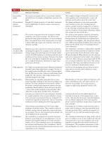
the prominent post-translational modifications, such as those listed in Table 5.5, can
act as “on–off switches” that regulate the function or cellular location of the pro-
tein. Approximately 500 post-translational modifications are listed in the RESID
database accessible through the National Cancer Institute Frederick Advanced Bio-
medical Computing Center at />A common form of post-translational modification not to be found in such a
database or in Table 5.5 is the removal of amino acids from the protein by prote-
olytic cleavage. Many proteins localized in specific subcellular compartments have
120 Chapter 5 Proteins:Their Primary Structure and Biological Functions
N-terminal signal sequences that stipulate their proper destination. Such signal se-
quences typically are clipped off during their journey. Other proteins, such as some
hormones or potentially destructive proteases, are synthesized in an inactive form
and converted into an active form through proteolytic removal of some of their
amino acids.
The general term for proteins containing nonprotein constituents is conjugated
proteins (Table 5.6). Because association of the protein with the conjugated group
does not occur until the protein has been synthesized, these associations are post-
translational as well, although such terminology is usually not applied to these pro-
teins (with the possible exception of glycoproteins). As Table 5.6 indicates, conju-
gated proteins are typically classified according to the chemistry of the nonprotein
part. If the nonprotein part participates in the protein’s function, it is referred to as
a prosthetic group. Conjugation of proteins with these different nonprotein con-
stituents dramatically enhances the repertoire of functionalities available to proteins.
5.8 What Are the Many Biological Functions of Proteins?
Proteins are the agents of biological function. Virtually every cellular activity is dependent
on one or more particular proteins. Thus, a convenient way to classify the enormous
number of proteins is to group them according to the biological roles they serve.
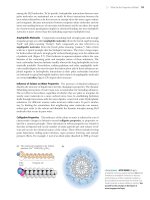
Figure 5.26 summarizes the classification of proteins found in the human proteome
according to their function.
Proteins fill essentially every biological role, with the exception of information
storage. The ability to bind other molecules (ligands) is common to many proteins.
Binding proteins typically interact noncovalently with their specific ligands. Trans-
port proteins are one class of binding proteins. Transport proteins include mem-
Amino Acid Side
Name Nonprotein Part Chain Modified Examples
Phosphorylation ϪPO
3
2Ϫ
S, T, Y Hormone receptors,
regulatory enzymes
Acetylation ϪCH
2
COO
Ϫ
K Histones
Methylation ϪCH
3
K, R Histones
Acylation Palmitic acid C G-protein-coupled
receptors
Prenylation Prenyl group C Ras p21
ADP-ribosylation ADP-ribose H, R G proteins, eukaryotic
elongation factors
Adenylylation AMP Y Glutamine synthetase
TABLE 5.5
Some Prominent Post-Translational Modifications Found in Proteins
Name Nonprotein Part Association Examples
Lipoproteins Lipids Noncovalent Blood lipoprotein complexes (HDL, LDL)
Nucleoproteins RNA, DNA Noncovalent Ribosomes, chromosomes
Glycoproteins Carbohydrate groups Covalent Immunoglobulins, LDL receptor
Metalloproteins and Ca
2ϩ
, K
ϩ
, Fe
2ϩ
, Zn
2ϩ
, Covalent to noncovalent Metabolic enzymes, kinases, phosphatases,
metal-activated proteins Co
2ϩ
, others among others
Hemoproteins Heme group Covalent or noncovalent Hemoglobin, cytochromes
Flavoproteins FMN, FAD Covalent or noncovalent Electron transfer enzymes
TABLE 5.6
Some Common Conjugated Proteins
Proteome is the complete catalog of proteins
encoded by a genome; in cell-specific terms, a
proteome is the complete set of proteins found
in a particular cell type at a particular time.
5.8 What Are the Many Biological Functions of Proteins? 121
brane proteins that transport substances across membranes, as well as soluble pro-
teins that deliver specific nutrients or waste products throughout the body. Scaffold
proteins are a class of binding proteins that uses protein–protein interactions to re-
cruit other proteins into multimeric assemblies whose purpose is to mediate and co-
ordinate the flow of information in cells. Catalytic proteins (enzymes) mediate al-
most every metabolic reaction. Regulatory proteins that bind to specific nucleotide
sequences within DNA control gene expression. Hormones are another kind of reg-
ulatory protein in that they convey information about the environment and deliver
this information to cells when they bind to specific receptors. Switch proteins such
as G-proteins can switch between two conformational states—an “on” state and an
“off” state—and act via this conformational switching, as regulatory proteins. Struc-
tural proteins give form to cells and subcellular structures. The great diversity in
function that characterizes biological systems is based on attributes that proteins
possess.
All Proteins Function through Specific Recognition and Binding of Some Target
Molecule Although the classification of proteins according to function has ad-
vantages, many proteins are not assigned readily to one of the traditional groupings.
Further, classification can be somewhat arbitrary, because many proteins fit more
than one category. However, for all categories, the protein always functions through
Chaperone (159, 0.5%)
Cell adhesion (577, 1.9%)
Miscellaneous (1318, 4.3%)
Viral protein (100, 0.3%)
Transfer/carrier protein (203, 0.7%)
Transcription factor (1850, 6.0%)
Nucleic acid enzyme (2308, 7.5%)
Signaling molecule (376, 1.2%)
Receptor (1543, 5.0%)
Kinase (868, 2.8%)
Select regulatory
molecule (988, 3.2%)
Transferase (610, 2.0%)
Synthase and synthetase
(313, 1.0%)
Oxidoreductase (656, 2.1%)
Lyase (117, 0.4%)
Ligase (56, 0.2%)
Isomerase (163, 0.5%)
Hydrolase (1227, 4.0%)
Cytoskeletal structural protein (876, 2.8%)
Extracellular matrix (437, 1.4%)
Immunoglobulin (264, 0.9%)
Ion channel (406, 1.3%)
Motor (376, 1.2%)
Structural protein of muscle (296, 1.0%)
Protooncogene (902, 2.9%)
Select calcium-binding protein (34, 0.1%)
Intracellular transporter (350, 1.1%)
Transporter (533, 1.7%)
Molecular function unknown (12809, 41.7%)
Nucleic acid
binding
Enzyme
None
Signal transduction
FIGURE 5.26 Proteins of the human genome grouped according to their molecular function.The numbers and
percentages within each functional category are enclosed in parentheses.Note that the function of more than
40% of the proteins encoded by the human genome remains unknown. Considering those of known function,
enzymes (including kinases and nucleic acid enzymes) account for about 20% of the total number of proteins;
nucleic acid–binding proteins of various kinds, about 14%, among which almost half are gene-regulatory proteins
(transcription factors).Transport proteins collectively constitute about 5% of the total; and structural proteins,
another 5%.
(Adapted from Figure 15 in Venter,J. C., et al., 2001. The sequence of the human genome. Science 291:1304–1351.)
122 Chapter 5 Proteins:Their Primary Structure and Biological Functions
specific recognition and binding of some other molecule, although for structural
proteins, it is usually self-recognition and assembly into stable multimeric arrays.
Protein behavior provides the cardinal example of molecular recognition through struc-
tural complementarity, a fundamental principle of biochemistry that was presented in
Chapter 1.
Protein Binding The interaction of a protein with its target usually can be de-
scribed in simple quantitative terms. Let’s explore the simplest situation in which a
protein has a single binding site for the molecule it binds (its ligand; Chapter 1). If
we treat the interaction between the protein (P) and the ligand (L) as a dissociation
reaction:
PL
34
P ϩ L.
The equilibrium constant for the reaction as written,
K
eq
ϭ [P][L]/[PL],
is a dissociation constant, because it describes the dissociation of the ligand from
the protein. Biochemists typically use dissociation constants (K
D
) to describe bind-
ing phenomena. Because brackets ([ ]) denote molar concentrations, dissociation
constants have the units of M.
Typically, the ligand concentration is much greater than the protein concentra-
tion. Under such conditions, a plot of the moles of ligand bound per mole of pro-
tein (defined as [PL]/([P] ϩ [PL])) versus [L] yields a hyperbolic curve known as
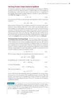
a saturation curve or binding isotherm (Figure 5.27).
If we define the fractional saturation of P with L, [PL]/([P] ϩ [PL]), as , a lit-
tle algebra yields
ϭ [L]/(K
D
ϩ [L]).
Thus, when ϭ 0.5,
[L] ϭ K
D
That is, the concentration of L where half the protein has L bound is equal to the
value of K
D
. The smaller this number is, the better the ligand binds to the protein;
that is, a small K
D
means that the protein is half-saturated with L at a low con-
centration of L. In other words, if K
D
is small, the protein binds the ligand avidly.
Typical K
D
values fall in a range from 10
Ϫ3
M to 10
Ϫ12
M.
The Ligand-Binding Site Ligand binding occurs through noncovalent interac-
tions between the protein and ligand. The lack of covalent interactions means that
binding is readily reversible. Proteins display specificity in ligand binding because
they possess a specific site, the binding site, within their structure that is comple-
Li
g
and concentration([L])
1.0
0
0
[L] = K
D
[PL]
[P]+[PL]
=
0.5
FIGURE 5.27 Saturation curve or binding isotherm.
