Biochemistry, 4th Edition P59 docx
Bạn đang xem bản rút gọn của tài liệu. Xem và tải ngay bản đầy đủ của tài liệu tại đây (658.9 KB, 10 trang )
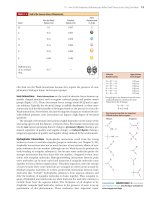
18.3 What Are the Chemical Principles and Features of the First Phase of Glycolysis? 543
Adenylate kinase rapidly interconverts ADP, ATP, and AMP to maintain this equi-
librium. ADP levels in cells are typically 10% of ATP levels, and AMP levels are
often less than 1% of the ATP concentration. Under such conditions, a small net
change in ATP concentration due to ATP hydrolysis results in a much larger rela-
tive increase in the AMP levels because of adenylate kinase activity.
Clearly, the activity of phosphofructokinase depends on both ATP and AMP levels
and is a function of the cellular energy status. Phosphofructokinase activity is in-
creased when the energy status falls and is decreased when the energy status is high.
The rate of glycolysis activity thus decreases when ATP is plentiful and increases when
more ATP is needed.
Glycolysis and the citric acid cycle (to be discussed in Chapter 19) are coupled via
phosphofructokinase, because citrate, an intermediate in the citric acid cycle, is an
allosteric inhibitor of phosphofructokinase. When the citric acid cycle reaches satu-
ration, glycolysis (which “feeds” the citric acid cycle under aerobic conditions) slows
down. The citric acid cycle directs electrons into the electron-transport chain (for the
purpose of ATP synthesis in oxidative phosphorylation) and also provides precursor
molecules for biosynthetic pathways. Inhibition of glycolysis by citrate ensures
that glucose will not be committed to these activities if the citric acid cycle is already
saturated.
Phosphofructokinase is also regulated by -
D-fructose-2,6-bisphosphate, a po-
tent allosteric activator that increases the affinity of phosphofructokinase for the
substrate fructose-6-phosphate (Figure 18.10). Stimulation of phosphofructo-
kinase is also achieved by decreasing the inhibitory effects of ATP (Figure 18.11).
Fructose-2,6-bisphosphate increases the net flow of glucose through glycolysis by
stimulating phosphofructokinase and, as we shall see in Chapter 22, by inhibiting
fructose-1,6-bisphosphatase, the enzyme that catalyzes this reaction in the opposite
direction.
Reaction 4: Cleavage by Fructose Bisphosphate Aldolase Creates
Two 3-Carbon Intermediates
Fructose bisphosphate aldolase cleaves fructose-1,6-bisphosphate between the
C-3 and C-4 carbons to yield two triose phosphates. The products are dihydroxy-
acetone phosphate (DHAP) and glyceraldehyde-3-phosphate. The reaction has an
equilibrium constant of approximately 10
Ϫ4
M, and a corresponding ⌬G°Ј of
ϩ23.9 kJ/mol. These values might imply that the reaction does not proceed ef-
fectively from left to right as written. However, the reaction makes two molecules
(glyceraldehyde-3-P and dihydroxyacetone-P) from one molecule (fructose-1,6-
bisphosphate), and the equilibrium is thus greatly influenced by concentration.
A DEEPER LOOK
Phosphoglucoisomerase—A Moonlighting Protein
When someone has a day job but also works at night (that is, un-
der the moon) at a second job, they are said to be “moonlight-
ing.” Similarly, a number of proteins have been found to have
two or more different functions, and Constance Jeffery at Bran-
deis University has dubbed these “moonlighting proteins.” Phos-
phoglucoisomerase catalyzes the second step of glycolysis but
also moonlights as a nerve growth factor outside animal cells. In
fact, outside the cell, this protein is known as neuroleukin (NL),
autocrine motility factor (AMF), and differentiation and matu-
ration mediator (DMM). Neuroleukin is secreted by (immune
system) T cells and promotes the survival of certain spinal neu-
rons and sensory nerves. AMF is secreted by tumor cells and
stimulates cancer cell migration. DMM causes certain leukemia
cells to differentiate.
How phosphoglucoisomerase is secreted by the cell for its
moonlighting functions is unknown, but there is evidence that the
organism itself may be harmed by this secretion. Diane Mathis and
Christophe Benoist at the University of Strasbourg have shown
that, in mice with disorders similar to rheumatoid arthritis, the im-
mune system recognizes extracellular phosphoglucoisomerase as
an antigen—that is, a protein that is “nonself.” That a protein can
be vital to metabolism inside the cell and also function as a growth
factor and occasionally act as an antigen outside the cell is indeed
remarkable.
OH H
HHO
CH
2
OH
2–
O
3
POCH
2
O
OPO
3
2
–
H
Fructose-2,6-bisphosphate
100
20
Relative velocity
12
[Fructose-6-phosphate] (
M)
40
60
80
0
0
345
0.1
M
1.0
M F-2,6-BP
FIGURE 18.10 Fructose-2,6-bisphosphate activates phos-
phofructokinase, increasing the affinity of the enzyme for
fructose-6-phosphate and restoring the hyperbolic de-
pendence of enzyme activity on substrate concentra-
tion.
544 Chapter 18 Glycolysis
The value of ⌬G in erythrocytes is actually Ϫ0.23 kJ/mol (see Table 18.1). At phys-
iological concentrations, the reaction is essentially at equilibrium.
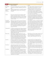
Two classes of aldolase enzymes are found in nature. Animal tissues produce a
Class I aldolase, characterized by the formation of a covalent Schiff base intermediate
between an active-site lysine and the carbonyl group of the substrate. Class I aldolases
do not require a divalent metal ion. Class II aldolases are produced mainly in bacte-
ria and fungi and do not form a covalent E-S intermediate, but they contain an active-
site metal (normally zinc, Zn
2ϩ
). Cyanobacteria and some other simple organisms
possess both classes of aldolase.
The aldolase reaction is merely the reverse of the aldol condensation well known
to organic chemists. The latter reaction involves an attack by a nucleophilic enolate
anion of an aldehyde or ketone on the carbonyl carbon of an aldehyde. The oppo-
site reaction, aldol cleavage, begins with removal of a proton from the -hydroxyl
group, which is followed by the elimination of the enolate anion. A mechanism for
the aldol cleavage reaction of fructose-1,6-bisphosphate in the Class I–type aldolases
is shown in Figure 18.12a. In Class II aldolases, an active-site metal such as Zn
2ϩ
be-
haves as an electrophile, polarizing the carbonyl group of the substrate and stabiliz-
ing the enolate intermediate (Figure 18.12b).
Reaction 5: Triose Phosphate Isomerase Completes
the First Phase of Glycolysis
Of the two products of the aldolase reaction, only glyceraldehyde-3-phosphate
goes directly into the second phase of glycolysis. The other triose phosphate,
dihydroxyacetone phosphate, must be converted to glyceraldehyde-3-phosphate by
the enzyme triose phosphate isomerase. This reaction thus permits both products of
the aldolase reaction to continue in the glycolytic pathway and in essence makes the
C-1, C-2, and C-3 carbons of the starting glucose molecule equivalent to the C-6,
C-5, and C-4 carbons, respectively. The reaction mechanism involves an enediol in-
termediate that can donate either of its hydroxyl protons to a basic residue on the
enzyme and thereby become either dihydroxyacetone phosphate or glyceraldehyde-
3-phosphate (Figure 18.13). Triose phosphate isomerase is one of the enzymes that
have evolved to a state of “catalytic perfection,” with a turnover number near the dif-
fusion limit (see Table 13.5).
The triose phosphate isomerase reaction completes the first phase of glycolysis,
each glucose that passes through being converted to two molecules of glyceralde-
hyde-3-phosphate. Although the last two steps of the pathway are energetically un-
favorable, the overall five-step reaction sequence has a net ⌬G°Ј of ϩ2.2 kJ/mol
(K
eq
≈ 0.43). It is the free energy of hydrolysis from the two priming molecules of
ATP that brings the overall equilibrium constant close to 1 under standard-state
12
[ATP] (
M)
0.1
M
3450
1.0
M F-2,6-BP
0
Relative velocity
FIGURE 18.11 Fructose-2,6-bisphosphate decreases the
inhibition of phosphofructokinase due to ATP.
CHO H
CHOH
CHOH
Aldol
cleavage
C O
PO
3
2
–
CH
2
O
C O
CH
2
OH
+
CHOH
C
OH
PO
3
2
–
CH
2
O
PO
3
2
–
CH
2
O
PO
3
2
–
CH
2
O
D-Fructose-1,6-bisphosphate
(FBP)
Fructose
bisphosphate
aldolase
Dihydroxyacetone
phosphate (DHAP)
D-Glyceraldehyde
3-phosphate (G-3-P)
ΔG°' = 23.9 kJ/mol
O
H
R
C
O
–
C
H
O
–
O
RЈ
H
H
R
H
RЈ = H (aldehyde)
RЈ = alkyl, etc. (ketone)
Aldol condensation
RЈ
H
HO
C
HCOH
CH
2
OPO
3
2
–
G-3-P
DHAP
CH
2
OPO
3
2
–
CO
CH
2
OH
ΔG°
Ј = +7.56 kJ/mol
Triose
phosphate
isomerase
18.3 What Are the Chemical Principles and Features of the First Phase of Glycolysis? 545
C
CH
2
OPO
3
2–
O
H
2
N
C H
H
C
C
O
O
–
O
H
H
C OH
C
N
HO HO C H
H
C O H
H
C OH
C
HOCH
H
N
H
C OH
C
O
C
O
G-3-P
C O
DHAP
Lys
229
–
H
+
+
CH
2
OPO
3
2–
H
2
O
Schiff base
formation
Schiff base
hydrolysis
CH
2
OPO
3
2–
CH
2
OPO
3
2–
CH
2
OPO
3
2–
CH
2
OPO
3
2–
CH
2
OPO
3
2–
CH
2
OH
H
2
O
Asp
33
C
O
O
–
O
Asp
33
Asp
33
Lys
229
H
C
HO
H
2
N
Lys
229
Asp
33
–
O
Enzyme main
chain
(a)
CH
2
OPO
3
2
–
C
O
C
–
HHO
Zn
2+
CH
2
OPO
3
2
–
C
O
–
C
HHO
Zn
2+
G-3-P
FBP
E
E
(b)
ACTIVE FIGURE 18.12 (a) A mecha-
nism for the fructose-1,6-bisphosphate aldolase reac-
tion.The Schiff base formed between the substrate
carbonyl and an active-site lysine acts as an electron
sink, increasing the acidity of the -hydroxyl group and
facilitating cleavage as shown. The catalytic residues in
the rabbit muscle enzyme are Lys
229
and Asp
33
. (b) In
Class II aldolases, an active-site Zn
2ϩ
stabilizes the eno-
late intermediate, leading to polarization of the sub-
strate carbonyl group. Test yourself on the concepts
in this figure at www.cengage.com/login.
H
CO
CH
2
OPO
3
2
–
O
O
HB
+
Glu
165
O
O
Glu
H
H
C
OH
CH
2
OPO
3
2
–
C
OH
C
HOH
B
E
C
H
C OHH
CH
2
OPO
3
2
–
C
O
O
O
_
Glu
C
–
E
E
E
E
Glyceraldehyde-3-P
Enediol
intermediate
DHAP
ACTIVE FIGURE 18.13 A reaction mecha-
nism for triose phosphate isomerase.In the enzyme from
yeast, the catalytic residue is Glu
165
. Test yourself on the
concepts in this figure at www.cengage.com/login.
Triose phosphate isomerase with substrate analog
2-phosphoglycerate shown in cyan (pdb id ϭ 1YPI).
Glu
165
546 Chapter 18 Glycolysis
conditions. The net ⌬G under cellular conditions is quite negative (Ϫ53.4 kJ/mol
in erythrocytes).
18.4 What Are the Chemical Principles and Features
of the Second Phase of Glycolysis?
The second half of the glycolytic pathway involves the reactions that convert the
metabolic energy in the glucose molecule into ATP. Altogether, four new ATP mol-
ecules are produced. If two are considered to offset the two ATPs consumed in
phase 1, a net yield of two ATPs per glucose is realized. Phase 2 starts with the oxi-
dation of glyceraldehyde-3-phosphate, a reaction with a large enough energy “kick”
to produce a high-energy phosphate, namely, 1,3-bisphosphoglycerate (see Figure
18.1). Phosphoryl transfer from 1,3-BPG to ADP to make ATP is highly favorable.
The product, 3-phosphoglycerate, is converted via several steps to phosphoenol-
pyruvate (PEP), another high-energy phosphate. PEP readily transfers its phos-
phoryl group to ADP in the pyruvate kinase reaction to make another ATP.
Reaction 6: Glyceraldehyde-3-Phosphate Dehydrogenase Creates
a High-Energy Intermediate
In the first glycolytic reaction to involve oxidation–reduction, glyceraldehyde-3-
phosphate is oxidized to 1,3-bisphosphoglycerate by glyceraldehyde-3-phosphate de-
hydrogenase. Although the oxidation of an aldehyde to a carboxylic acid is a highly
exergonic reaction, the overall reaction involves both formation of a carboxylic–
phosphoric anhydride and the reduction of NAD
ϩ
to NADH and is therefore slightly
endergonic at standard state, with a ⌬G°Ј of ϩ6.30 kJ/mol. The free energy that
might otherwise be released as heat in this reaction is directed into the formation of
a high-energy phosphate compound, 1,3-bisphosphoglycerate, and the reduction of
NAD
ϩ
. The reaction mechanism involves nucleophilic attack by a cysteine OSH
group on the carbonyl carbon of glyceraldehyde-3-phosphate to form a hemithioac-
etal (Figure 18.14). The hemithioacetal intermediate decomposes by hydride (HϺ
Ϫ
)
transfer to NAD
ϩ
to form a high-energy thioester. Nucleophilic attack by phosphate
displaces the product, 1,3-bisphosphoglycerate, from the enzyme. The enzyme can be
inactivated by reaction with iodoacetate, which reacts with and blocks the essential cys-
teine sulfhydryl.
O
HO
C
HCOH
CH
2
OPO
3
2
–
+ + HPO
4
2
–
HCOH
CH
2
OPO
3
2
–
+
COPO
3
2
–
H
+
+
NADH
NAD
+
Glyceraldehyde-
3-phosphate
(G-3-P)
1,3-Bisphosphoglycerate
(1,3-BPG)
ΔGЊ' = +6.3 kJ/mol
The glyceraldehyde-3-phosphate dehydrogenase reaction is the site of action of
arsenate (AsO
4
3Ϫ
), an anion analogous to phosphate. Arsenate is an effective sub-
strate in this reaction, forming 1-arseno-3-phosphoglycerate, but acyl arsenates are
quite unstable and are rapidly hydrolyzed. 1-Arseno-3-phosphoglycerate breaks
down to yield 3-phosphoglycerate, the product of the seventh reaction of glycolysis.
The result is that glycolysis continues in the presence of arsenate, but the molecule
of ATP formed in reaction 7 (phosphoglycerate kinase) is not made because this
step has been bypassed. The lability of 1-arseno-3-phosphoglycerate effectively un-
couples the oxidation and phosphorylation events, which are normally tightly cou-
pled in the glyceraldehyde-3-phosphate dehydrogenase reaction.
CH
2
OPO
3
2
–
O
C
OAsO
–
O
O
–
CH OH
1-Arseno-3-phosphoglycerate
18.4 What Are the Chemical Principles and Features of the Second Phase of Glycolysis? 547
Reaction 7: Phosphoglycerate Kinase Is the Break-Even Reaction
The glycolytic pathway breaks even in terms of ATPs consumed and produced
with this reaction. The enzyme phosphoglycerate kinase transfers a phosphoryl
group from 1,3-bisphosphoglycerate to ADP to form an ATP. Because each glu-
cose molecule sends two molecules of glyceraldehyde-3-phosphate into the sec-
ond phase of glycolysis and because two ATPs were consumed per glucose in the
first-phase reactions, the phosphoglycerate kinase reaction “pays off” the ATP
debt created by the priming reactions. As might be expected for a phosphoryl
transfer enzyme, Mg
2ϩ
ion is required for activity and the true nucleotide sub-
strate for the reaction is MgADP
Ϫ
. It is appropriate to view the sixth and seventh
reactions of glycolysis as a coupled pair, with 1,3-bisphosphoglycerate as an in-
termediate. The phosphoglycerate kinase reaction is sufficiently exergonic at
standard state to pull the G-3-P dehydrogenase reaction along. (In fact, the
H
C
HCOH
O
SHCys
149
C
HCOH
CH
2
OPO
3
2
–
S
O
H
O
H
2
N
N
R
+
N
R
HH
O
NH
2
+
C
HCOH
CH
2
OPO
3
2
–
S
O
C
HCOH
CH
2
OPO
3
2
–
S
O
–
OPO
3
2
–
H
+
C
HCOH
CH
2
OPO
3
2
–
CH
2
OPO
3
2
–
OPO
3
2
–
O
E
E
E
E
1,3-Bisphosphoglycerate
OH
O
–
H
+
P
O
–
O
H
ACTIVE FIGURE 18.14 A mechanism for the glyceraldehyde-
3-phosphate dehydrogenase reaction. Reaction of an enzyme sulfhydryl with
the carbonyl carbon of glyceraldehyde-3-P forms a thiohemiacetal, which
loses a hydride to NAD
ϩ
to become a thioester. Phosphorolysis of this
thioester releases 1,3-bisphosphoglycerate. In the enzyme from rabbit
muscle, the catalytic residue is Cys
149
. Test yourself on the concepts in this
figure at www.cengage.com/login.
C
O
OPO
3
2
–
HCOH
CH
2
OPO
3
2
–
+
Mg
2+
COO
HCOH
+
CH
2
OPO
3
2
–
–
ATP
ADP
1,3-Bisphosphoglycerate
(1,3-BPG)
3-Phosphoglycerate
(3-PG)
ΔGЊ' = –18.9 kJ/mol
Phosphoglycerate
kinase
(a)
(b)
The open (a) and closed (b) forms of phosphoglycerate
kinase. ATP (cyan), 3-phosphoglycerate (purple), and
Mg
2H
(gold) (a: pdb id ϭ 3PGK; b: pdb id ϭ 1VPE).
548 Chapter 18 Glycolysis
aldolase and triose phosphate isomerase are also pulled forward by phospho-
glycerate kinase.) The net result of these coupled reactions is
Glyceraldehyde-3-phosphate ϩ ADP ϩ P
i
ϩ NAD
ϩ
⎯→
3-phosphoglycerate ϩ ATP ϩ NADH ϩ H
ϩ
⌬G°ЈϭϪ12.6 kJ/mol (18.8)
Another reflection of the coupling between these reactions lies in their values
of ⌬G under cellular conditions (Table 18.1). Despite its strongly negative ⌬G°Ј,
the phosphoglycerate kinase reaction operates at equilibrium in the erythrocyte
(⌬G ϭ 0.1 kJ/mol). In essence, the free energy available in the phosphoglycerate
kinase reaction is used to bring the three previous reactions closer to equilib-
rium. Viewed in this context, it is clear that ADP has been phosphorylated to
form ATP at the expense of a substrate, namely, glyceraldehyde-3-phosphate.
This is an example of substrate-level phosphorylation, a concept that will be en-
countered again. (The other kind of phosphorylation, oxidative phosphorylation,
is driven energetically by the transport of electrons from appropriate coen-
zymes and substrates to oxygen. Oxidative phosphorylation will be covered in
detail in Chapter 20.) Even though the coupled reactions exhibit a very favorable
⌬G°Ј, there are conditions (that is, high ATP and 3-phosphog lycerate levels)
under which the phosphoglycerate kinase reaction can be reversed so that 3-
phosphoglycerate is phosphorylated from ATP.
An important regulatory molecule, 2,3-bisphosphoglycerate, is synthesized and
metabolized by a pair of reactions that make a detour around the phosphoglycerate
kinase reaction. 2,3-BPG, which stabilizes the deoxy form of hemoglobin and is pri-
marily responsible for the cooperative nature of oxygen binding by hemoglobin (see
Chapter 15), is formed from 1,3-bisphosphoglycerate by bisphosphoglycerate mutase
(Figure 18.15). Interestingly, 3-phosphoglycerate is required for this reaction, which
involves phosphoryl transfer from the C-1 position of 1,3-bisphosphoglycerate to the
C-2 position of 3-phosphoglycerate (Fig
ure 18.16). Hydrolysis of 2,3-BPG is carried
out by 2,3-bisphosphoglycerate phosphatase. Although other cells contain only a trace of
2,3-BPG, erythrocytes typically contain 4 to 5 mM 2,3-BPG.
Reaction 8: Phosphoglycerate Mutase Catalyzes a Phosphoryl Transfer
The remaining steps in the glycolytic pathway prepare for synthesis of the second ATP
equivalent. This begins with the phosphoglycerate mutase reaction, in which the
phosphoryl group of 3-phosphoglycerate is moved from C-3 to C-2. (The term mutase
is applied to enzymes that catalyze migration of a functional group within a substrate
molecule.) The free energy change for this reaction is very small under cellular con-
ditions (⌬G ϭ 0.83 kJ/mol in erythrocytes). Phosphoglycerate mutase enzymes iso-
lated from different sources exhibit different reaction mechanisms. As shown in Fig-
ure 18.17, the enzymes isolated from yeast and from rabbit muscle form phosphoenzyme
intermediates, use 2,3-bisphosphoglycerate as a cofactor, and undergo intermolecular
phosphoryl group transfers (in which the phosphate of the product 2-phosphoglyc-
erate is not that from the 3-phosphoglycerate substrate). The prevalent form of phos-
C
H
OPO
3
2
–
O
C
H
C
H
OH
OPO
3
2
–
H
+
C
H
O
C
H
C
H
H
2
O
P
i
+ H
+
C
H
O
C
H
C
H
O
–
OH
OPO
3
2
–
OPO
3
2
–
O
–
OPO
3
2
–
1,3-Bisphosphoglycerate
(1,3-BPG)
Bisphosphoglycerate
mutase
2,3-Bisphosphoglycerate
(2,3-BPG)
3-Phosphoglycerate
2,3-Bisphosphoglycerate
phosphatase
FIGURE 18.15 Formation and decomposition of 2,3-bisphosphoglycerate.
3-Phosphoglycerate
(3-PG)
2-Phosphoglycerate
(2-PG)
Phosphoglycerate mutase
ΔGЊ' = +4.4 kJ/mol
COO
–
HCOH
CH
2
OPO
3
2
–
HCOPO
3
2
–
CH
2
OH
COO
–
18.4 What Are the Chemical Principles and Features of the Second Phase of Glycolysis? 549
phoglycerate mutase is a phosphoenzyme, with a phosphoryl group covalently bound to
a histidine residue at the active site. This phosphoryl group is transferred to the C-2
position of the substrate to form a transient, enzyme-bound 2,3-bisphosphoglycerate,
which then decomposes by a second phosphoryl transfer from the C-3 position of the
intermediate to the histidine residue on the enzyme. About once in every 100 enzyme
turnovers, the intermediate, 2,3-bisphosphoglycerate, dissociates from the active site,
leaving an inactive, unphosphorylated enzyme. The unphosphorylated enzyme can
be reactivated by binding 2,3-BPG. For this reason, maximal activity of phosphoglyc-
erate mutase requires the presence of small amounts of 2,3-BPG.
Reaction 9: Dehydration by Enolase Creates PEP
Recall that prior to synthesizing ATP in the phosphoglycerate kinase reaction, it was
necessary to first make a substrate having a high-energy phosphate. Reaction 9 of
glycolysis similarly makes a high-energy phosphate in preparation for ATP synthesis.
Enolase catalyzes the formation of phosphoenolpyruvate from 2-phosphoglycerate. The
reaction involves the removal of a water molecule to form the enol structure of PEP.
The ⌬G°Ј for this reaction is relatively small at 1.8 kJ/mol (K
eq
ϭ 0.5); and, under
cellular conditions, ⌬G is very close to zero. In light of this condition, it may be dif-
ficult at first to understand how the enolase reaction transforms a substrate with a
relatively low free energy of hydrolysis into a product (PEP) with a very high free
+
1
2
3
1
2
3
1
2
3
+
3
1
2
P
P
P
P
P
P
FIGURE 18.16 The mutase that forms 2,3-BPG from 1,3-BPG requires 3-phosphoglycerate.The reaction is actu-
ally an intermolecular phosphoryl transfer from C-1 of 1,3-BPG to C-2 of 3-PG.
P
O
N
–
O
+
+
NH
NNH
B
Phosphohistidine
H
2
C
HC
H
P
P
O
O
O
O
–
O
–
O
–
O
–
O
COO
–
H
2
C
HC
OH
O
COO
–
H
2
C
HC
P
O
O
O
O
–
O
–
O
–
P
O
O
–
O
–
COO
–
+
BH
P
O
N
–
O
NH
O
–
FIGURE 18.17 A mechanism for the phosphoglycerate mutase
reaction in rabbit muscle and in yeast. Zelda Rose of the Institute
for Cancer Research in Philadelphia showed that the enzyme
requires a small amount of 2,3-BPG to phosphorylate the histidine
residue before the mechanism can proceed.Prior to her work,
the role of the phosphohistidine in this mechanism was not
understood.
His
10
His
183
Gly
11
Gly
184
Glu
88
Asn
16
Arg
61
Arg
9
SO
4
–
The catalytic histidine (His
183
) at the active site of
Escherichia coli phosphoglycerate mutase (pdb id ϭ
1E58). Note that His
10
is phosphorylated.
HC O
COO
–
CH
2
OH
C O
COO
–
CH
2
+
Mg
2+
PO
3
2
–
H
2
O
PO
3
2
–
2-Phosphoglycerate
(2-PG)
Phosphoenolpyruvate
(PEP)
ΔGЊ' = +1.8 kJ/mol
550 Chapter 18 Glycolysis
energy of hydrolysis. This puzzle is clarified by realizing that 2-phosphoglycerate and
PEP contain about the same amount of potential metabolic energy, with respect to
decomposition to P
i
, CO
2
, and H
2
O. What the enolase reaction does is rearrange the
substrate into a form from which more of this potential energy can be released upon
hydrolysis. The enzyme is strongly inhibited by fluoride ion in the presence of phos-
phate. Thomas Nowak has shown that fluoride, phosphate, and a divalent cation
form a transition-state–like complex in the enzyme active site, with fluoride appar-
ently mimicking the hydroxide ion nucleophile in the enolase reaction.
Yeast enolase is a dimer of identical subunits. However, if the enzyme is crystal-
lized in the presence of a mixture of the substrate (2-phosphoglycerate) and the
product (phosphoenolpyruvate), the crystallized dimer is asymmetric! One subunit
active site contains 2-phosphoglycerate, and the other contains PEP (Figure 18.18),
thus providing a “before-and-after” picture of this glycolytic enzyme.
Reaction 10: Pyruvate Kinase Yields More ATP
The second ATP-synthesizing reaction of glycolysis is catalyzed by pyruvate kinase,
which brings the pathway at last to its pyruvate branch point. Pyruvate kinase me-
diates the transfer of a phosphoryl group from phosphoenolpyruvate to ADP to
make ATP and pyruvate. The reaction requires Mg
2ϩ
ion and is stimulated by K
ϩ
and certain other monovalent cations.
The corresponding K
eq
at 25°C is 3.63 ϫ 10
5
, and it is clear that the pyruvate ki-
nase reaction equilibrium lies very far to the right. Concentration effects reduce the
magnitude of the free energy change somewhat in the cellular environment, but
the ⌬G in erythrocytes is still quite favorable at Ϫ23.0 kJ/mol. The high free energy
(a) (b)
His
159
His
159
Mg
Li
Phosphoglycerate
Mg
PEP
H
2
O
FIGURE 18.18 The yeast enolase dimer is asymmetric.The active site of one subunit (a) contains
2-phosphoglycerate, the enolase substrate. Also shown are a Mg
2ϩ
ion (blue), a Li
ϩ
ion (purple), and His
159
,
which participates in catalysis. The other subunit (b) binds phosphoenolpyruvate, the product of the enolase
reaction. An active site water molecule (yellow), Mg
2ϩ
(blue), and His
159
are also shown (pdb id ϭ 2ONE).
C O
COO
–
CH
2
PO
3
2
–
++ADP
3–
Mg
2+
K
+
C O
COO
–
CH
3
+
H
+
ATP
4–
PEP Pyruvate
ΔGЊ' = –31.7 kJ/mol
18.4 What Are the Chemical Principles and Features of the Second Phase of Glycolysis? 551
change for the conversion of PEP to pyruvate is due largely to the highly favorable
and spontaneous conversion of the enol tautomer of pyruvate to the more stable
keto form (Figure 18.19) following the phosphoryl group transfer step.
The large negative ⌬G of this reaction makes pyruvate kinase a suitable target site
for regulation of glycolysis. For each glucose molecule in the glycolysis pathway, two
ATPs are made at the pyruvate kinase stage (because two triose molecules were pro-
duced per glucose in the aldolase reaction). Because the pathway broke even in terms
of ATP at the phosphoglycerate kinase reaction (two ATPs consumed and two ATPs
produced), the two ATPs produced by pyruvate kinase represent the “payoff” of
glycolysis—a net yield of two ATP molecules.
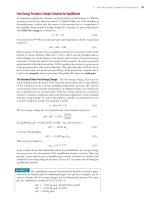
Pyruvate kinase possesses allosteric sites for numerous effectors. It is activated by
AMP and fructose-1,6-bisphosphate and inhibited by ATP, acetyl-CoA, and alanine.
(Note that alanine is the ␣-amino acid counterpart of the ␣-keto acid, pyruvate.)
Furthermore, liver pyruvate kinase is regulated by covalent modification. Hormones
such as glucagon activate a cAMP-dependent protein kinase, which transfers a phos-
phoryl group from ATP to the enzyme. The phosphorylated form of pyruvate kinase
is more strongly inhibited by ATP and alanine and has a higher K
m
for PEP, so in
the presence of physiological levels of PEP, the enzyme is inactive. Then PEP is used
as a substrate for glucose synthesis in the gluconeogenesis pathway (to be described in
Chapter 22), instead of going on through glycolysis and the citric acid cycle (or fer-
mentation routes). A suggested active-site geometry for pyruvate kinase, based on
NMR and EPR studies by Albert Mildvan and colleagues, is presented in Figure
18.20. The carbonyl oxygen of pyruvate and the ␥-phosphorus of ATP lie within
0.3 nm of each other at the active site, consistent with direct transfer of the phos-
phoryl group without formation of a phosphoenzyme intermediate.
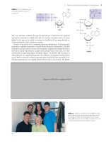
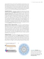
The structure of the pyruvate kinase tetramer is sensitive to bound ligands.The inactive E.coli enzyme in the
absence of ligands (left, pdb id ϭ 1E0U).The active form of the yeast dimer, with fructose-1,6-bisphosphate (an
allosteric regulator, blue), substrate analog (red), and K
ϩ
(gold) (pdb id ϭ 1A3W).
C
C
–
OOC
–
OOC
–
OOC
O PO
3
2
–
HH
PEP
C
C
O H
HH
H
HH
H
+
C
C
O
ATP
ADP
Enol
tautomer
Keto
tautomer
Pyruvate
FIGURE 18.19 The conversion of phosphoenolpyruvate
(PEP) to pyruvate may be viewed as involving two steps:
phosphoryl transfer followed by an enol–keto tautomer-
ization.The tautomerization is spontaneous (⌬G°Ј Ϸ
Ϫ35–40 kJ/mol) and accounts for much of the free en-
ergy change for PEP hydrolysis.
552 Chapter 18 Glycolysis
18.5 What Are the Metabolic Fates of NADH and Pyruvate
Produced in Glycolysis?
In addition to ATP, the products of glycolysis are NADH and pyruvate. Their pro-
cessing depends upon other cellular pathways. NADH must be recycled to NAD
ϩ
,
lest NAD
ϩ
become limiting in glycolysis. NADH can be recycled by both aerobic and
anaerobic paths, either of which results in further metabolism of pyruvate. What a
given cell does with the pyruvate produced in glycolysis depends in part on the avail-
ability of oxygen. Under aerobic conditions, pyruvate can be sent into the citric acid
cycle (also known as the TCA cycle; see Chapter 19), where it is oxidized to CO
2
with the production of additional NADH (and FADH
2
). Under aerobic conditions,
the NADH produced in glycolysis and the citric acid cycle is reoxidized to NAD
ϩ
in
the mitochondrial electron-transport chain (see Chapter 20).
Anaerobic Metabolism of Pyruvate Leads to Lactate or Ethanol
Under anaerobic conditions, the pyruvate produced in glycolysis is processed differ-
ently. In yeast, it is reduced to ethanol; in other microorganisms and in animals, it is
reduced to lactate. These processes are examples of fermentation—the production of
ATP energy by reaction pathways in which organic molecules function as donors and
acceptors of electrons. In either case, reduction of pyruvate provides a means of re-
oxidizing the NADH produced in the glyceraldehyde-3-phosphate dehydrogenase
reaction of glycolysis (Figure 18.21). In yeast, alcoholic fermentation is a two-step
M+
O
O
O
O
O
P
P
O
M+
O
O
O
B
H
H
H
H
C
C
C
C
C
C
Mg
2+
H
B
–
O
P
P
O
P
O
O
O
O
O
O
Mg
2+
O
O
O
O
O
O
H
Mg
2+
H
H
O
P
O
O
Mg
2+
O
Adenine
Ribose
Adenine
Ribose
H
2
O
FIGURE 18.20 A mechanism for the pyruvate kinase
reaction, based on NMR and EPR studies by Albert
Mildvan and colleagues. Phosphoryl transfer from phos-
phoenolpyruvate (PEP) to ADP occurs in four steps:
(1) A water on the Mg
2ϩ
ion coordinated to ADP is
replaced by the phosphoryl group of PEP, (2) Mg
2ϩ
dis-
sociates from the ␣-P of ADP, (3) the phosphoryl group
is transferred, and (4) the enolate of pyruvate is proto-
nated. (Adapted from Mildvan, A., 1979.The role of metals in
enzyme-catalyzed substitutions at each of the phosphorus
atoms of ATP. Advances in Enzymology 49:103–126.)
