Biochemistry, 4th Edition P86 ppsx
Bạn đang xem bản rút gọn của tài liệu. Xem và tải ngay bản đầy đủ của tài liệu tại đây (622.4 KB, 10 trang )
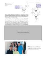
© Adam Woolfitt/CORBIS
26
Synthesis and Degradation
of Nucleotides
Nucleotides are ubiquitous constituents of life, actively participating in the majority
of biochemical reactions. Recall that ATP is the “energy currency” of the cell, that
uracil nucleotide derivatives of carbohydrates are common intermediates in cellu-
lar transformations of carbohydrates (see Chapter 22), and that biosynthesis of
phospholipids proceeds via cytosine nucleotide derivatives (see Chapter 24). In
Chapter 30, we will see that GTP serves as the immediate energy source driving the
endergonic reactions of protein synthesis. Many of the coenzymes (such as coen-
zyme A, NAD, NADP, and FAD) are derivatives of nucleotides. Nucleotides also act
in metabolic regulation, as in the response of key enzymes of intermediary metabo-
lism to the relative concentrations of AMP, ADP, and ATP (PFK is a prime example
here; see also Chapter 18). Furthermore, cyclic derivatives of purine nucleotides
such as cAMP and cGMP have no other role in metabolism than regulation. Last but
not least, nucleotides are the monomeric units of nucleic acids. Deoxynucleoside
triphosphates (dNTPs) and nucleoside triphosphates (NTPs) serve as the immedi-
ate substrates for the biosynthesis of DNA and RNA, respectively (see Part 4).
26.1 Can Cells Synthesize Nucleotides?
Nearly all organisms can make the purine and pyrimidine nucleotides via so-called de
novo biosynthetic pathways. (De novo means “anew”; a less literal but more apt transla-
tion might be “from scratch” because de novo pathways are metabolic sequences that
form complex end products from rather simple precursors.) Many organisms also have
salvage pathways to recover purine and pyrimidine compounds obtained in the diet or
released during nucleic acid turnover and degradation. Whereas the ribose of nu-
cleotides can be catabolized to generate energy, the nitrogenous bases do not serve as
energy sources; their catabolism does not lead to products used by pathways of energy
conservation. Compared to slowly dividing cells, rapidly proliferating cells synthesize
larger amounts of DNA and RNA per unit time. To meet the increased demand for nu-
cleic acid synthesis, substantially greater quantities of nucleotides must be produced.
The pathways of nucleotide biosynthesis thus become attractive targets for the clinical
control of rapidly dividing cells such as cancers or infectious bacteria. Many antibiotics
and anticancer drugs are inhibitors of purine or pyrimidine nucleotide biosynthesis.
26.2 How Do Cells Synthesize Purines?
Substantial insight into the de novo pathway for purine biosynthesis was provided in
1948 by John Buchanan, who cleverly exploited the fact that birds excrete excess ni-
trogen principally in the form of uric acid, a water-insoluble purine analog. Buchanan
fed isotopically labeled compounds to pigeons and then examined the distribution of
the labeled atoms in uric acid (Figure 26.1). By tracing the metabolic source of the var-
ious atoms in this end product, he showed that the nine atoms of the purine ring
Pigeon drinking at Gaia Fountain, Siena, Italy.The basic
features of purine biosynthesis were elucidated ini-
tially from metabolic studies of nitrogen metabolism
in pigeons. Pigeons excrete excess N as uric acid, a
purine analog.
Guano, a substance found on some coasts
frequented by sea birds, is composed chiefly of
the birds’ partially decomposed excrement. . . .
The name for the purine guanine derives
from the abundance of this base in guano.
J. C. Nesbit
On Agricultural Chemistry and the Nature
and Properties of Peruvian Guano (1850)
KEY QUESTIONS
26.1 Can Cells Synthesize Nucleotides?
26.2 How Do Cells Synthesize Purines?
26.3 Can Cells Salvage Purines?
26.4 How Are Purines Degraded?
26.5 How Do Cells Synthesize Pyrimidines?
26.6 How Are Pyrimidines Degraded?
26.7 How Do Cells Form the Deoxyribo-
nucleotides That Are Necessary for DNA
Synthesis?
26.8 How Are Thymine Nucleotides Synthesized?
ESSENTIAL QUESTION
Virtually all cells are capable of synthesizing purine and pyrimidine nucleotides.
These compounds then serve as essential intermediates in metabolism and as the
building blocks for DNA and RNA synthesis.
How do cells synthesize purines and pyrimidines?
Create your own study path for
this chapter with tutorials, simulations, animations,
and Active Figures at www.cengage.com/login.
814 Chapter 26 Synthesis and Degradation of Nucleotides
system (Figure 26.2) are contributed by aspartic acid (N-1), glutamine (N-3 and N-9),
glycine (C-4, C-5, and N-7), CO
2
(C-6), and THF one-carbon derivatives (C-2 and
C-8). THF is tetrahydrofolate, a coenzyme serving as a one-carbon transfer agent, not
only in purine ring synthesis but also in amino acid metabolism (see Figures 25.27
and 25.32) and in synthesis of the pyrimidine thymine (see Figure 26.26). The for-
mation and function of THF is summarized in A Deeper Look on pages 816–817.
IMP Is the Immediate Precursor to GMP and AMP
The de novo synthesis of purines occurs in an interesting manner: The atoms form-
ing the purine ring are successively added to ribose-5-phosphate; thus, purines are di-
rectly synthesized as nucleotide derivatives by assembling the atoms that comprise
the purine ring system directly on the ribose. In step 1, ribose-5-phosphate is acti-
vated via the direct transfer of a pyrophosphoryl group from ATP to C-1 of the
ribose, yielding 5-phosphoribosyl-␣-pyrophosphate (PRPP) (Figure 26.3). The enzyme
is ribose-5-phosphate pyrophosphokinase. PRPP is the limiting substance in purine
biosynthesis. The two major purine nucleoside diphosphates, ADP and GDP, are
negative effectors of ribose-5-phosphate pyrophosphokinase. However, because
PRPP serves additional metabolic needs, the next reaction is actually the committed
step in the pathway.
Step 2 (Figure 26.3) is catalyzed by glutamine phosphoribosyl pyrophosphate
amidotransferase. The anomeric carbon atom of the substrate PRPP is in the
␣-configuration; the product is a -glycoside (recall that all the biologically impor-
tant nucleotides are -glycosides). The N atom of this N-glycoside becomes N-9 of
the nine-membered purine ring; it is the first atom added in the construction of this
ring. Glutamine phosphoribosyl pyrophosphate amidotransferase is subject to feed-
back inhibition by GMP, GDP, and GTP as well as AMP, ADP, and ATP. The G series
of nucleotides interacts at a guanine-specific allosteric site on the enzyme, whereas
the adenine nucleotides act at an A-specific site. The pattern of inhibition by these
nucleotides is such that residual enzyme activity is expressed until sufficient
amounts of both adenine and guanine nucleotides are synthesized. Glutamine
phosphoribosyl pyrophosphate amidotransferase is also sensitive to inhibition by
the glutamine analog azaserine (Figure 26.4). Azaserine has been used as an anti-
tumor agent because it irreversibly inactivates glutamine-dependent enzymes by re-
acting with nucleophilic groups at the glutamine-binding site. Two such enzymes
are found at steps 2 and 5 of the purine biosynthetic pathway.
Step 3 is carried out by
glycinamide ribonucleotide synthetase (GAR synthetase) via
its ATP-dependent condensation of the glycine carboxyl group with the amine of
5-phosphoribosyl--amine (see Figure 26.3). The reaction proceeds in two stages. First,
the glycine carboxyl group is activated via ATP-dependent phosphorylation. Next, an
amide bond is formed between the activated carboxyl group of glycine and the
-amine. Glycine contributes C-4, C-5, and N-7 of the purine.
Step 4 is the first of two THF-dependent reactions in the purine pathway. GAR
transformylase transfers the N
10
-formyl group of N
10
-formyl-THF to the free amino
H
O
H
N
N
O
H
N
N
H
O
Uric acid
FIGURE 26.1 Nitrogen waste is excreted by birds princi-
pally as the purine analog, uric acid.
Human phosphoribosyl-
pyrophosphate synthetase I
(
p
db id = 2H06)
B. subtilis glutamine phosphosphoribosyl-
pyrophosphate amidotransferase
(iron-sulfur clusters in red, AMP in
orange) (pdb id =1GPH)
C
6
N
1
C
2
C
4
C
5
C
8
N
3
N
7
N
9
Glutamine (amide-N)
CO
2
Aspartate
N
10
-formyl-THF
N
10
-formyl-THF
Glycine
FIGURE 26.2 The metabolic origin of the nine atoms in the purine ring system.
ᮣ
ACTIVE FIGURE 26.3 The de novo pathway for purine synthesis. IMP (inosine monophos-
phate or inosinic acid) serves as a precursor to AMP and GMP. Test yourself on the concepts in this figure
at www.cengage.com/login.
26.2 How Do Cells Synthesize Purines? 815
C
+
R
P
P
P CH
2
P CH
2
P CH
2
P
CH
2
H
HH
O
HO OH
H
OH
H
HH
O
HO OH
H
OO
O
P
O
–
O
P
O
–
O
–
+Glutamate
Glutamine H
2
O
CO
2
H
2
O
H
2
O
+
P
NH
2
HH
O
HO
ADP
OH
H
H
+
Glycine
NH
HH
O
HO OH
H
H
O
CH
2
NH
2
N
10
-formyl -THF
THF
C
NH
CH
O
H
2
C
O
ADP
ADP
H
N
P
+
+
Glutamate
Glutamine
+
+
R
C
NH
CH
O
H
2
C
HN
H
N
+
P
H
2
N
C
HC
N
CH
N
R
H
2
N
C
C
N
CH
N
R
5
4
5
–
OOC
H
2
N
C
C
N
CH
N
R
4
5
C
+
P
+
Aspartate
O
ADP
P
+
ADP
NHHC
CH
2
COO
–
COO
–
Fumarate
H
2
N
C
C
N
CH
N
R
4
5
C
O
H
2
N
N
10
-formyl -THF
THF
N
H
C
C
N
CH
N
R
C
O
H
2
N
CHO
HN
C
O
C
C
N
HC
N
N
CH
CH
2
HH
O
HO OH
H
H
P
ATP
ATP
ATP
ATP
AMP
ATP
ATP
1
7
2
3
10
11
4
9
5
8
6
␣-D-Ribose-5-phosphate
Ribose-5-phosphate
pyrophosphokinase
5-Phosphoribosyl-␣-pyrophosphate (PRPP)
Phosphoribosyl--amine
Gln: PRPP amido-
transferase
GAR synthetase
Glycinamide ribonucleotide (GAR)
GAR transformylase
Formylglycinamide ribonucleotide (FGAR)
FGAM synthetase
Formylglycinamidine ribonucleotide (FGAM)
AIR synthetase
N-succinylo-5-aminoimidazole-4-carboxamide
ribonucleotide (SAICAR)
AIR carboxylase
Carboxyaminoimidazole ribonucleotide (CAIR)
SAICAR synthetase
Adenylosuccinate lyase
5-Aminoimidazole-4-carboxamide ribonucleotide (AICAR)
AICAR transformylase
N-formylaminoimidazole-4-carboxamide
ribonucleotide (FAICAR)
IMP synthase
Inosine monophosphate (IMP)
5-Aminoimidazole ribonucleotide (AIR)
816 Chapter 26 Synthesis and Degradation of Nucleotides
group of GAR to yield ␣-N-formylglycinamide ribonucleotide (FGAR). Thus, C-8 of the
purine is “formyl-ly” introduced. Although all of the atoms of the imidazole portion
of the purine ring are now present, the ring is not closed until Reaction 6.
Step 5 is catalyzed by FGAR amidotransferase (also known as FGAM synthetase). ATP-
dependent transfer of the glutamine amido group to the C-4-carbonyl of FGAR yields
formylglycinamidine ribonucleotide (FGAM). The imino-N becomes N-3 of the purine.
Step 6 is an ATP-dependent dehydration that leads to formation of the imidazole
ring. ATP is used to phosphorylate the oxygen atom of the formyl group, activating
it for the ring closure step that follows. Because the product is 5-aminoimidazole ribo-
nucleotide, or AIR, this enzyme is called AIR synthetase. In avian liver, the enzymatic
activities for steps 3, 4, and 6 (GAR synthetase, GAR transformylase, and AIR syn-
thetase) reside on a single, 110-kD multifunctional polypeptide.
In step 7, carbon dioxide is added at the C-4 position of the imidazole ring by AIR
carboxylase in an ATP-dependent reaction; the carbon of CO
2
will become C-6 of the
purine ring. The product is carboxyaminoimidazole ribonucleotide (CAIR).
In step 8, the amino-N of aspartate provides N-1 through linkage to the C-6 carboxyl
function of CAIR. ATP hydrolysis drives the condensation of Asp with CAIR. The
product is N-succinylo-5-aminoimidazole-4-carboxamide ribonucleotide (SAICAR). SAICAR
A DEEPER LOOK
Tetrahydrofolate and One-Carbon Units
Folic acid, a B vitamin found in green plants, fresh fruits, yeast,
and liver, takes its name from folium, Latin for “leaf.” Folic acid is
a pterin (the 2-amino-4-oxo derivative of pteridine); pterins are
named from the Greek word pté ryj, for “wing,” because these sub-
stances were first identified as the pigments in insect wings. Mam-
mals cannot synthesize pterins and thus cannot make folates; they
derive folates from their diet or from microorganisms in their in-
testines. (See A Deeper Look on page 818 for the complete struc-
ture of folate.)
Folates are acceptors and donors of one-carbon units for all ox-
idation levels of carbon except CO
2
(for which biotin is the rele-
vant carrier). The active form is tetrahydrofolate (THF). THF is
formed through two successive reductions of folate by dihydrofolate
reductase (panel a of figure). One-carbon units in three different
oxidation states may be bound to THF at the N
5
or N
10
nitrogens
(table and panel b of figure). The one-carbon unit carried by THF
can come from formate (HCOO
Ϫ
), the ␣-carbon of glycine, the
-carbon of serine (see Figure 25.32), or the 3-position carbon
in the imidazole ring of histidine. NADPH-dependent reactions
interconvert the oxidation states of the various THF-bound one-
carbon units.
N
HN
NADPH
+ H
+
(a)
NADPH
+ H
+
NADP
+
NADP
+
N
N
O
H
2
N
CH
2
N
H
H
N
HN
N
N
O
H
2
N
CH
2
N
H
H
H
H
R
N
HN
N
N
O
H
2
N
CH
2
N
H
H
H
H
R
H
H
R
109
Folate
Dihydrofolate
Tetrahydrofolate
Oxidation One-Carbon
Number* Oxidation Level Form† Tetrahydrofolate Form
Ϫ2 Methanol (most reduced) OCH
3
N
5
-Methyl-THF
Ϫ0 Formaldehyde OCH
2
O N
5
,N
10
-Methylene-THF
Ϫ 2 Formate (most oxidized) OCHPO N
5
-Formyl-THF
OCHPO N
10
-Formyl-THF
OCHPNH N
5
-Formimino-THF
OCHP N
5
,N
10
-Methenyl-THF
*Calculated by assigning valence bond electrons to the more electronegative atom and then counting the charge on the
quasi ion. A carbon assigned four valence electrons would have an oxidation number of 0.The carbon in N
5
-methyl-THF
is assigned six electrons from the three COH bonds and thus has an oxidation number of Ϫ2.
†Note: All vacant bonds in the structures shown are to atoms more electronegative than C.
Oxidation States of Carbon in One-Carbon Units Carried by Tetrahydrofolate
CH
2
–
N N CH C
O
O CH C
O
O
–
+
NH
3
H
2
N C
O
CH
2
CH
2
CH C
O
O
–
+
NH
3
+
Azaserine
Glutamine
FIGURE 26.4 The structure of azaserine. Azaserine acts
as an irreversible inhibitor of glutamine-dependent
enzymes by covalently attaching to nucleophilic
groups in the glutamine-binding site.
26.2 How Do Cells Synthesize Purines? 817
synthetase catalyzes the reaction. The enzymatic activities for steps 7 and 8 reside on a
single, bifunctional polypeptide in avian liver.
Step 9 removes the four carbons of Asp as fumarate in a nonhydrolytic cleavage.
The product is 5-aminoimidazole-4-carboxamide ribonucleotide (AICAR); the enzyme is
adenylosuccinase (adenylosuccinate lyase). Adenylosuccinase acts again in that part of
the purine pathway leading from IMP to AMP and takes its name from this latter re-
action (see following). AICAR is also a byproduct of the histidine biosynthetic path-
way (see Chapter 25), but because ATP is the precursor to AICAR in that pathway,
no net purine synthesis is achieved.
Step 10 adds the formyl carbon of N
10
-formyl-THF as the ninth and last atom nec-
essary for forming the purine nucleus. The enzyme is called AICAR transformylase;
the products are THF and N-formylaminoimidazole-4-carboxamide ribonucleotide (FAICAR).
Step 11 involves dehydration and ring closure to form the purine nucleotide IMP
(inosine-5Ј-monophosphate or inosinic acid); this completes the initial phase of purine
biosynthesis. The enzyme is IMP cyclohydrolase (also known as IMP synthase and
inosinicase). Unlike step 6, this ring closure does not require ATP. In avian liver, the en-
zymatic activities catalyzing steps 10 and 11 (AICAR transformylase and inosinicase) ac-
tivities reside on 67-kD bifunctional polypeptides organized into 135-kD dimers.
N
HN
H
2
N
O
H
N
N
CH
3
H
CH
2
H
H
NH
R
N
HN
H
2
N
O
H
N
N
H
2
C
H
H
H
N
R
N
HN
H
2
N
O
H
N
N
CH
H
CH
2
H
H
NH
R
NH
N
HN
H
2
N
O
H
N
N
CH
H
CH
2
H
H
NH
R
O
N
HN
H
2
N
O
H
N
N
H
HC
H
CH
2
H
H
N
R
O
N
HN
H
2
N
O
H
N
N
HC
H
H
H
N
R
+
CH
2
Methanol
1-Carbon unit
oxidation
level:
(b)
–2
N
5
,N
10
-methylene THF
Formaldehyde
0
Formate
N
5
,N
10
-methenyl THF
N
5
-formimino THF N
5
-formyl THF
N
10
-formyl THF
+2
818 Chapter 26 Synthesis and Degradation of Nucleotides
Note that 6 ATPs are required in the purine biosynthetic pathway from ribose-5-
phosphate to IMP: one each at steps 1, 3, 5, 6, 7, and 8. However, 7 high-energy
phosphate bonds (equal to 7 ATP equivalents) are consumed because ␣-PRPP for-
mation in Reaction 1 followed by PP
i
release in Reaction 2 represents the loss of
2 ATP equivalents.
HUMAN BIOCHEMISTRY
Folate Analogs as Antimicrobial and Anticancer Agents
The dependence of de novo purine biosynthesis on folic acid com-
pounds at steps 4 and 10 means that antagonists of folic acid me-
tabolism indirectly inhibit purine formation and, in turn, nucleic
acid synthesis, cell growth, and cell division. Clearly, rapidly divid-
ing cells such as malignancies or infective bacteria are more sus-
ceptible to these antagonists than slower-growing normal cells.
Among the folic acid antagonists are sulfonamides (see accompa-
nying figure). Folic acid is a vitamin for animals and is obtained in
the diet. In contrast, bacteria synthesize folic acid from precursors,
including p-aminobenzoic acid (PABA), and thus are more suscep-
tible to sulfonamides than are animal cells.
Formation of THF, the functional folate form, depends on re-
duction of folate (and dihydrofolate or DHF) by dihydrofolate
reductase, or DHFR (see A Deeper Look on page 816). Metho-
trexate (amethopterin), aminopterin, and trimethoprim are three
analogs of folic acid. The first two have been used in cancer
chemotherapy and the treatment of autoimmune disorders. Each
binds to DHFR with about 1000-fold greater affinity than folate or
DHF, thus acting as a virtually irreversible inhibitor of THF for-
mation. Trimethoprim acts more effectively on bacterial DHFR
and is prescribed for infections of the urinary tract.
H
2
NSN
O
O
H
R
H
2
N C
O
OH
H
2
N
2
H
N
O
N
1
3
4
N
H
5
H
CH
2
9
H
H
6
7
N
H
8
N
H
10
C
O
N
H
CH C
O
CH
2
CH
2
C
OO
_
O
_
Additional ␥-glutamyl
residues (up to a maximum
of seven) may add here
O
–
O
O
R
H
2
N
N
N
N
N
CH
2
N
NH
2
C N
H
C H
C
CH
2
CH
2
C
O
–
O
H
2
N
N
N
NH
2
CH
2
OCH
3
OCH
3
OCH
3
Sulfonamides have the generic structure:
PABA (p-aminobenzoic acid)
THF (tetrahydrofolate)
6-Methyl pterin
PABA Glutamate
(a)
(b)
R
= H Aminopterin
R
= CH
3
Amethopterin (methotrexate)
2-Amino, 4-amino analogs of folic acid
Trimethoprim
ᮡ
(a) Sulfa drugs, or sulfonamides, owe their antibiotic properties to their similarity to p-aminobenzoate
(PABA), an important precursor in folic acid synthesis. Sulfonamides block folic acid formation by com-
peting with PABA. (b) Precursors and analogs of folic acid employed as antimetabolites include metho-
trexate, aminopterin, and trimethoprim, as well as sulfonamides.
26.2 How Do Cells Synthesize Purines? 819
AMP and GMP Are Synthesized from IMP
IMP is the precursor to both AMP and GMP. These major purine nucleotides are
formed via distinct two-step metabolic pathways that diverge from IMP. The branch
leading to AMP (adenosine 5Ј-monophosphate) involves the displacement of the 6-O
group of inosine with aspartate (Figure 26.5) in a GTP-dependent reaction, followed
by the nonhydrolytic removal of the four-carbon skeleton of Asp as fumarate; the Asp
amino group remains as the 6-amino group of AMP. Adenylosuccinate synthetase and
adenylosuccinase are the two enzymes. Recall that adenylosuccinase also acted at step
9 in the pathway from ribose-5-phosphate to IMP. Fumarate production provides a con-
nection between purine synthesis and the citric acid cycle.
The formation of GMP from IMP requires oxidation at C-2 of the purine ring, fol-
lowed by a glutamine-dependent amidotransferase reaction that replaces the oxygen
on C-2 with an amino group to yield 2-amino,6-oxy purine nucleoside monophosphate, or
as this compound is commonly known, guanosine monophosphate (Figure 26.5). The
enzymes in the GMP branch are IMP dehydrogenase and GMP synthetase. Note
that, starting from ribose-5-phosphate, 8 ATP equivalents are consumed in the syn-
thesis of AMP and 9 in the synthesis of GMP.
The Purine Biosynthetic Pathway Is Regulated at Several Steps
The regulatory network that controls purine synthesis is schematically represented
in Figure 26.6. To recapitulate, the purine biosynthetic pathway from ribose-5-
phosphate to IMP is allosterically regulated at the first two steps. Ribose-5-phosphate
pyrophosphokinase, although not the committed step in purine synthesis, is subject
to feedback inhibition by ADP and GDP. The enzyme catalyzing the next step, gluta-
mine phosphoribosyl pyrophosphate amidotransferase, has two allosteric sites, one
where the “A” series of nucleoside phosphates (AMP, ADP, and ATP) binds and feed-
back-inhibits, and another where the corresponding “G” series binds and inhibits.
Furthermore, PRPP is a “feed-forward” activator of this enzyme. Thus, the rate of IMP
+
P
R
P
P
N
N
N
O
NH
+
+
Aspartate
+
CH
2
CCHC
O
O
–
O
–
O
R
N
N
N
N
NH
Fumarate
R
N
N
N
N
NH
2
R
N
N
N
N
O
H
O
++
+ Glutamine
Glutamate
R
N
N
N
N
H
O
H
2
N
GMP
synthetase
+
ATP
NAD
+
H
2
O
H
2
O
NADH H
+
AMP
GTP
GDP
Step 1
IMP
Step 1
Adenylosuccinate
Step 2
AMP
XMP
Step 2
GMP
Adenylosuccinate
synthetase
IMP
dehydrogenase
Adenylosuccinate
lyase
(a)(b)
ANIMATED FIGURE 26.5 The synthesis
of AMP and GMP from IMP. (a) AMP synthesis:The two
reactions of AMP synthesis mimic steps 8 and 9 in the
purine pathway leading to IMP. (b) GMP synthesis. See
this figure animated at www.cengage.com/login.
Human adenylosuccinate
lyase (AMP in red)
(
p
db id =2J91)
820 Chapter 26 Synthesis and Degradation of Nucleotides
formation by this pathway is governed by the levels of the final end products, the ade-
nine and guanine nucleotides.
The purine pathway splits at IMP. The first enzyme in the AMP branch, adenyl-
osuccinate synthetase, is competitively inhibited by AMP. Its counterpart in the GMP
branch, IMP dehydrogenase, is inhibited in a similar fashion by GMP. Thus, the fate of
IMP is determined by the relative levels of AMP and GMP, so any deficiency in the
amount of either of the principal purine nucleotides is self-correcting. This reciproc-
ity of regulation is an effective mechanism for balancing the formation of AMP and
GMP to satisfy cellular needs. Note also that reciprocity is even manifested at the level
of energy input: GTP provides the energy to drive AMP synthesis, whereas ATP serves
this role in GMP synthesis (Figure 26.6).
ATP-Dependent Kinases Form Nucleoside Diphosphates
and Triphosphates from the Nucleoside Monophosphates
The products of de novo purine biosynthesis are the nucleoside monophosphates
AMP and GMP. These nucleotides are converted by successive phosphorylation re-
actions into their metabolically prominent triphosphate forms, ATP and GTP. The
first phosphorylation, to give the nucleoside diphosphate forms, is carried out by two
base-specific, ATP-dependent kinases, adenylate kinase and guanylate kinase.
Adenylate kinase: AMP ϩ ATP ⎯⎯→2 ADP
Guanylate kinase: GMP ϩ ATP ⎯⎯→GDP ϩ ADP
These nucleoside monophosphate kinases also act on deoxynucleoside mono-
phosphates to give dADP or dGDP.
Oxidative phosphorylation (see Chapter 20) is primarily responsible for the con-
version of ADP into ATP. ATP then serves as the phosphoryl donor for synthesis of
the other nucleoside triphosphates from their corresponding NDPs in a reaction
catalyzed by nucleoside diphosphate kinase, a nonspecific enzyme. For example,
GDP ϩ ATP 34 GTP ϩ ADP
Because this enzymatic reaction is readily reversible and nonspecific with respect to
both phosphoryl acceptor and donor, in effect any NDP can be phosphorylated by
Ribose-5-phosphate
pyrophosphokinase
Gln-PRPP amidotransferase
Phosphoribosyl--1-amine
IMP
XMP
Adenylosuccinate
AMP
ADP
ATP
␣-PRPP
GMP
GDP
GTP
Ribose-5-P
IMP dehydrogenase
Adenylosuccinate
synthetase
FIGURE 26.6 The regulatory circuit controlling purine
biosynthesis.
GMP synthetase tetramer with Mg
2ϩ
(yellow) adjacent
to AMP (red) (pdb id ϭ 1GPM).
26.4 How Are Purines Degraded? 821
any NTP, and vice versa. The preponderance of ATP over all other nucleoside
triphosphates means that, in quantitative terms, it is the principal nucleoside
diphosphate kinase substrate. The enzyme does not discriminate between the ri-
bose moieties of nucleotides and thus functions in phosphoryl transfers involving
deoxy-NDPs and deoxy-NTPs as well.
26.3 Can Cells Salvage Purines?
Nucleic acid turnover (synthesis and degradation) is an ongoing metabolic process
in most cells. Messenger RNA in particular is actively synthesized and degraded.
These degradative processes can lead to the release of free purines in the form of
adenine, guanine, and hypoxanthine (the base in IMP). Purines represent a meta-
bolic investment by cells. So-called salvage pathways exist to recover them in useful
form. Salvage reactions involve resynthesis of nucleotides from bases via phospho-
ribosyltransferases.
Base ϩ PRPP 34 nucleoside-5Ј-phosphate ϩ PP
i
The subsequent hydrolysis of PP
i
to inorganic phosphate by pyrophosphatases ren-
ders the phosphoribosyltransferase reaction effectively irreversible.
The purine phosphoribosyltransferases are adenine phosphoribosyltransferase
(APRT), which mediates AMP formation, and hypoxanthine-guanine phosphoribosyl-
transferase (HGPRT), which can act on either hypoxanthine to form IMP or guanine
to form GMP (Figure 26.7).
26.4 How Are Purines Degraded?
Because nucleic acids are ubiquitous in cellular material, significant amounts are in-
gested in the diet. Nucleic acids are degraded in the digestive tract to nucleotides by
various nucleases and phosphodiesterases. Nucleotides are then converted to nucleo-
sides by base-specific nucleotidases and nonspecific phosphatases.
NMP ϩ H
2
O ⎯⎯→nucleoside ϩ P
i
+
P
P
P
O
HN
N
N
H
N
O
CH
2
OH OH
O
P
O
O
O P
O
O
–
O
–
O
HN
N
N
N
P
O
CH
2
OH OH
P
P
O
HN
N
N
N
P
O
CH
2
OH OH
H
2
N
+
P
O
HN
N
N
H
N
O
CH
2
OH OH
O P
O
O
–
O P
O
O
–
O
–
H
2
N
O
–
Hypoxanthine ␣-PRPP
HGPRT
IMP
HGPRT
GMP
Guanine ␣-PRPP
FIGURE 26.7 Purine salvage by the HGPRT
reaction.
Human HGPRT (Mg
2
+
in yellow,
␣-PRPP in blue, purine analog in red)
(
p
db id = 1D6N)
822 Chapter 26 Synthesis and Degradation of Nucleotides
Nucleosides are hydrolyzed by nucleosidases or nucleoside phosphorylases to re-
lease the purine base:
The pentoses liberated in these reactions provide the only source of metabolic en-
ergy available from purine nucleotide degradation.
Feeding experiments using radioactively labeled nucleic acids as metabolic tracers
have demonstrated that little of the nucleotide ingested in the diet is incorporated
into cellular nucleic acids. Dietary purines are converted to uric acid (see following
discussion) in the gut and excreted, and pyrimidine nucleosides are inefficiently ab-
sorbed into the bloodstream. These findings confirm the de novo pathways of nu-
cleotide biosynthesis as the primary source of nucleic acid precursors. Ingested bases
are, for the most part, excreted. Nevertheless, cellular nucleic acids do undergo
degradation in the course of the continuous recycling of cellular constituents.
The Major Pathways of Purine Catabolism Lead to Uric Acid
The major pathways of purine catabolism in animals lead to uric acid formation (Fig-
ure 26.8). The various nucleotides are first converted to nucleosides by intracellular
nucleotidases. These nucleotidases are under strict metabolic regulation to ensure
that their substrates, which act as intermediates in many vital processes, are not
depleted below critical levels. Nucleosides are then degraded by the enzyme purine
nucleoside phosphorylase (PNP) to release the purine base and ribose-l-P. Note that
neither adenosine nor deoxyadenosine is a substrate for PNP. Instead, these nucleo-
sides are first converted to inosine by adenosine deaminase. The PNP products are
merged into xanthine by guanine deaminase and xanthine oxidase, and xanthine is
then oxidized to uric acid by this latter enzyme.
888n8888888
Nucleoside ϩ H
2
O base ϩ ribose
nucleosidase
888n8888888888888888
Nucleoside ϩ P
i
base ϩ ribose-1-P
nucleoside phosphorylase
HUMAN BIOCHEMISTRY
Lesch-Nyhan Syndrome—HGPRT Deficiency Leads to a Severe Clinical Disorder
The symptoms of Lesch-Nyhan syndrome are tragic: a crippling
gouty arthritis due to excessive uric acid accumulation (uric acid
is a purine degradation product, discussed in the next section)
and, worse, severe malfunctions in the nervous system that lead
to mental retardation, spasticity, aggressive behavior, and self-
mutilation. Lesch-Nyhan syndrome results from a complete defi-
ciency in HGPRT activity. The structural gene for HGPRT is
located on the X chromosome, and the disease is a congenital, re-
cessive, sex-linked trait manifested only in males. The severe con-
sequences of HGPRT deficiency argue that purine salvage has
greater metabolic importance than simply the energy-saving re-
covery of bases. Although HGPRT might seem to play a minor
role in purine metabolism, its absence has profound conse-
quences: De novo purine biosynthesis is dramatically increased,
and uric acid levels in the blood are elevated. Presumably, these
changes ensue because lack of consumption of PRPP by HGPRT
elevates its availability for glutamine-PRPP amidotransferase, en-
hancing overall de novo purine synthesis and, ultimately, uric
acid production (see accompanying figure). The dramatically el-
evated uric acid levels lead to the particular neurological aberra-
tions characteristic of the syndrome. Fortunately, deficiencies in
HGPRT activity in fetal cells can be detected following amniocen-
tesis. However, no medication ameliorates the neurological and
behavioral consequences of this disease.
Ribose-5-P
␣-PRPP
Nucleotides
Hypoxanthine
Guanine
Uric acid
de novo pathway
Purine turnover
Purine catabolism
Allosteric
activation
HGPRT
RNA
RNA turnover
␣-PRPP
Blocked by HGPRT genetic
deficiency in Lesch-Nyhan
syndrome
