Chapter 030. Disorders of Smell, Taste, and Hearing (Part 7) pps
Bạn đang xem bản rút gọn của tài liệu. Xem và tải ngay bản đầy đủ của tài liệu tại đây (22.12 KB, 9 trang )
Chapter 030. Disorders of Smell,
Taste, and Hearing
(Part 7)
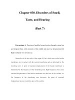
Ear anatomy. A. Drawing of modified coronal section through external ear
and temporal bone, with structures of the middle and inner ear demonstrated. B.
High-resolution view of inner ear.
Stereocilia of the hair cells of the organ of Corti, which rests on the basilar
membrane, are in contact with the tectorial membrane and are deformed by the
traveling wave. A point of maximal displacement of the basilar membrane is
determined by the frequency of the stimulating tone. High-frequency tones cause
maximal displacement of the basilar membrane near the base of the cochlea. As
the frequency of the stimulating tone decreases, the point of maximal
displacement moves toward the apex of the cochlea.
The inner and outer hair cells of the organ of Corti have different
innervation patterns, but both are mechanoreceptors. The afferent innervation
relates principally to the inner hair cells, and the efferent innervation relates
principally to outer hair cells. The motility of the outer hair cells alters the
micromechanics of the inner hair cells, creating a cochlear amplifier, which
explains the exquisite sensitivity and frequency selectivity of the cochlea.
Beginning in the cochlea, the frequency specificity is maintained at each
point of the central auditory pathway: dorsal and ventral cochlear nuclei, trapezoid
body, superior olivary complex, lateral lemniscus, inferior colliculus, medial
geniculate body, and auditory cortex. At low frequencies, individual auditory
nerve fibers can respond more or less synchronously with the stimulating tone. At
higher frequencies, phase-locking occurs so that neurons alternate in response to
particular phases of the cycle of the sound wave. Intensity is encoded by the
amount of neural activity in individual neurons, the number of neurons that are
active, and the specific neurons that are activated.
Genetic Causes of Hearing Loss
More than half of childhood hearing impairment is thought to be
hereditary; hereditary hearing impairment (HHI) can also manifest later in life.
HHI may be classified as either nonsyndromic, when hearing loss is the only
clinical abnormality, or syndromic, when hearing loss is associated with anomalies
in other organ systems. Nearly two-thirds of HHIs are nonsyndromic, and the
remaining one-third are syndromic. Between 70 and 80% of nonsyndromic HHI is
inherited in an autosomal recessive manner and designated DFNB; another 15–
20% is autosomal dominant (DFNA). Less than 5% is X-linked or maternally
inherited via the mitochondria.
Nearly 100 loci harboring genes for nonsyndromic HHI have been mapped,
with equal numbers of dominant and recessive modes of inheritance; numerous
genes have now been cloned (Table 30-3). The hearing genes fall into the
categories of structural proteins (MYH9, MYO7A, MYO15, TECTA, DIAPH1),
transcription factors (POU3F4, POU4F3), ion channels (KCNQ4, SLC26A4), and
gap junction proteins (GJB2, GJB3, GJB6). Several of these genes, including
connexin 26 (GJB2), TECTA, and TMC1, cause both autosomal dominant and
recessive forms of nonsyndromic HHI. In general, the hearing loss associated with
dominant genes has its onset in adolescence or adulthood and varies in severity,
whereas the hearing loss associated with recessive inheritance is congenital and
profound. Connexin 26 is particularly important because it is associated with
nearly 20% of cases of childhood deafness. Two frame-shift mutations, 35delG
and 167delT, account for >50% of the cases; however, screening for these two
mutations alone is insufficient to diagnose GJB2-related recessive deafness. The
167delT mutation is highly prevalent in Ashkenazi Jews; ~1 in 1765 individuals in
this population are homozygous and affected. The hearing loss can also vary
among the members of the same family, suggesting that other genes or factors
influence the auditory phenotype.
Table 30-3 Hereditary Hearing Impairment Genes
Designation Gene Function
Autosomal Dominant
CRYM Thyroid hormone binding
protein
DFNA1 DIAPH1 Cytoskeletal protein
DFNA2 GJB3 (Cx31) Gap junctions
DFNA2 KCNQ4 Potassium channel
DFNA3 GJB2 (Cx26) Gap junctions
DFNA3 GJB6 (Cx30) Gap junctions
DFNA4 MYH14 Class II nonmuscle myosin
DFNA5 DFNA5 Unknown
DFNA6/14/38
WFS Transmembrane protein
DFNA8/12 TECTA Tectorial membrane
protein
DFNA9 COCH Unknown
DFNA10 EYA4 Developmental gene
DFNA11 MYO7A Cytoskeletal protein
DFNA13 COL11A2 Cytoskeletal protein
DFNA15 POU4F3 Transcription factor
DFNA17 MYH9 Cytoskeletal protein
DFNA20/26 ACTG1 Cytoskeletal protein
DFNA22 MYO6 Unconventional myosin
DFNA28 TFCP2L3 Transcription factor
DFNA36 TMC1 Transmembrane protein
DFNA48 MYO1A Unconventional myosin
Autosomal Recessive
SLC26A5
(Prestin)
Motor protein
DFNB1 GJB2 (CX26) Gap junction
GJB6(CX30) Gap junction
DFNB2 MYO7A Cytoskeletal protein
DFNB3 MYO15 Cytoskeletal protein
DFNB4 PDS(SLC26A4)
Chloride/iodide transporter
DFNB6 TMIE Transmembrane protein
DFNB7/B11 TMC1 Transmembrane protein
DFNB9 OTOF Trafficking of membrane
vesicles
DFNB8/10 TMPRSS3 Transmembrane serine
protease
DFNB12 CDH23 Intercellular adherence
protein
DFNB16 STRC Stereocilia protein
DFNB18 USH1C Unknown
DFNB21 TECTA Tectorial membrane
protein
DFNB22 OTOA Gel attachement to
nonsensory cell
DFNB23 PCDH15 Morphogenesis and
cohesion
DFNB28 TRIOBP Cytoskeletal-organizing
protein
DFNB29 CLDN14 Tight junctions
DFNB30 MYO3A Hybrid motor-signaling
myosin
DFNB31 WHRN PDZ domain–containing
protein
DFNB36 ESPN Ca-insensitive actin-
bundling protein
DFNB37 MYO6 Unconventional myosin
DFNB67 TMHS Unknown function;
tetraspan protein