Chapter 029. Disorders of the Eye (Part 8) ppsx
Bạn đang xem bản rút gọn của tài liệu. Xem và tải ngay bản đầy đủ của tài liệu tại đây (103.39 KB, 5 trang )
Chapter 029. Disorders of the Eye
(Part 8)
Episcleritis
This is an inflammation of the episclera, a thin layer of connective tissue
between the conjunctiva and sclera. Episcleritis resembles conjunctivitis but is a
more localized process and discharge is absent. Most cases of episcleritis are
idiopathic, but some occur in the setting of an autoimmune disease. Scleritis refers
to a deeper, more severe inflammatory process, frequently associated with a
connective tissue disease such as rheumatoid arthritis, lupus erythematosus,
polyarteritis nodosa, Wegener's granulomatosis, or relapsing polychondritis. The
inflammation and thickening of the sclera can be diffuse or nodular. In anterior
forms of scleritis, the globe assumes a violet hue and the patient complains of
severe ocular tenderness and pain. With posterior scleritis the pain and redness
may be less marked, but there is often proptosis, choroidal effusion, reduced
motility, and visual loss. Episcleritis and scleritis should be treated with NSAIDs.
If these agents fail, topical or even systemic glucocorticoid therapy may be
necessary, especially if an underlying autoimmune process is active.
Uveitis
Involving the anterior structures of the eye, this is also called iritis or
iridocyclitis. The diagnosis requires slit-lamp examination to identify
inflammatory cells floating in the aqueous humor or deposited upon the corneal
endothelium (keratic precipitates). Anterior uveitis develops in sarcoidosis,
ankylosing spondylitis, juvenile rheumatoid arthritis, inflammatory bowel disease,
psoriasis, Reiter's syndrome, and Behçet's disease. It is also associated with herpes
infections, syphilis, Lyme disease, onchocerciasis, tuberculosis, and leprosy.
Although anterior uveitis can occur in conjunction with many diseases, no cause is
found to explain the majority of cases. For this reason, laboratory evaluation is
usually reserved for patients with recurrent or severe anterior uveitis. Treatment is
aimed at reducing inflammation and scarring by judicious use of topical
glucocorticoids. Dilation of the pupil reduces pain and prevents the formation of
synechiae.
Posterior Uveitis
This is diagnosed by observing inflammation of the vitreous, retina, or
choroid on fundus examination. It is more likely than anterior uveitis to be
associated with an identifiable systemic disease. Some patients have panuveitis, or
inflammation of both the anterior and posterior segments of the eye. Posterior
uveitis is a manifestation of autoimmune diseases such as sarcoidosis, Behçet's
disease, Vogt-Koyanagi-Harada syndrome, and inflammatory bowel disease (Fig.
29-4). It also accompanies diseases such as toxoplasmosis, onchocerciasis,
cysticercosis, coccidioidomycosis, toxocariasis, and histoplasmosis; infections
caused by organisms such as Candida, Pneumocystis carinii, Cryptococcus,
Aspergillus, herpes, and cytomegalovirus (see Fig. 175-1); and other diseases such
as syphilis, Lyme disease, tuberculosis, cat-scratch disease, Whipple's disease, and
brucellosis. In multiple sclerosis, chronic inflammatory changes can develop in the
extreme periphery of the retina (pars planitis or intermediate uveitis).
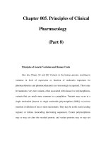
Figure 29-4
Retinal vasculitis, uveitis, and hemorrhage in a 32-year-old woman with
Crohn's disease. Note that the veins are frosted with a white exudate. Visual acuity
improved from 20/400 to 20/20 following treatment with intravenous
methylprednisolone.
Acute Angle-Closure Glaucoma
This is a rare and frequently misdiagnosed cause of a red, painful eye.
Susceptible eyes have a shallow anterior chamber, either because the eye has a
short axial length (hyperopia) or a lens enlarged by the gradual development of
cataract. When the pupil becomes mid-dilated, the peripheral iris blocks aqueous
outflow via the anterior chamber angle and the intraocular pressure rises abruptly,
producing pain, injection, corneal edema, obscurations, and blurred vision. In
some patients, ocular symptoms are overshadowed by nausea, vomiting, or
headache, prompting a fruitless workup for abdominal or neurologic disease. The
diagnosis is made by measuring the intraocular pressure during an acute attack or
by observing a narrow chamber angle by means of a specially mirrored contact
lens. Acute angle closure is treated with acetazolamide (PO or IV), topical beta
blockers, prostaglandin analogues, α
2
-adrenergic agonists, and pilocarpine to
induce miosis. If these measures fail, a laser can be used to create a hole in the
peripheral iris to relieve pupillary block. Many physicians are reluctant to dilate
patients routinely for fundus examination because they fear precipitating an angle-
closure glaucoma. The risk is actually remote and more than outweighed by the
potential benefit to patients of discovering a hidden fundus lesion visible only
through a fully dilated pupil. Moreover, a single attack of angle closure after
pharmacologic dilation rarely causes any permanent damage to the eye and serves
as an inadvertent provocative test to identify patients with narrow angles who
would benefit from prophylactic laser iridectomy.