Chapter 029. Disorders of the Eye (Part 9) potx
Bạn đang xem bản rút gọn của tài liệu. Xem và tải ngay bản đầy đủ của tài liệu tại đây (52.92 KB, 5 trang )
Chapter 029. Disorders of the Eye
(Part 9)
Endophthalmitis
This occurs from bacterial, viral, fungal, or parasitic infection of the
internal structures of the eye. It is usually acquired by hematogenous seeding from
a remote site.
Chronically ill, diabetic, or immunosuppressed patients, especially those
with a history of indwelling IV catheters or positive blood cultures, are at greatest
risk for endogenous endophthalmitis.
Although most patients have ocular pain and injection, visual loss is
sometimes the only symptom. Septic emboli, from a diseased heart valve or a
dental abscess, that lodge in the retinal circulation can give rise to
endophthalmitis.
White-centered retinal hemorrhages (Roth's spots) are considered
pathognomonic for subacute bacterial endocarditis, but they also appear in
leukemia, diabetes, and many other conditions.
Endophthalmitis also occurs as a complication of ocular surgery,
occasionally months or even years after the operation. An occult penetrating
foreign body or unrecognized trauma to the globe should be considered in any
patient with unexplained intraocular infection or inflammation.[newpage]
Transient or Sudden Visual Loss
Amaurosis Fugax
This term refers to a transient ischemic attack of the retina (Chap. 364).
Because neural tissue has a high rate of metabolism, interruption of blood flow to
the retina for more than a few seconds results in transient monocular blindness, a
term used interchangeably with amaurosis fugax.
Patients describe a rapid fading of vision like a curtain descending,
sometimes affecting only a portion of the visual field. Amaurosis fugax usually
occurs from an embolus that becomes stuck within a retinal arteriole (Fig. 29-5).
If the embolus breaks up or passes, flow is restored and vision returns
quickly to normal without permanent damage. With prolonged interruption of
blood flow, the inner retina suffers infarction.
Ophthalmoscopy reveals zones of whitened, edematous retina following the
distribution of branch retinal arterioles. Complete occlusion of the central retinal
artery produces arrest of blood flow and a milky retina with a cherry-red fovea
(Fig. 29-6).
Emboli are composed of either cholesterol (Hollenhorst plaque), calcium,
or platelet-fibrin debris. The most common source is an atherosclerotic plaque in
the carotid artery or aorta, although emboli can also arise from the heart,
especially in patients with diseased valves, atrial fibrillation, or wall motion
abnormalities.
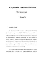
Figure 29-5
Hollenhorst plaque lodged at the bifurcation of a retinal arteriole
proves that a patient is shedding emboli from either the carotid artery, great
vessels, or heart.
Figure 29-6