Chapter 045. Azotemia and Urinary Abnormalities (Part 5) pps
Bạn đang xem bản rút gọn của tài liệu. Xem và tải ngay bản đầy đủ của tài liệu tại đây (117.48 KB, 5 trang )
Chapter 045. Azotemia and
Urinary Abnormalities
(Part 5)
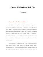
Approach to the patient with hematuria. RBC, red blood cell; WBC,
white blood cell; GBM, glomerular basement membrane; ANCA, antineutrophil
cytoplasmic antibody; VDRL, venereal disease research laboratory; ASLO,
antistreptolysin O; UA, urinalysis; IVP, intravenous pyelography; CT, computed
tomography.
A detailed discussion of glomerulonephritis and diseases of the
microvasculature can be found in Chap. 277.
OLIGURIA AND ANURIA
Oliguria refers to a 24-h urine output of <500 mL, and anuria is the
complete absence of urine formation (<50 mL). Anuria can be caused by total
urinary tract obstruction, total renal artery or vein occlusion, and shock
(manifested by severe hypotension and intense renal vasoconstriction). Cortical
necrosis, ATN, and rapidly progressive glomerulonephritis can occasionally cause
anuria. Oliguria can accompany any cause of acute renal failure and carries a more
serious prognosis for renal recovery in all conditions except prerenal azotemia.
Nonoliguria refers to urine output >500 mL/d in patients with acute or chronic
azotemia. With nonoliguric ATN, disturbances of potassium and hydrogen
balance are less severe than in oliguric patients, and recovery to normal renal
function is usually more rapid.
Proteinuria
The evaluation of proteinuria is shown schematically in Fig. 45-3 and is
typically initiated after detection of proteinuria by dipstick examination. The
dipstick measurement detects mostly albumin and gives false-positive results
when pH > 7.0 and the urine is very concentrated or contaminated with blood. A
very dilute urine may obscure significant proteinuria on dipstick examination, and
proteinuria that is not predominantly albumin will be missed. This is particularly
important for the detection of Bence-Jones proteins in the urine of patients with
multiple myeloma. Tests to measure total urine concentration accurately rely on
precipitation with sulfosalicylic or trichloracetic acids. Currently, ultrasensitive
dipsticks are available to measure microalbuminuria (30–300 mg/d), an early
marker of glomerular disease that has been shown to predict glomerular injury in
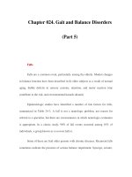
early diabetic nephropathy (Fig. 45-3).
Figure 45-3
[newpage]
Approach to the patient with proteinuria. Investigation of proteinuria is
often initiated by a positive dipstick on routine urinalysis. Conventional dipsticks
detect predominantly albumin and cannot detect urinary albumin levels of 30–300
mg/d. However, more exact determination of proteinuria should employ a 24-h
urine collection or a spot morning protein/creatinine ratio (mg/g). The pattern of
proteinuria on UPEP (urine protein electrophoresis) can be classified as
"glomerular," "tubular," or "abnormal" depending upon the origin of the urine
proteins. Glomerular proteinuria is due to abnormal glomerular permeability.
"Tubular proteins" such as Tamm-Horsfall are normally produced by the renal
tubule and shed into the urine. Abnormal circulating proteins such as kappa or
lambda light chains are readily filtered because of their small size. RBC, red blood
cell; FSGS, focal segmental glomerulosclerosis; MPGN, membranoproliferative
glomerulonephritis.
The magnitude of proteinuria and the protein composition of the urine
depend upon the mechanism of renal injury leading to protein losses. Both charge
and size selectivity normally prevent virtually all plasma albumin, globulins, and
other large-molecular-weight proteins from crossing the glomerular wall.
However, if this barrier is disrupted, there can be leakage of plasma proteins into
the urine (glomerular proteinuria; Fig. 45-3). Smaller proteins (<20 kDa) are
freely filtered but are readily reabsorbed by the proximal tubule. Normal
individuals excrete <150 mg/d of total protein and <30 mg/d of albumin. The
remainder of the protein in the urine is secreted by the tubules (Tamm-Horsfall,
IgA, and urokinase) or represents small amounts of filtered β
2
-microglobulin,
apoproteins, enzymes, and peptide hormones. Another mechanism of proteinuria
occurs when there is excessive production of an abnormal protein that exceeds the
capacity of the tubule for reabsorption. This most commonly occurs with plasma
cell dyscrasias such as multiple myeloma, amyloidosis, and lymphomas that are
associated with monoclonal production of immunoglobulin light chains.