Chapter 052. Approach to the Patient with a Skin Disorder (Part 1) ppt
Bạn đang xem bản rút gọn của tài liệu. Xem và tải ngay bản đầy đủ của tài liệu tại đây (51.52 KB, 5 trang )
Chapter 052. Approach to the Patient
with a Skin Disorder
(Part 1)
Harrison's Internal Medicine > Chapter 52. Approach to the Patient with
a Skin Disorder
APPROACH TO THE PATIENT WITH A SKIN DISORDER:
INTRODUCTION
The challenge of examining the skin lies in distinguishing normal from
abnormal, significant findings from trivial ones, and in integrating pertinent signs
and symptoms into an appropriate differential diagnosis. The fact that the largest
organ in the body is visible is both an advantage and a disadvantage to those who
examine it. It is advantageous because no special instrumentation is necessary and
because the skin can be biopsied with little morbidity. However, the casual
observer can be misled by a variety of stimuli and overlook important, subtle signs
of skin or systemic disease. For instance, the sometimes minor differences in color
and shape that distinguish a melanoma (Fig. 52-1) from a benign nevomelanocytic
nevus (Fig. 52-2) can be difficult to recognize. To aid in the interpretation of skin
lesions, a variety of descriptive terms have been developed to characterize
cutaneous lesions (Tables 52-1, 52-2, and 52-3 as well as Fig. 52-3) and to
formulate a differential diagnosis (Table 52-4). For instance, the finding of scaling
papules (present in patients with psoriasis or atopic dermatitis) places the patient
in a different diagnostic category than would hemorrhagic papules, which may
indicate vasculitis or sepsis (Figs. 52-4 and 52-5, respectively). It is also important
to differentiate primary from secondary skin lesions. If the examiner focuses on
linear erosions overlying an area of erythema and scaling, he or she may
incorrectly assume that the erosion is the primary lesion and the redness and scale
are secondary, while the correct interpretation would be that the patient has a
pruritic eczematous dermatitis with erosions caused by scratching.
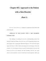
Figure 52-1
Superficial spreading melanoma. This is the most common type of
melanoma. Such lesions usually demonstrate asymmetry, border irregularity, color
variegation (black, blue, brown, pink, and white), a diameter >6 mm, and a history
of change (e.g., an increase in size or development of associated symptoms such as
pruritus or pain).
Figure 52-2
Table 52-1 Description of Primary Skin Lesions
Macule: A flat, colored lesion, <2 cm in diameter, not raised above the
surface of the surrounding skin. A "freckle," or ephelid, is a prototype pigmented
macule.
Patch: A large (>2 cm) flat lesion with a color different from the
surrounding skin. This differs from a macule only in size.
Papule: A small, solid lesion, <0.5 cm in diameter, raised above the surface
of the surrounding skin and hence palpable (e.g., a closed comedone, or
whitehead, in acne).
Nodule: A larger (0.5–5.0 cm), firm lesion raised above the surface of the
surrounding skin. This differs from a papule only in size (e.g., a dermal
nevomelanocytic nevus).
Tumor: A solid, raised growth >5 cm in diameter.
Plaque: A large (>1 cm), flat-topped, raised lesion; edges may either be
distinct (e.g., in psoriasis) or gradually blend with surrounding skin (e.g., in
eczematous dermatitis).
Vesicle: A small, fluid-filled lesion, <0.5 cm in diameter, raised above the
plane of surrounding skin. Fluid is often visible, and the lesions are translucent
[e.g., vesicles in allergic contact dermatitis caused by Toxicodendron (poison
ivy)].
Pustule: A vesicle filled with leukocytes. Note: The presence of pustules
does not necessarily signify the existence of an infection.
Bulla: A fluid-filled, raised, often translucent lesion >0.5 cm in diameter.
Wheal: A raised, erythematous, edematous papule or plaque, usually
representing short-lived vasodilatation and vasopermeability.
Telangiectasia: A dilated, superficial blood vessel.