The Insects - Outline of Entomology 3th Edition - Chapter 3 potx
Bạn đang xem bản rút gọn của tài liệu. Xem và tải ngay bản đầy đủ của tài liệu tại đây (2.15 MB, 36 trang )
Chapter 3
INTERNAL ANATOMY
AND PHYSIOLOGY
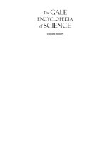
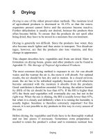
Internal structures of a locust. (After Uvarov 1966.)
TIC03 5/20/04 4:48 PM Page 49
50 Internal anatomy and physiology
What you see if you dissect open the body of an insect
is a complex and compact masterpiece of functional
design. Figure 3.1 shows the “insides” of two omnivor-
ous insects, a cockroach and a cricket, which have
relatively unspecialized digestive and reproductive
systems. The digestive system, which includes salivary
glands as well as an elongate gut, consists of three
main sections. These function in storage, biochemical
breakdown, absorption, and excretion. Each gut sec-
tion has more than one physiological role and this
may be reflected in local structural modifications,
such as thickening of the gut wall or diverticula (exten-
sions) from the main lumen. The reproductive systems
depicted in Fig. 3.1 exemplify the female and male
organs of many insects. These may be dominated in
males by very visible accessory glands, especially as
the testes of many adult insects are degenerate or
absent. This is because the spermatozoa are produced
in the pupal or penultimate stage and stored. In gravid
female insects, the body cavity may be filled with eggs
at various stages of development, thereby obscuring
other internal organs. Likewise, the internal structures
(except the gut) of a well-fed, late-stage caterpillar may
be hidden within the mass of fat body tissue.
The insect body cavity, called the hemocoel
(haemocoel) and filled with fluid hemolymph (haemo-
lymph), is lined with endoderm and ectoderm. It is not
a true coelom, which is defined as a mesoderm-lined
cavity. Hemolymph (so-called because it combines
many roles of vertebrate blood (hem/haem) and lymph)
bathes all internal organs, delivers nutrients, removes
metabolites, and performs immune functions. Unlike
vertebrate blood, hemolymph rarely has respiratory
pigments and therefore has little or no role in gaseous
exchange. In insects this function is performed by
the tracheal system, a ramification of air-filled tubes
(tracheae), which sends fine branches throughout the
body. Gas entry to and exit from tracheae is controlled
by sphincter-like structures called spiracles that open
through the body wall. Non-gaseous wastes are filtered
from the hemolymph by filamentous Malpighian
tubules (named after their discoverer), which have
free ends distributed through the hemocoel. Their con-
tents are emptied into the gut from which, after further
modification, wastes are eliminated eventually via the
anus.
All motor, sensory, and physiological processes in
insects are controlled by the nervous system in con-
junction with hormones (chemical messengers). The
brain and ventral nerve cord are readily visible in
dissected insects, but most endocrine centers, neuro-
secretion sites, numerous nerve fibers, muscles, and
other tissues cannot be seen by the unaided eye.
This chapter describes insect internal structures
and their functions. Topics covered are the muscles
and locomotion (walking, swimming, and flight), the
nervous system and co-ordination, endocrine centers
and hormones, the hemolymph and its circulation, the
tracheal system and gas exchange, the gut and diges-
tion, the fat body, nutrition and microorganisms, the
excretory system and waste disposal, and lastly the
reproductive organs and gametogenesis. A full account
of insect physiology cannot be provided in one chapter,
and we direct readers to Chapman (1998) for a com-
prehensive treatment, and to relevant chapters in the
Encyclopedia of Insects (Resh & Cardé 2003).
3.1 MUSCLES AND LOCOMOTION
As stated in section 1.3.4, much of the success of insects
relates to their ability to sense, interpret, and move
around their environment. Although the origin of
flight at least 340 million years ago was a major
innovation, terrestrial and aquatic locomotion also is
well developed. Power for movement originates from
muscles operating against a skeletal system, either the
rigid cuticular exoskeleton or, in soft-bodied larvae, a
hydrostatic skeleton.
3.1.1 Muscles
Vertebrates and many non-insect invertebrates have
striated and smooth muscles, but insects have only
striated muscles, so-called because of overlapping
thicker myosin and thinner actin filaments giving a
microscopic appearance of cross-banding. Each striated
muscle fiber comprises many cells, with a common
plasma membrane and sarcolemma, or outer sheath.
The sarcolemma is invaginated, but not broken, where
an oxygen-supplying tracheole (section 3.5, Fig. 3.10b)
contacts the muscle fiber. Contractile myofibrils run
the length of the fiber, arranged in sheets or cylinders.
When viewed under high magnification, a myofibril
comprises a thin actin filament sandwiched between
a pair of thicker myosin filaments. Muscle contrac-
tion involves the sliding of filaments past each other,
stimulated by nerve impulses. Innervation comes from
one to three motor axons per bundle of fibers, each
TIC03 5/20/04 4:48 PM Page 50
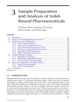
Fig. 3.1 Dissections of (a) a female American cockroach, Periplaneta americana (Blattodea: Blattidae), and (b) a male black field
cricket, Teleogryllus commodus (Orthoptera: Gryllidae). The fat body and most of the tracheae have been removed; most details of
the nervous system are not shown.
TIC03 5/20/04 4:48 PM Page 51
52 Internal anatomy and physiology
separately tracheated and referred to as one muscle
unit, with several units grouped in a functional
muscle.
There are several different muscle types. The most
important division is between those that respond syn-
chronously, with a contraction cycle once per impulse,
and fibrillar muscles that contract asynchronously,
with multiple contractions per impulse. Examples of
the latter include some flight muscles (see below) and
the tymbal muscle of cicadas (section 4.1.4).
There is no inherent difference in action between
muscles of insects and vertebrates, although insects
can produce prodigious muscular feats, such as the
leap of a flea or the repetitive stridulation of the cicada
tympanum. Reduced body size benefits insects because
of the relationship between (i) power, which is pro-
portional to muscle cross-section and decreases with
reduction in size by the square root, and (ii) the body
mass, which decreases with reduction in size by the
cube root. Thus the power : mass ratio increases as
body size decreases.
3.1.2 Muscle attachments
Vertebrates’ muscles work against an internal skeleton,
but the muscles of insects must attach to the inner
surface of an external skeleton. As musculature is
mesodermal and the exoskeleton is of ectodermal ori-
gin, fusion must take place. This occurs by the growth
of tonofibrillae, fine connecting fibrils that link the
epidermal end of the muscle to the epidermal layer
(Fig. 3.2a,b). At each molt tonofibrillae are discarded
along with the cuticle and therefore must be regrown.
At the site of tonofibrillar attachment, the inner cut-
icle often is strengthened through ridges or apodemes,
which, when elongated into arms, are termed apophy-
ses (Fig. 3.2c). These muscle attachment sites, particu-
larly the long, slender apodemes for individual muscle
attachments, often include resilin to give an elasticity
that resembles that of vertebrate tendons.
Some insects, including soft-bodied larvae, have
mainly thin, flexible cuticle without the rigidity to
anchor muscles unless given additional strength. The
body contents form a hydrostatic skeleton, with tur-
gidity maintained by criss-crossed body wall “turgor”
muscles that continuously contract against the incom-
pressible fluid of the hemocoel, giving a strengthened
foundation for other muscles. If the larval body wall
is perforated, the fluid leaks, the hemocoel becomes
compressible and the turgor muscles cause the larva
to become flaccid.
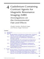
Fig. 3.2 Muscle attachments to body wall: (a) tonofibrillae traversing the epidermis from the muscle to the cuticle; (b) a muscle
attachment in an adult beetle of Chrysobothrus femorata (Coleoptera: Buprestidae); (c) a multicellular apodeme with a muscle
attached to one of its thread-like, cuticular “tendons” or apophyses. (After Snodgrass 1935.)
TIC03 5/20/04 4:48 PM Page 52
3.1.3 Crawling, wriggling, swimming,
and walking
Soft-bodied larvae with hydrostatic skeletons move
by crawling. Muscular contraction in one part of the
body gives equivalent extension in a relaxed part else-
where on the body. In apodous (legless) larvae, such as
dipteran “maggots”, waves of contractions and relaxa-
tion run from head to tail. Bands of adhesive hooks
or tubercles successively grip and detach from the
substrate to provide a forward motion, aided in some
maggots by use of their mouth hooks to grip the sub-
strate. In water, lateral waves of contraction against
the hydrostatic skeleton can give a sinuous, snake-like,
swimming motion, with anterior-to-posterior waves
giving an undulating motion.
Larvae with thoracic legs and abdominal prolegs,
like caterpillars, develop posterior-to-anterior waves of
turgor muscle contraction, with as many as three waves
visible simultaneously. Locomotor muscles operate in
cycles of successive detachment of the thoracic legs,
reaching forwards and grasping the substrate. These
cycles occur in concert with inflation, deflation, and
forward movement of the posterior prolegs.
Insects with hard exoskeletons can contract and
relax pairs of agonistic and antagonistic muscles that
attach to the cuticle. Compared to crustaceans and
myriapods, insects have fewer (six) legs that are located
more ventrally and brought close together on the
thorax, allowing concentration of locomotor muscles
(both flying and walking) into the thorax, and pro-
viding more control and greater efficiency. Motion with
six legs at low to moderate speed allows continuous
contact with the ground by a tripod of fore and hind
legs on one side and mid leg of the opposite side thrust-
ing rearwards (retraction), whilst each opposite leg is
moved forwards (protraction) (Fig. 3.3). The center of
gravity of the slow-moving insect always lies within
this tripod, giving great stability. Motion is imparted
through thoracic muscles acting on the leg bases, with
transmission via internal leg muscles through the leg
to extend or flex the leg. Anchorage to the substrate,
Muscles and locomotion 53
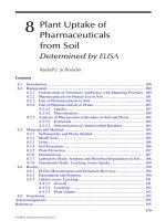
Fig. 3.3 (right) A ground beetle (Coleoptera: Carabidae:
Carabus) walking in the direction of the broken line. The
three blackened legs are those in contact with the ground
in the two positions illustrated – (a) is followed by (b).
(After Wigglesworth 1972.)
TIC03 5/20/04 4:48 PM Page 53
54 Internal anatomy and physiology
needed to provide a lever to propel the body, is through
pointed claws and adhesive pads (the arolium or, in
flies and some beetles, pulvilli). Claws such as those
illustrated in the vignette to Chapter 2 can obtain pur-
chase on the slightest roughness in a surface, and the
pads of some insects can adhere to perfectly smooth
surfaces through the application of lubricants to the
tips of numerous fine hairs and the action of close-
range molecular forces between the hairs and the
substrate.
When faster motion is required there are several
alternatives – increasing the frequency of the leg move-
ment by shortening the retraction period; increasing
the stride length; altering the triangulation basis of
support to adopt quadrupedy (use of four legs); or even
hind-leg bipedality with the other legs held above
the substrate. At high speeds even those insects that
maintain triangulation are very unstable and may
have no legs in contact with the substrate at intervals.
This instability at speed seems to cause no difficulty for
cockroaches, which when filmed with high-speed video
cameras have been shown to maintain speeds of up to
1ms
−1
whilst twisting and turning up to 25 times
per second. This motion was maintained by sensory
information received from one antenna whose tip
maintained contact with an experimentally provided
wall, even when it had a zig-zagging surface.
Many insects jump, some prodigiously, usually
using modified hind legs. In orthopterans, flea beetles
(Alticinae), and a range of weevils, an enlarged hind
(meta-) femur contains large muscles whose slow con-
traction produces energy stored by either distortion of
the femoro-tibial joint or in some spring-like sclerotiza-
tion, for example the meta-tibial extension tendon. In
fleas, the energy is produced by the trochanter levator
muscle raising the femur and is stored by compression
of an elastic resilin pad in the coxa. In all these jumpers,
release of tension is sudden, resulting in propulsion
of the insect into the air – usually in an uncontrolled
manner, but fleas can attain their hosts with some con-
trol over the leap. It has been suggested that the main
benefit for flighted jumpers is to get into the air and
allow the wings to be opened without damage from the
surrounding substrate.
In swimming, contact with the water is maintained
during protraction, so it is necessary for the insect to
impart more thrust to the rowing motion than to the
recovery stroke to progress. This is achieved by expand-
ing the effective leg area during retraction by extending
fringes of hairs and spines (Fig. 10.8). These collapse
onto the folded leg during the recovery stroke. We have
seen already how some insect larvae swim using con-
tractions against their hydrostatic skeleton. Others,
including many nymphs and the larvae of caddisflies,
can walk underwater and, particularly in running
waters, do not swim routinely.
The surface film of water can support some specialist
insects, most of which have hydrofuge (water-repelling)
cuticles or hair fringes and some, such as gerrid water-
striders (Fig. 5.7), move by rowing with hair-fringed
legs.
3.1.4 Flight
The development of flight allowed insects much greater
mobility, which helped in food and mate location and
gave much improved powers of dispersal. Importantly,
flight opened up many new environments for exploita-
tion. Plant microhabitats such as flowers and foliage
are more accessible to winged insects than to those
without flight.
Fully developed, functional, flying wings occur only
in adult insects, although in nymphs the developing
wings are visible as wing buds in all but the earliest
instars. Usually two pairs of functional wings arise
dorsolaterally, as fore wings on the second and hind
wings on the third thoracic segment. Some of the many
derived variations are described in section 2.4.2.
To fly, the forces of weight (gravity) and drag (air
resistance to movement) must be overcome. In gliding
flight, in which the wings are held rigidly outstretched,
these forces are overcome through the use of passive air
movements – known as the relative wind. The insect
attains lift by adjusting the angle of the leading edge
of the wing when orientated into the wind. As this
angle (the attack angle) increases, so lift increases until
stalling occurs, i.e. when lift is catastrophically lost. In
contrast to aircraft, nearly all of which stall at around
20°, the attack angle of insects can be raised to more
than 30°, even as high as 50°, giving great maneu-
verability. Aerodynamic effects such as enhanced
lift and reduced drag can come from wing scales and
hairs, which affect the boundary layer across the wing
surface.
Most insects glide a little, and dragonflies (Odonata)
and some grasshoppers (Orthoptera), notably locusts,
glide extensively. However, most winged insects fly
by beating their wings. Examination of wing beat is
difficult because the frequency of even a large slow-
TIC03 5/20/04 4:48 PM Page 54
flying butterfly may be five times a second (5 Hz), a bee
may beat its wings at 180 Hz, and some midges emit an
audible buzz with their wing-beat frequency of greater
than 1000 Hz. However, through the use of slowed-
down, high-speed cine film, the insect wing beat can be
slowed from faster than the eye can see until a single
beat can be analyzed. This reveals that a single beat
comprises three interlinked movements. First is a cycle
of downward, forward motion followed by an upward
and backward motion. Second, during the cycle each
wing is rotated around its base. The third component
occurs as various parts of the wing flex in response to
local variations in air pressure. Unlike gliding, in which
the relative wind derives from passive air movement, in
true flight the relative wind is produced by the moving
wings. The flying insect makes constant adjustments,
so that during a wing beat, the air ahead of the insect is
thrown backwards and downwards, impelling the
insect upwards (lift) and forwards (thrust). In climbing,
the emergent air is directed more downwards, reducing
thrust but increasing lift. In turning, the wing on the
inside of the turn is reduced in power by decrease in the
amplitude of the beat.
Despite the elegance and intricacy of detail of insect
flight, the mechanisms responsible for beating the
wings are not excessively complicated. The thorax of
the wing-bearing segments can be envisaged as a box
with the sides (pleura) and base (sternum) rigidly fused,
and the wings connected where the rigid tergum is
attached to the pleura by flexible membranes. This
membranous attachment and the wing hinge are com-
posed of resilin (section 2.1), which gives crucial elas-
ticity to the thoracic box. Flying insects have one of two
kinds of arrangements of muscles powering their flight:
1 direct flight muscles connected to the wings;
2 an indirect system in which there is no muscle-to-
wing connection, but rather muscle action deforms the
thoracic box to move the wing.
A few old groups such as Odonata and Blattodea
appear to use direct flight muscles to varying degrees,
although at least some recovery muscles may be indir-
ect. More advanced insects use indirect muscles for
flight, with direct muscles providing wing orientation
rather than power production.
Direct flight muscles produce the upward stroke by
contraction of muscles attached to the wing base inside
the pivotal point (Fig. 3.4a). The downward wing
stroke is produced through contraction of muscles that
extend from the sternum to the wing base outside the
pivot point (Fig. 3.4b). In contrast, indirect flight mus-
cles are attached to the tergum and sternum. Contrac-
tion causes the tergum, and with it the very base of the
wing, to be pulled down. This movement levers the
outer, main part of the wing in an upward stroke
(Fig. 3.4c). The down beat is powered by contraction of
the second set of muscles, which run from front to back
of the thorax, thereby deforming the box and lifting the
tergum (Fig. 3.4d). At each stage in the cycle, when
the flight muscles relax, energy is conserved because
the elasticity of the thorax restores its shape.
Primitively, the four wings may be controlled inde-
pendently with small variation in timing and rate
allowing alteration in direction of flight. However,
excessive variation impedes controlled flight and the
beat of all wings is usually harmonized, as in butterflies,
bugs, and bees, for example, by locking the fore and
hind wings together, and also by neural control. For
insects with slower wing-beat frequencies (<100 Hz),
such as dragonflies, one nerve impulse for each beat
can be maintained by synchronous muscles. How-
ever, in faster-beating wings, which may attain a fre-
quency of 100 to over 1000 Hz, one impulse per beat is
impossible and asynchronous muscles are required.
In these insects, the wing is constructed such that
only two wing positions are stable – fully up and fully
down. As the wing moves from one extreme to the
alternate one, it passes through an intermediate un-
stable position. As it passes this unstable (“click”) point,
thoracic elasticity snaps the wing through to the altern-
ate stable position. Insects with this asynchronous
mechanism have peculiar fibrillar flight muscles with
the property that, on sudden release of muscle tension,
as at the click point, the next muscle contraction is
induced. Thus muscles can oscillate, contracting at a
much higher frequency than the nerve impulses,
which need be only periodic to maintain the insect in
flight. Harmonization of the wing beat on each side is
maintained through the rigidity of the thorax – as the
tergum is depressed or relaxed, what happens to one
wing must happen identically to the other. However,
insects with indirect flight muscles retain direct mus-
cles that are used in making fine adjustments in wing
orientation during flight.
Direction and any deviations from course, perhaps
caused by air movements, are sensed by insects pre-
dominantly through their eyes and antennae. However,
the true flies (Diptera) have extremely sophisticated
sensory equipment, with their hind wings modified as
balancing organs. These halteres, which each comprise
a base, stem, and apical knob (Fig. 2.22f ), beat in time
Muscles and locomotion 55
TIC03 5/20/04 4:48 PM Page 55
56 Internal anatomy and physiology
but out of phase with the fore wings. The knob, which
is heavier than the rest of the organ, tends to keep
the halteres beating in one plane. When the fly alters
direction, whether voluntarily or otherwise, the haltere
is twisted. The stem, which is richly endowed with
sensilla, detects this movement, and the fly can respond
accordingly.
Initiation of flight, for any reason, may involve the
legs springing the insect into the air. Loss of tarsal con-
tact with the ground causes neural firing of the direct
flight muscles. In flies, flight activity originates in con-
traction of a mid-leg muscle, which both propels the leg
downwards (and the fly upwards) and simultaneously
pulls the tergum downwards to inaugurate flight. The
legs are also important when landing because there is
no gradual braking by running forwards – all the shock
is taken on the outstretched legs, endowed with pads,
spines, and claws for adhesion.
3.2 THE NERVOUS SYSTEM AND
CO-ORDINATION
The complex nervous system of insects integrates a
diverse array of external sensory and internal physio-
logical information and generates some of the beha-
viors discussed in Chapter 4. In common with other
animals, the basic component is the nerve cell, or
neuron (neurone), composed of a cell body with two
projections (fibers) – the dendrite, which receives
stimuli; and the axon, which transmits information,
either to another neuron or to an effector organ such
as a muscle. Insect neurons release a variety of chem-
icals at synapses to either stimulate or inhibit effector
neurons or muscles. In common with vertebrates,
particularly important neurotransmitters include
acetylcholine and catecholamines such as dopamine.
Neurons (Fig. 3.5) are of at least four types:
Fig. 3.4 Direct flight mechanisms: thorax during (a) upstroke and (b) downstroke of the wings. Indirect flight mechanisms:
thorax during (c) upstroke and (d) downstroke of the wings. Stippled muscles are those contracting in each illustration.
(After Blaney 1976.)
TIC03 5/20/04 4:48 PM Page 56
1 sensory neurons receive stimuli from the insect’s
environment and transmit them to the central nervous
system (see below);
2 interneurons (or association neurons) receive
information from and transmit it to other neurons;
3 motor neurons receive information from inter-
neurons and transmit it to muscles;
4 neuroendocrine cells (section 3.3.1).
The cell bodies of interneurons and motor neurons
are aggregated with the fibers interconnecting all types
of nerve cells to form nerve centers called ganglia.
Simple reflex behavior has been well studied in insects
(described further in section 4.5), but insect behavior
can be complex, involving integration of neural infor-
mation within the ganglia.
The central nervous system (CNS) (Fig. 3.6) is the
principal division of the nervous system and consists of
series of ganglia joined by paired longitudinal nerve
cords called connectives. Primitively there are a pair
of ganglia per body segment but usually the two
ganglia of each thoracic and abdominal segment are
fused into a single structure and the ganglia of all head
segments are coalesced to form two ganglionic centers
– the brain and the suboesophageal (subesophageal)
ganglion (seen in Fig. 3.7). The chain of thoracic and
abdominal ganglia found on the floor of the body cavity
is called the ventral nerve cord. The brain, or the
dorsal ganglionic center of the head, is composed of
three pairs of fused ganglia (from the first three head
segments):
1 protocerebrum, associated with the eyes and thus
bearing the optic lobes;
2 deutocerebrum, innervating the antennae;
3 tritocerebrum, concerned with handling the sig-
nals that arrive from the body.
Coalesced ganglia of the three mouthpart-bearing seg-
ments form the suboesophageal ganglion, with nerves
emerging that innervate the mouthparts.
The visceral (or sympathetic) nervous system
consists of three subsystems – the stomodeal (or sto-
matogastric) (which includes the frontal ganglion); the
ventral visceral; and the caudal visceral. Together
the nerves and ganglia of these subsystems innervate
the anterior and posterior gut, several endocrine organs
(corpora cardiaca and corpora allata), the reproductive
organs, and the tracheal system including the spiracles.
The peripheral nervous system consists of all of
the motor neuron axons that radiate to the muscles
from the ganglia of the CNS and stomodeal nervous
system plus the sensory neurons of the cuticular
sensory structures (the sense organs) that receive
mechanical, chemical, thermal, or visual stimuli from
an insect’s environment. Insect sensory systems are
discussed in detail in Chapter 4.
The nervous system and co-ordination 57
Fig. 3.5 Diagram of a simple reflex mechanism of an insect. The arrows show the paths of nerve impulses along nerve fibers
(axons and dendrites). The ganglion, with its outer cortex and inner neuropile, is shown on the right. (After various sources.)
TIC03 5/20/04 4:48 PM Page 57
58 Internal anatomy and physiology
Fig. 3.7 Mediolongitudinal section of an immature cockroach of Periplaneta americana (Blattodea: Blattidae) showing internal
organs and tissues.
Fig. 3.6 The central nervous system of various insects showing the diversity of arrangement of ganglia in the ventral nerve cord.
Varying degrees of fusion of ganglia occur from the least to the most specialized: (a) three separate thoracic and eight abdominal
ganglia, as in Dictyopterus (Coleoptera: Lycidae) and Pulex (Siphonaptera: Pulicidae); (b) three thoracic and six abdominal, as in
Blatta (Blattodea: Blattidae) and Chironomus (Diptera: Chironomidae); (c) two thoracic and considerable abdominal fusion of
ganglia, as in Crabro and Eucera (Hymenoptera: Crabronidae and Anthophoridae); (d) highly fused with one thoracic and no
abdominal ganglia, as in Musca, Calliphora, and Lucilia (Diptera: Muscidae and Calliphoridae); (e) extreme fusion with no separate
suboesophageal ganglion, as in Hydrometra (Hemiptera: Hydrometridae) and Rhizotrogus (Scarabaeidae). (After Horridge 1965.)
TIC03 5/20/04 4:48 PM Page 58
The endocrine system and the function of hormones 59
3.3 THE ENDOCRINE SYSTEM AND
THE FUNCTION OF HORMONES
Hormones are chemicals produced within an organ-
ism’s body and transported, generally in body fluids,
away from their point of synthesis to sites where they
influence a remarkable variety of physiological pro-
cesses, even though present in extremely small quant-
ities. Insect hormones have been studied in detail in
only a handful of species but similar patterns of pro-
duction and function are likely to apply to all insects.
The actions and interrelationships of these chemical
messengers are varied and complex but the role of
hormones in the molting process is of overwhelming
importance and will be discussed more fully in this con-
text in section 6.3. Here we provide a general picture
of the endocrine centers and the hormones that they
export.
Historically, the implication of hormones in the
processes of molting and metamorphosis resulted
from simple but elegant experiments. These utilized
techniques that removed the influence of the brain
(decapitation), isolated the hemolymph of different
parts of the body (ligation), or artificially connected
the hemolymph of two or more insects by joining their
bodies. Ligation and decapitation of insects enabled
researchers to localize the sites of control of develop-
mental and reproductive processes and to show that
substances are released that affect tissues at sites
distant from the point of release. In addition, critical
developmental periods for the action of these con-
trolling substances have been identified. The blood-
sucking bug Rhodnius prolixus (Hemiptera: Reduviidae)
and various moths and flies were the principal experi-
mental insects. More refined technologies allowed
microsurgical removal or transplant of various tissues,
hemolymph transfusion, hormone extraction and puri-
fication, and radioactive labeling of hormone extracts.
Today, molecular biological (Box 3.1) and advanced
chemical analytical techniques allow hormone isola-
tion, characterization, and manipulation.
3.3.1 Endocrine centers
The hormones of the insect body are produced by neu-
ronal, neuroglandular, or glandular centers (Fig. 3.8).
Hormonal production by some organs, such as the
ovaries, is secondary to their main function, but several
tissues and organs are specialized for an endocrine role.
Neurosecretory cells
Neurosecretory cells (NSC) (also called neuroendocrine
cells) are modified neurons found throughout the nerv-
ous system (within the CNS, peripheral nervous sys-
tem, and the stomodeal nervous system), but they
occur in major groups in the brain. These cells produce
most of the known insect hormones, the notable excep-
tions being the production by non-neural tissues of
ecdysteroids and juvenile hormones. However, the syn-
thesis and release of the latter hormones are regulated
by neurohormones from NSC.
Corpora cardiaca
The corpora cardiaca (singular: corpus cardiacum)
are a pair of neuroglandular bodies located on either
side of the aorta and behind the brain. As well as
producing their own neurohormones, they store and
release neurohormones, including prothoracicotropic
hormone (PTTH, formerly called brain hormone or
ecdysiotropin), originating from the NSC of the brain.
PTTH stimulates the secretory activity of the prothor-
acic glands.
Prothoracic glands
The prothoracic glands are diffuse, paired glands gener-
ally located in the thorax or the back of the head. In
cyclorrhaphous Diptera they are part of the ring gland,
which also contains the corpora cardiaca and corpora
allata. The prothoracic glands secrete an ecdysteroid,
usually ecdysone (sometimes called molting hormone),
which, after hydroxylation, elicits the molting process
of the epidermis (section 6.3).
Corpora allata
The corpora allata (singular: corpus allatum) are small,
discrete, paired glandular bodies derived from the epi-
thelium and located on either side of the foregut. In some
insects they fuse to form a single gland. Their function
is to secrete juvenile hormone ( JH), which has regu-
latory roles in both metamorphosis and reproduction.
3.3.2 Hormones
Three hormones or hormone types are integral to the
growth and reproductive functions in insects. These
TIC03 5/20/04 4:48 PM Page 59
60 Internal anatomy and physiology
are the ecdysteroids, the juvenile hormones, and the
neurohormones (also called neuropeptides).
Ecdysteroid is a general term applied to any steroid
with molt-promoting activity. All ecdysteroids are
derived from sterols, such as cholesterol, which insects
cannot synthesize de novo and must obtain from their
diet. Ecdysteroids occur in all insects and form a large
group of compounds, of which ecdysone and 20-
hydroxyecdysone are the most common members.
Ecdysone (also called α-ecdysone) is released from the
prothoracic glands into the hemolymph and usually
is converted to the more active hormone 20-
hydroxyecdysone in several peripheral tissues. The
20-hydroxyecdysone (often referred to as ecdysterone
or β-ecdysone in older literature) is the most wide-
spread and physiologically important ecdysteroid in
insects. The action of ecdysteroids in eliciting molting
has been studied extensively and has the same function
in different insects. Ecdysteroids also are produced by
the ovary of the adult female insect and may be
involved in ovarian maturation (e.g. yolk deposition) or
be packaged in the eggs to be metabolized during the
formation of embryonic cuticle.
Juvenile hormones form a family of related sesquiter-
penoid compounds, so that the symbol JH may denote
one or a mixture of hormones, including JH-I, JH-II, JH-
III, and JH-0. The occurrence of mixed-JH-producing
insects (such as the tobacco hornworm, Manduca sexta)
Box 3.1 Molecular genetic techniques and their application to
neuropeptide research*
Molecular biology is essentially a set of techniques for
the isolation, analysis, and manipulation of DNA and
its RNA and protein products. Molecular genetics is
concerned primarily with the nucleic acids, whereas
research on the proteins and their constituent amino
acids involves chemistry. Thus, genetics and chemistry
are integral to molecular biology. Molecular biological
tools provide:
• a means of cutting DNA at specific sites using restric-
tion enzymes and of rejoining naked ends of cut frag-
ments with ligase enzymes;
• techniques, such as the polymerase chain reaction
(PCR), that produce numerous identical copies by
repeated cycles of amplification of a segment of DNA;
• methods for rapid sequencing of nucleotides of DNA
or RNA, and amino acids of proteins;
• the ability to synthesize short sequences of DNA or
proteins;
• DNA–DNA affinity hybridization to compare the match
of the synthesized DNA with the original sequence;
• the ability to search a genome for a specific nucleo-
tide sequence using oligonucleotide probes, which are
defined nucleic acid segments that are complementary
to the sequence being sought;
• site-directed mutation of specific DNA segments in
vitro;
• genetic engineering – the isolation and transfer of
intact genes into other organisms, with subsequent
stable transmission and gene expression;
• cytochemical techniques to identify how, when, and
where genes are actually transcribed;
• immunochemical and histochemical techniques to
identify how, when, and where a specific gene product
functions.
Insect peptide hormones have been difficult to study
because of the minute quantities produced by individual
insects and their structural complexity and occasional
instability. Currently, neuropeptides are the subject of
an explosion of studies because of the realization that
these proteins play crucial roles in most aspects of
insect physiology (see Table 3.1), and the availability of
appropriate technologies in chemistry (e.g. gas-phase
sequencing of amino acids in proteins) and genetics.
Knowledge of neuropeptide amino acid sequences
provides a means of using the powerful capabilities of
molecular genetics. Nucleotide sequences deduced
from primary protein structures allow construction of
oligonucleotide probes for searching out peptide genes
in other parts of the genome or, more importantly, in
other organisms, especially pests. Methods such as
PCR and its variants facilitate the production of probes
from partial amino acid sequences and trace amounts
of DNA. Genetic amplification methods, such as PCR,
allow the production of large quantities of DNA and thus
allow easier sequencing of genes. Of course, these
uses of molecular genetic methods depend on the initial
chemical characterization of the neuropeptides. Fur-
thermore, appropriate bioassays are essential for
assessing the authenticity of any product of molecular
biology. The possible application of neuropeptide
research to control of insect pests is discussed in sec-
tion 16.4.3.
*After Altstein 2003; Hoy 2003.
TIC03 5/20/04 4:48 PM Page 60
adds to the complexity of unraveling the functions of
the homologous JHs. These hormones have two major
roles – the control of metamorphosis and regulation
of reproductive development. Larval characteristics
are maintained and metamorphosis is inhibited by JH;
adult development requires a molt in the absence of JH
(see section 6.3 for details). Thus JH controls the degree
and direction of differentiation at each molt. In the
adult female insect, JH stimulates the deposition of yolk
in the eggs and affects accessory gland activity and
pheromone production (section 5.11).
Neurohormones constitute the third and largest
class of insect hormones. They are generally peptides
(small proteins) and hence have the alternative name
neuropeptides. These protein messengers are the
master regulators of many aspects of insect devel-
opment, homeostasis, metabolism, and reproduction,
including the secretion of the JHs and ecdysteroids.
Nearly 150 neuropeptides have been recognized, and
some (perhaps many) exist in multiple forms encoded
by the same gene following gene duplication events.
From this diversity, Table 3.1 summarizes a represent-
ative range of physiological processes reportedly affected
by neurohormones in various insects. The diversity
and vital co-ordinating roles of these small molecules
continue to be revealed thanks to technological devel-
opments in peptide molecular chemistry (Box 3.1)
allowing characterization and functional interpreta-
tion. Structural diversity among peptides of equivalent
or related biological activity is a consequence of synthe-
sis from large precursors that are cleaved and modified
to form the active peptides. Neuropeptides either reach
terminal effector sites directly along nerve axons or
via the hemolymph, or indirectly exert control via their
action on other endocrine glands (corpora allata and
prothoracic glands). Both inhibitory and stimulatory
signals are involved in neurohormone regulation. The
effectiveness of regulatory neuropeptides depends on
stereospecific high-affinity binding sites located in the
plasma membrane of the target cells.
Hormones reach their target tissues by transport
(even over short distances) by the body fluid or hemo-
lymph. Hormones are often water-soluble but some
may be transported bound to proteins in the hemo-
lymph; for example, ecdysteroid-binding proteins and
JH-binding proteins are known in a number of insects.
These hemolymph-binding proteins may contribute to
the regulation of hormone levels by facilitating uptake
by target tissues, reducing non-specific binding, or pro-
tecting from degradation or excretion.
3.4 THE CIRCULATORY SYSTEM
Hemolymph, the insect body fluid (with properties
and functions as described in section 3.4.1), circulates
freely around the internal organs. The pattern of flow
is regular between compartments and appendages,
assisted by muscular contractions of parts of the
body, especially the peristaltic contractions of a lon-
gitudinal dorsal vessel, part of which is sometimes
called the heart. Hemolymph does not directly contact
the cells because the internal organs and the epidermis
are covered in a basement membrane, which may
regulate the exchange of materials. This open circulat-
ory system has only a few vessels and compartments
to direct hemolymph movement, in contrast to the
The circulatory system 61
Fig. 3.8 The main endocrine centers in a generalized insect.
(After Novak 1975.)
TIC03 5/20/04 4:48 PM Page 61
Table 3.1 Examples of some important insect physiological processes mediated by neuropeptides. (After Keeley & Hayes
1987; Holman et al. 1990; Gäde et al. 1997; Altstein 2003.)
Neuropeptide Action
Growth and development
Allatostatins and allatotropins Induce/regulate juvenile hormone (JH) production
Bursicon Controls cuticular sclerotization
Crustacean cardioactive peptide (CCAP) Switches on ecdysis behavior
Diapause hormone (DH) Causes dormancy in silkworm eggs
Pre-ecdysis triggering hormone (PETH) Stimulates pre-ecdysis behavior
Ecdysis triggering hormone (ETH) Initiates events at ecdysis
Eclosion hormone (EH) Controls events at ecdysis
JH esterase inducing factor Stimulates JH degradative enzyme
Prothoracicotropic hormone (PTTH) Induces ecdysteroid secretion from prothoracic gland
Puparium tanning factor Accelerates fly puparium tanning
Reproduction
Antigonadotropin (e.g. oostatic hormone, OH) Suppresses oocyte development
Ovarian ecdysteroidogenic hormone (OEH = EDNH) Stimulates ovarian ecdysteroid production
Ovary maturing peptide (OMP) Stimulates egg development
Oviposition peptides Stimulate egg deposition
Prothoracicotropic hormone (PTTH) Affects egg development
Pheromone biosynthesis activating neuropeptide Regulates pheromone production
(PBAN)
Homeostasis
Metabolic peptides (= AKH/RPCH family)
Adipokinetic hormone (AKH) Releases lipid from fat body
Hyperglycemic hormone Releases carbohydrate from fat body
Hypoglycemic hormone Enhances carbohydrate uptake
Protein synthesis factors Enhance fat body protein synthesis
Diuretic and antidiuretic peptides
Antidiuretic peptide (ADP) Suppresses water excretion
Diuretic peptide (DP) Enhances water excretion
Chloride-transport stimulating hormone Stimulates Cl
−
absorption (rectum)
Ion-transport peptide (ITP) Stimulates Cl
−
absorption (ileum)
Myotropic peptides
Cardiopeptides Increase heartbeat rate
Kinin family (e.g. leukokinins and myosuppressins) Regulate gut contraction
Proctolin Modifies excitation response of some muscles
Chromatotropic peptides
Melanization and reddish coloration hormone (MRCH) Induces darkening
Pigment-dispersing hormone (PDH) Disperses pigment
Corazonin Darkens pigment
62 Internal anatomy and physiology
TIC03 5/20/04 4:48 PM Page 62
closed network of blood-conducting vessels seen in
vertebrates.
3.4.1 Hemolymph
The volume of the hemolymph may be substantial
(20–40% of body weight) in soft-bodied larvae, which
use the body fluid as a hydrostatic skeleton, but is less
than 20% of body weight in most nymphs and adults.
Hemolymph is a watery fluid containing ions, mole-
cules, and cells. It is often clear and colorless but may be
variously pigmented yellow, green, or blue, or rarely, in
the immature stages of a few aquatic and endoparasitic
flies, red owing to the presence of hemoglobin. All
chemical exchanges between insect tissues are medi-
ated via the hemolymph – hormones are transported,
nutrients are distributed from the gut, and wastes are
removed to the excretory organs. However, insect
hemolymph only rarely contains respiratory pigments
and hence has a very low oxygen-carrying capacity.
Local changes in hemolymph pressure are important
in ventilation of the tracheal system (section 3.5.1), in
thermoregulation (section 4.2.2), and at molting to aid
splitting of the old and expansion of the new cuticle.
The hemolymph serves also as a water reserve, as its
main constituent, plasma, is an aqueous solution of
inorganic ions, lipids, sugars (mainly trehalose), amino
acids, proteins, organic acids, and other compounds.
High concentrations of amino acids and organic phos-
phates characterize insect hemolymph, which also is
the site of deposition of molecules associated with cold
protection (section 6.6.1). Hemolymph proteins include
those that act in storage (hexamerins) and those that
transport lipids (lipophorin) or complex with iron (fer-
ritin) or juvenile hormone (JH-binding protein).
The blood cells, or hemocytes (haemocytes), are
of several types (mainly plasmatocytes, granulocytes,
and prohemocytes) and all are nucleate. They have
four basic functions:
1 phagocytosis – the ingestion of small particles and
substances such as metabolites;
2 encapsulation of parasites and other large foreign
materials;
3 hemolymph coagulation;
4 storage and distribution of nutrients.
The hemocoel contains two additional types of cells.
Nephrocytes (sometimes called pericardial cells) gen-
erally occur near the dorsal vessel and appear to func-
tion as ductless glands by sieving the hemolymph of
certain substances and metabolizing them for use or
excretion elsewhere. Oenocytes may occur in the
hemocoel, fat body, or epidermis and, although their
functions are unclear in most insects, they appear to
have a role in cuticle lipid (hydrocarbon) synthesis and,
in some chironomids, they produce hemoglobins.
3.4.2 Circulation
Circulation in insects is maintained mostly by a system
of muscular pumps moving hemolymph through com-
partments separated by fibromuscular septa or mem-
branes. The main pump is the pulsatile dorsal vessel.
The anterior part may be called the aorta and the poster-
ior part may be called the heart, but the two terms
are inconsistently applied. The dorsal vessel is a simple
tube, generally composed of one layer of myocardial
cells and with segmentally arranged openings, or ostia.
The lateral ostia typically permit the one-way flow of
hemolymph into the dorsal vessel as a result of valves
that prevent backflow. In many insects there also are
more ventral ostia that permit hemolymph to flow out
of the dorsal vessel, probably to supply adjacent active
muscles. There may be up to three pairs of thoracic
ostia and nine pairs of abdominal ostia, although there
is an evolutionary tendency towards reduction in num-
ber of ostia. The dorsal vessel lies in a compartment,
the pericardial sinus, above a dorsal diaphragm
(a fibromuscular septum – a separating membrane)
formed of connective tissue and segmental pairs of
alary muscles. The alary muscles support the dorsal
vessel but their contractions do not affect heartbeat.
Hemolymph enters the pericardial sinus via segmental
openings in the diaphragm and/or at the posterior
border and then moves into the dorsal vessel via the
ostia during a muscular relaxation phase. Waves of
contraction, which normally start at the posterior end
of the body, pump the hemolymph forwards in the
dorsal vessel and out via the aorta into the head. Next,
the appendages of the head and thorax are supplied
with hemolymph as it circulates posteroventrally and
eventually returns to the pericardial sinus and the
dorsal vessel. A generalized pattern of hemolymph cir-
culation in the body is shown in Fig. 3.9a; however, in
adult insects there also may be a periodic reversal of
hemolymph flow in the dorsal vessel (from thorax
posteriorly) as part of normal circulatory regulation.
Another important component of the circulation of
many insects is the ventral diaphragm (Fig. 3.9b) – a
The circulatory system 63
TIC03 5/20/04 4:48 PM Page 63
64 Internal anatomy and physiology
fibromuscular septum that lies in the floor of the
body cavity and is associated with the ventral nerve
cord. Circulation of the hemolymph is aided by active
peristaltic contractions of the ventral diaphragm,
which direct the hemolymph backwards and laterally
in the perineural sinus below the diaphragm.
Hemolymph flow from the thorax to the abdomen also
may be dependent, at least partially, on expansion of
the abdomen, thus “sucking” hemolymph posteriorly.
Hemolymph movements are especially important in
insects that use the circulation in thermoregulation
(some Odonata, Diptera, Lepidoptera, and Hymenoptera).
Another function of the diaphragm may be to facilitate
rapid exchange of chemicals between the ventral nerve
cord and the hemolymph by either actively moving the
hemolymph and/or moving the cord itself.
Hemolymph generally is circulated to appendages
unidirectionally by various tubes, septa, valves, and
pumps (Fig. 3.9c). The muscular pumps are termed
accessory pulsatile organs and occur at the base
of the antennae, at the base of the wings, and some-
times in the legs. Furthermore, the antennal pulsatile
organs may release neurohormones that are carried to
the antennal lumen to influence the sensory neurons.
Wings have a definite but variable circulation, although
it may be apparent only in the young adult. At least in
Fig. 3.9 Schematic diagram of a well-developed circulatory system: (a) longitudinal section through body; (b) transverse section
of the abdomen; (c) transverse section of the thorax. Arrows indicate directions of hemolymph flow. (After Wigglesworth 1972.)
TIC03 5/20/04 4:48 PM Page 64
some Lepidoptera, circulation in the wing occurs by the
reciprocal movement of hemolymph (in the wing vein
sinuses) and air (within the elastic wing tracheae) into
and from the wing, brought about by pulsatile organ
activity, reversals of heartbeat, and tracheal volume
changes.
The insect circulatory system displays an impressive
degree of synchronization between the activities of the
dorsal vessel, fibromuscular diaphragms, and accessory
pumps, mediated by both nervous and neurohormonal
regulation. The physiological regulation of many body
functions by the neurosecretory system occurs via
neurohormones transported in the hemolymph.
3.4.3 Protection and defense by
the hemolymph
Hemolymph provides various kinds of protection and
defense from (i) physical injury; (ii) the entry of disease
organisms, parasites, or other foreign substances; and
sometimes (iii) the actions of predators. In some insects
the hemolymph contains malodorous or distasteful
chemicals, which are deterrent to predators (Chapter
14). Injury to the integument elicits a wound-healing
process that involves hemocytes and plasma coagula-
tion. A hemolymph clot is formed to seal the wound and
reduce further hemolymph loss and bacterial entry. If
disease organisms or particles enter an insect’s body,
then immune responses are invoked. These include the
cellular defense mechanisms of phagocytosis, encap-
sulation, and nodule formation mediated by the hemo-
cytes, as well as the actions of humoral factors such as
enzymes or other proteins (e.g. lysozymes, propheno-
loxidase, lectins, and peptides).
The immune system of insects bears little resem-
blance to the complex immunoglobulin-based ver-
tebrate system. However, insects sublethally infected
with bacteria can rapidly develop greatly increased
resistance to subsequent infection. Hemocytes are
involved in phagocytosing bacteria but, in addition,
immunity proteins with antibacterial activity appear in
the hemolymph after a primary infection. For example,
lytic peptides called cecropins, which disrupt the cell
membranes of bacteria and other pathogens, have been
isolated from certain moths. Furthermore, some neuro-
peptides may participate in cell-mediated immune
responses by exchanging signals between the neuro-
endocrine system and the immune system, as well as
influencing the behavior of cells involved in immune
reactions. The insect immune system is much more
complicated than once thought.
3.5 THE TRACHEAL SYSTEM AND
GAS EXCHANGE
In common with all aerobic animals, insects must
obtain oxygen from their environment and eliminate
carbon dioxide respired by their cells. This is gas
exchange, distinguished from respiration, which
strictly refers to oxygen-consuming, cellular metabolic
processes. In almost all insects, gas exchange occurs
by means of internal air-filled tracheae. These tubes
branch and ramify through the body (Fig. 3.10). The
finest branches contact all internal organs and tissues,
and are especially numerous in tissues with high
oxygen requirements. Air usually enters the tracheae
via spiracular openings that are positioned laterally on
the body, primitively with one pair per post-cephalic
segment. No extant insect has more than 10 pairs (two
thoracic and eight abdominal) (Fig. 3.11a), most have
eight or nine, and some have one (Fig. 3.11c), two, or
none (Fig. 3.11d–f ). Typically, spiracles (Fig. 3.10a)
have a chamber, or atrium, with an opening-and-
closing mechanism, or valve, either projecting extern-
ally or at the inner end of the atrium. In the latter type,
a filter apparatus sometimes protects the outer open-
ing. Each spiracle may be set in a sclerotized cuticular
plate called a peritreme.
The tracheae are invaginations of the epidermis and
thus their lining is continuous with the body cuticle.
The characteristic ringed appearance of the tracheae
seen in tissue sections (as in Fig. 3.7) is due to the spiral
ridges or thickenings of the cuticular lining, the taeni-
dia, which allow the tracheae to be flexible but resist
compression (analogous to the function of the ringed
hose of a vacuum cleaner). The cuticular linings of the
tracheae are shed with the rest of the exoskeleton when
the insect molts. Usually even the linings of the finest
branches of the tracheal system are shed at ecdysis but
linings of the fluid-filled blind endings, the tracheoles,
may or may not be shed. Tracheoles are less than 1 µm
in diameter and closely contact the respiring tissues
(Fig. 3.10b), sometimes indenting into the cells that
they supply. However, the tracheae that supply oxygen
to the ovaries of many insects have very few tracheoles,
the taenidia are weak or absent, and the tracheal sur-
face is evaginated as tubular spirals projecting into the
hemolymph. These aptly named aeriferous tracheae
The tracheal system and gas exchange 65
TIC03 5/20/04 4:48 PM Page 65
66 Internal anatomy and physiology
have a highly permeable surface that allows direct
aeration of the surrounding hemolymph from tracheae
that may exceed 50 µm in diameter.
In terrestrial and many aquatic insects the tracheae
open to the exterior via the spiracles (an open tracheal
system) (Fig. 3.11a–c). In contrast, in some aquatic
and many endoparasitic larvae spiracles are absent (a
closed tracheal system) and the tracheae divide
Fig. 3.10 Schematic diagram of a generalized tracheal system seen in a transverse section of the body at the level of a pair of
abdominal spiracles. Enlargements show: (a) an atriate spiracle with closing valve at inner end of atrium; (b) tracheoles running
to a muscle fiber. (After Snodgrass 1935.)
TIC03 5/20/04 4:48 PM Page 66
peripherally to form a network. This covers the general
body surface (allowing cutaneous gas exchange) (Fig.
3.11d) or lies within specialized filaments or lamellae
(tracheal gills) (Fig. 3.11e,f ). Some aquatic insects with
an open tracheal system carry gas gills with them (e.g.
bubbles of air); these may be temporary or permanent
(section 10.3.4).
The volume of the tracheal system ranges between
5% and 50% of the body volume depending on species
and stage of development. The more active the insect,
the more extensive is the tracheal system. In many
insects, parts of tracheae are dilated or enlarged to
increase the reservoir of air, and in some species the
dilations form air sacs (Fig. 3.11b), which collapse
readily because the taenidia of the cuticular lining are
reduced or absent. Sometimes the tracheal volume
may decrease within a developmental stage as air sacs
are occluded by growing tissues. Air sacs reach their
The tracheal system and gas exchange 67
Fig. 3.11 Some basic variations in the
open (a–c) and closed (d–f ) tracheal
systems of insects. (a) Simple tracheae
with valved spiracles, as in cockroaches.
(b) Tracheae with mechanically
ventilated air sacs, as in honey bees. (c)
Metapneustic system with only terminal
spiracles functional, as in mosquito
larvae. (d) Entirely closed tracheal system
with cutaneous gas exchange, as in most
endoparasitic larvae. (e) Closed tracheal
system with abdominal tracheal gills, as
in mayfly nymphs. (f ) Closed tracheal
system with rectal tracheal gills, as in
dragonfly nymphs. (After Wigglesworth
1972; details in (a) after Richards &
Davies 1977, (b) after Snodgrass 1956,
(c) after Snodgrass 1935, (d) after
Wigglesworth 1972.)
TIC03 5/20/04 4:48 PM Page 67
68 Internal anatomy and physiology
greatest development in very active flying insects, such
as bees and cyclorrhaphous Diptera. They may assist
flight by increasing buoyancy, but their main function
is in ventilation of the tracheal system.
3.5.1 Diffusion and ventilation
Oxygen enters the spiracle, passes through the length
of the tracheae to the tracheoles and into the target
cells by a combination of ventilation and diffusion
along a concentration gradient, from high in the exter-
nal air to low in the tissue. Whereas the net movement
of oxygen molecules in the tracheal system is inward,
the net movement of carbon dioxide and (in terrestrial
insects) water vapor molecules is outward. Hence gas
exchange in most terrestrial insects is a compromise
between securing sufficient oxygen and reducing water
loss via the spiracles. During periods of inactivity, the
spiracles in many insects are kept closed most of the
time, opening only periodically. In insects of xeric envir-
onments, the spiracles may be small with deep atria or
have a mesh of cuticular projections in the orifice.
In insects without air sacs, such as most holome-
tabolous larvae, diffusion appears to be the primary
mechanism for the movement of gases in the tracheae
and is always the sole mode of gas exchange at the
tissues. The efficiency of diffusion is related to the dis-
tance of diffusion and perhaps to the diameter of the
tracheae (Box 3.2). Recently, rapid cycles of tracheal
compression and expansion have been observed in the
head and thorax of some insects using X-ray videoing.
Movements of the hemolymph and body could not
explain these cycles, which appear to be a new mech-
anism of gas exchange in insects. In addition, large or
dilated tracheae may serve as an oxygen reserve when
the spiracles are closed. In very active insects, espe-
cially large ones, active pumping movements of the
thorax and/or abdomen ventilate (pump air through)
the outer parts of the tracheal system and so the
diffusion pathway to the tissues is reduced. Rhythmic
thoracic movements and/or dorsoventral flattening or
telescoping of the abdomen expels air, via the spiracles,
from extensible or some partially compressible tracheae
or from air sacs. Co-ordinated opening and closing of
the spiracles usually accompanies ventilatory move-
ments and provides the basis for the unidirectional air
flow that occurs in the main tracheae of larger insects.
Anterior spiracles open during inspiration and poster-
ior ones open during expiration. The presence of air
sacs, especially if large or extensive, facilitates ventila-
tion by increasing the volume of tidal air that can be
changed as a result of ventilatory movements. If the
main tracheal branches are strongly ventilated, diffu-
sion appears sufficient to oxygenate even the most
actively respiring tissues, such as flight muscles. How-
ever, the design of the gas-exchange system of insects
places an upper limit on size because, if oxygen has to
diffuse over a considerable distance, the requirements
of a very large and active insect either could not be met,
even with ventilatory movements and compression
and expansion of tracheae, or would result in substan-
tial loss of water through the spiracles. Interestingly,
many large insects are long and thin, thereby minimiz-
ing the diffusion distance from the spiracle along the
trachea to any internal organ.
3.6 THE GUT, DIGESTION, AND
NUTRITION
Insects of different groups consume an astonishing
variety of foods, including watery xylem sap (e.g.
nymphs of spittle bugs and cicadas), vertebrate blood
(e.g. bed bugs and female mosquitoes), dry wood (e.g.
some termites), bacteria and algae (e.g. black fly and
many caddisfly larvae), and the internal tissues of other
insects (e.g. endoparasitic wasp larvae). The diverse
range of mouthpart types (section 2.3.1) correlates
with the diets of different insects, but gut structure and
function also reflect the mechanical properties and the
nutrient composition of the food eaten. Four major
feeding specializations can be identified depending on
whether the food is solid or liquid or of plant or animal
origin (Fig. 3.12). Some insect species clearly fall into a
single category, but others with generalized diets may
fall between two or more of them, and most endoptery-
gotes will occupy different categories at different stages
of their life (e.g. moths and butterflies switch from solid-
plant as larvae to liquid-plant as adults). Gut morpho-
logy and physiology relate to these dietary differences in
the following ways. Insects that take solid food typically
have a wide, straight, short gut with strong musculat-
ure and obvious protection from abrasion (especially
in the midgut, which has no cuticular lining). These
features are most obvious in solid-feeders with rapid
throughput of food as in plant-feeding caterpillars. In
contrast, insects feeding on blood, sap, or nectar usu-
ally have long, narrow, convoluted guts to allow max-
imal contact with the liquid food; here, protection from
TIC03 5/20/04 4:48 PM Page 68
Running head 69
Resistance to diffusion of gases in insect tracheal sys-
tems arises from the spiracular valves when they are
partially or fully closed, the tracheae, and the cytoplasm
supplied by the tracheoles at the end of the tracheae.
Air-filled tracheae will have a much lower resistance per
unit length than the watery cytoplasm because oxygen
diffuses several orders of magnitude faster in air than in
cytoplasm for the same gradient of oxygen partial pres-
sure. Until recently, the tracheal system was believed
to provide more than sufficient oxygen (at least in
non-flying insects that lack air sacs), with the tracheae
offering trivial resistance to the passage of oxygen.
Experiments on mealworm larvae, Tenebrio molitor
(Coleoptera: Tenebrionidae), that were reared in differ-
ent levels of oxygen (all at the same total gas pressure)
showed that the main tracheae that supply oxygen to
the tissues in the larvae hypertrophy (increase in size)
at lower oxygen levels. The dorsal (D), ventral (V), and
visceral (or gut, G) tracheae were affected but not the
lateral longitudinal tracheae that interconnect the spir-
acles (the four tracheal categories are illustrated in an
inset on the graph). The dorsal tracheae supply the
dorsal vessel and dorsal musculature, the ventral tra-
cheae supply the nerve cord and ventral musculature,
whereas the visceral tracheae supply the gut, fat body,
and gonads. The graph shows that the cross-sectional
areas of the dorsal, ventral, and visceral tracheae were
greater when the larvae were reared in 10.5% oxygen
(᭹) than when they were reared in 21% oxygen (as
in normal air) (᭺) (after Loudon 1989). Each point on
the graph is for a single larva and is the average of
the summed areas of the dorsal, ventral, and visceral
tracheae for six pairs of abdominal spiracles. This
hypertrophy appears to be inconsistent with the widely
accepted hypothesis that tracheae contribute an insig-
nificant resistance to net oxygen movement in insect
tracheal systems. Alternatively, hypertrophy may simply
increase the amount of air (and thus oxygen) that can be
stored in the tracheal system, rather than reduce resist-
ance to air flow. This might be particularly important for
mealworms because they normally live in a dry environ-
ment and may minimize the opening of their spiracles.
Whatever the explanation, the observations suggest
that some adjustment can be made to the size of the
tracheae in mealworms (and perhaps other insects) to
match the requirements of the respiring tissues.
Box 3.2 Tracheal hypertrophy in mealworms at low oxygen concentrations
TIC03 5/20/04 4:48 PM Page 69
70 Internal anatomy and physiology
abrasion is unnecessary. The most obvious gut special-
ization of liquid-feeders is a mechanism for removing
excess water to concentrate nutrient substances prior
to digestion, as seen in hemipterans (Box 3.3). From a
nutritional viewpoint, most plant-feeding insects need
to process large amounts of food because nutrient levels
in leaves and stems are often low. The gut is usually
short and without storage areas, as food is available
continuously. By comparison, a diet of animal tissue is
nutrient-rich and, at least for predators, well balanced.
However, the food may be available only intermittently
(such as when a predator captures prey or a blood meal
is obtained) and the gut normally has large storage
capacity.
3.6.1 Structure of the gut
There are three main regions to the insect gut (or
alimentary canal), with sphincters (valves) controlling
food–fluid movement between regions (Fig. 3.13). The
foregut (stomodeum) is concerned with ingestion,
storage, grinding, and transport of food to the next
region, the midgut (mesenteron). Here digestive
enzymes are produced and secreted and absorption of
the products of digestion occurs. The material remain-
ing in the gut lumen together with urine from the
Malpighian tubules then enters the hindgut (proc-
todeum), where absorption of water, salts, and other
valuable molecules occurs prior to elimination of the
Fig. 3.12 The four major categories of insect feeding specialization. Many insects are typical of one category, but others cross
two categories (or more, as in generalist cockroaches). (After Dow 1986.)
TIC03 5/20/04 4:48 PM Page 70
The gut, digestion, and nutrition 71
Box 3.3 The filter chamber of Hemiptera
Most Hemiptera have an unusual arrangement of the
midgut which is related to their habit of feeding on plant
fluids. An anterior and a posterior part of the gut (typ-
ically involving the midgut) are in intimate contact to
allow concentration of the liquid food. This filter cham-
ber allows excess water and relatively small molecules,
such as simple sugars, to be passed quickly and
directly from the anterior gut to the hindgut, thereby
short-circuiting the main absorptive portion of the mid-
gut. Thus, the digestive region is not diluted by water
nor congested by superabundant food molecules. Well-
developed filter chambers are characteristic of cicadas
and spittle bugs, which feed on xylem (sap that is rich in
ions, low in organic compounds, and with low osmotic
TIC03 5/20/04 4:48 PM Page 71
72 Internal anatomy and physiology
feces through the anus. The gut epithelium is one cell
layer thick throughout the length of the canal and rests
on a basement membrane surrounded by a variably
developed muscle layer. Both the foregut and hind-
gut have a cuticular lining, whereas the midgut does
not.
Each region of the gut displays several local special-
izations, which are variously developed in different
Fig. 3.13 Generalized insect alimentary canal showing division into three regions. The cuticular lining of the foregut and
hindgut are indicated by thicker black lines. (After Dow 1986.)
pressure), and leafhoppers and coccoids, which feed
on phloem (sap that is rich in nutrients, especially sug-
ars, and with high osmotic pressure). The gut physi-
ology of such sap-suckers has been rather poorly studied
because accurate recording of gut fluid composition
and osmotic pressure depends on the technically dif-
ficult task of taking readings from an intact gut.
Adult female coccoids of gall-inducing Apiomorpha
species (Eriococcidae) (section 11.2.4) tap the vascular
tissue of the gall wall to obtain phloem sap. Some
species have a highly developed filter chamber formed
from loops of the anterior midgut and anterior hindgut
enclosed within the membranous rectum. Depicted
here is the gut of an adult female of A. munita viewed
from the ventral side of the body. The thread-like suck-
ing mouthparts (Fig. 11.4c) in series with the cibarial
pump connect to a short oesophagus, which can be
seen here in both the main drawing and the enlarged
lateral view of the filter chamber. The oesophagus
terminates at the anterior midgut, which coils upon itself
as three loops of the filter chamber. It emerges ventrally
and forms a large midgut loop lying free in the hemo-
lymph. Absorption of nutrients occurs in this free loop.
The Malpighian tubules enter the gut at the commence-
ment of the ileum, before the ileum enters the filter
chamber where it is closely apposed to the much nar-
rower anterior midgut. Within the irregular spiral of the
filter chamber, the fluids in the two tubes move in oppos-
ite directions (as indicated by the arrows).
The filter chamber of these coccoids apparently
transports sugar (perhaps by active pumps) and water
(passively) from the anterior midgut to the ileum and
then via the narrow colo-rectum to the rectum, from
which it is eliminated as honeydew. In A. munita, other
than water, the honeydew is mostly sugar (accounting
for 80% of the total osmotic pressure of about
550 mOsm kg
−1
*). Remarkably, the osmotic pressure of
the hemolymph (about 300 mOsm kg
−1
) is much lower
than that within the filter chamber (about 450 mOsm
kg
−1
) and rectum. Maintenance of this large osmotic
difference may be facilitated by the impermeability of
the rectal wall.
*Osmolarity values are from the unpublished data of P.D.
Cooper & A.T. Marshall.
TIC03 5/20/04 4:48 PM Page 72
insects, depending on diet. Typically the foregut is sub-
divided into a pharynx, an oesophagus (esophagus),
and a crop (food storage area), and in insects that ingest
solid food there is often a grinding organ, the proven-
triculus (or gizzard). The proventriculus is especially
well developed in orthopteroid insects, such as cock-
roaches, crickets, and termites, in which the epithelium
is folded longitudinally to form ridges on which the
cuticle is armed with spines or teeth. At the anterior
end of the foregut the mouth opens into a preoral cavity
bounded by the bases of the mouthparts and often divided
into an upper area, or cibarium, and a lower part, or
salivarium (Fig. 3.14a). The paired labial or salivary
glands vary in size and arrangement from simple elon-
gated tubes to complex branched or lobed structures.
Complicated glands occur in many Hemiptera that
produce two types of saliva (see section 3.6.2). In
Lepidoptera, the labial glands produce silk, whereas
mandibular glands secrete the saliva. Several types of
secretory cell may occur in the salivary glands of one
insect. The secretions from these cells are transported
along cuticular ducts and emptied into the ventral part
of the preoral cavity. In insects that store meals in their
foregut, the crop may take up the greater portion of the
food and often is capable of extreme distension, with a
posterior sphincter controlling food retention. The crop
may be an enlargement of part of the tubular gut
(Fig. 3.7) or a lateral diverticulum.
The generalized midgut has two main areas – the
tubular ventriculus and blind-ending lateral diver-
ticula called caeca (ceca). Most cells of the midgut are
structurally similar, being columnar with microvilli
(finger-like protrusions) covering the inner surface. The
distinction between the almost indiscernible foregut
epithelium and the thickened epithelium of the midgut
usually is visible in histological sections (Fig. 3.15). The
midgut epithelium mostly is separated from the food
by a thin sheath called the peritrophic membrane,
consisting of a network of chitin fibrils in a protein–
glycoprotein matrix. These proteins, called peritro-
phins, may have evolved from gastrointestinal mucus
proteins by acquiring the ability to bind chitin. The
peritrophic membrane either is delaminated from the
whole midgut or produced by cells in the anterior
region of the midgut. Exceptionally Hemiptera and
Thysanoptera lack a peritrophic membrane, as do just
the adults of several other orders.
Typically, the beginning of the hindgut is defined by
The gut, digestion, and nutrition 73
Fig. 3.14 Preoral and anterior foregut morphology in (a) a generalized orthopteroid insect and (b) a xylem-feeding cicada.
Musculature of the mouthparts and the (a) pharyngeal or (b) cibarial pump are indicated but not fully labeled. Contraction of
the respective dilator muscles causes dilation of the pharynx or cibarium and fluid is drawn into the pump chamber. Relaxation
of these muscles results in elastic return of the pharynx or cibarial walls and expels food upwards into the oesophagus.
(After Snodgrass 1935.)
TIC03 5/20/04 4:48 PM Page 73