Chapter 061. Disorders of Granulocytes and Monocytes (Part 2) docx
Bạn đang xem bản rút gọn của tài liệu. Xem và tải ngay bản đầy đủ của tài liệu tại đây (101.9 KB, 4 trang )
Chapter 061. Disorders of Granulocytes
and Monocytes
(Part 2)
Figure 61-2
Stages of neutrophil development shown schematically.
G-CSF (granulocyte colony-stimulating factor) and GM-CSF (granulocyte-
macrophage colony-stimulating factor) are critical to this process. Identifying
cellular characteristics and specific cell-surface markers are listed for each
maturational stage.
Figure 61-3
Neutrophil band with Döhle body.
The neutrophil with a sausage-shaped nucleus in the center of the field is a
band form. Döhle bodies are discrete, blue-staining nongranular areas found in the
periphery of the cytoplasm of the neutrophil in infections and other toxic states.
They represent aggregates of rough endoplasmic reticulum.
Figure 61-4
Normal granulocyte.
The normal granulocyte has a segmented nucleus with heavy, clumped
chromatin; fine neutrophilic granules are dispersed throughout the cytoplasm.
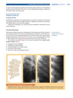
Figure 61-5