Chapter 080. Cancer Cell Biology and Angiogenesis (Part 4) ppsx
Bạn đang xem bản rút gọn của tài liệu. Xem và tải ngay bản đầy đủ của tài liệu tại đây (74.89 KB, 5 trang )
Chapter 080. Cancer Cell Biology
and Angiogenesis
(Part 4)
Telomerase
DNA polymerase is unable to replicate the tips of chromosomes, resulting
in the loss of DNA at the specialized ends of chromosomes (called telomeres) with
each replication cycle. At birth, human telomeres are 15- to 20-kb pairs long and
are composed of tandem repeats of a six-nucleotide sequence (TTAGGG) that
associate with specialized telomere-binding proteins to form a T-loop structure
that protects the ends of chromosomes from being mistakenly recognized as
damaged. The loss of telomeric repeats with each cell division cycle causes
gradual telomere shortening, leading to growth arrest (called replicative
senescence) when one or more critically short telomeres triggers a p53-regulated
DNA-damage checkpoint response. Cells can bypass this growth arrest if pRB and
p53 are nonfunctional, but cell death ensues when the unprotected ends of
chromosomes precipitate chromosome fusions or other catastrophic DNA
rearrangements (termed crisis). The ability to bypass telomere-based growth
limitations is thought to be a critical step in the evolution of most malignancies.
This occurs by the reactivation of telomerase expression in cancer cells.
Telomerase is an enzyme that adds TTAGGG repeats onto the 3' ends of
chromosomes. It contains a catalytic subunit with reverse transcriptase activity
(hTERT) and an RNA component that provides the template for telomere
extension. Most normal somatic cells do not express sufficient telomerase to
prevent telomere attrition with each cell division. Exceptions include stem cells
(such as those found in hematopoietic tissues, gut and skin epithelium, and germ
cells) that require extensive cell division to maintain tissue homeostasis. More
than 90% of human cancers express high levels of telomerase that prevent
telomere exhaustion and allow indefinite cell proliferation. In vitro experiments
indicate that inhibition of telomerase activity leads to tumor cell apoptosis. Major
efforts are underway to develop methods to inhibit telomerase activity in cancer
cells. The reverse transcriptase activity of telomerase is a prime target for small-
molecule pharmaceuticals. The protein component of telomerase (hTERT) can act
as a tumor-associated antigen recognized by antigen-specific cytotoxic T
lymphocytes (CTL) that lyse human melanoma, prostate, lung, breast, and colon
cancer cells in vitro. Clinical trials of telomerase vaccines are underway.
Signal Transduction Pathways as Therapeutic Targets in Cancer Cells
Since the discovery that the v-src oncogene has protein tyrosine kinase
activity, the central role of tyrosine phosphorylation in cellular responses to
growth factors has become apparent. Many tyrosine kinases act at the apex of
signaling pathways and are transmembrane proteins (receptor tyrosine kinases,
RTK) or are associated with structures at the plasma membrane (Src-, Janus-, and
Fak-family kinases). RTKs are transmembrane glycoproteins that undergo
dimerization upon ligand binding, with activation of their cytoplasmic tyrosine
kinase domains by proximity-induced trans-phosphorylation of the activation loop.
Tyrosine residues of the receptor or adaptor proteins (such as IRS-1 or Shc) are
phosphorylated and act as docking sites for proteins containing SH2 (Src-
homology 2) or PTB (phosphotyrosine binding) domains, thus initiating multiple
signal transduction pathways (Fig. 80-2). Normally, tyrosine kinase activity is
short-lived and reversed by protein tyrosine phosphatases (PTP). However, in
many human cancers, tyrosine kinases or components of their downstream
pathways are activated by mutation, gene amplification, or chromosomal
translocations. Because these pathways regulate proliferation, survival, migration,
and angiogenesis, they have been identified as important targets for cancer
therapeutics.
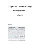
Figure 80-2
Therapeutic targeting of signal transduction pathways in cancer cells.
Three major signal transduction pathways are activated by receptor tyrosine
kinases (RTK). 1. The protooncogene Ras is activated by the Grb2/mSOS guanine
nucleotide exchange factor, which induces an association with Raf and activation
of downstream kinases (MEK and ERK1/2). 2. Activated PI3K phosphorylates the
membrane lipid PIP
2
to generate PIP
3
, which acts as a membrane-docking site for
a number of cellular proteins including the serine/threonine kinases PDK1 and
Akt. PDK1 has numerous cellular targets including Akt and mTOR. Akt
phosphorylates target proteins that promote resistance to apoptosis and enhance
cell cycle progression, while mTOR and its target p70S6K upregulate protein
synthesis to potentiate cell growth. 3. Activation of PLC-γ leads the formation of
diacylglycerol (DAG) and increased intracellular calcium, with activation of
multiple isoforms of PKC and other enzymes regulated by the calcium/calmodulin
system. Other important signaling pathways involve non-RTKs that are activated
by cytokine or integrin receptors. Janus kinases (JAK) phosphorylate STAT
(signal transducer and activator of transcription) transcription factors, which
translocate to the nucleus and activate target genes. Integrin receptors mediate
cellular interactions with the extracellular matrix (ECM), inducing activation of
FAK (focal adhesion kinase) and c-Src, which activate multiple downstream
pathways, including modulation of the cell cytoskeleton. Many activated kinases
and transcription factors migrate into the nucleus where they regulate gene
transcription, thus completing the path from extracellular signals, such as growth
factors, to a change in cell phenotype, such as induction of differentiation or cell
proliferation. The nuclear targets of these processes include transcription factors
(e.g., Myc, AP-1, and serum response factor) and the cell cycle machinery (CDKs
and cyclins). Inhibitors of many of these pathways have been developed for the
treatment of human cancers. Examples of inhibitors that are currently being
evaluated in clinical trials are shown in purple.