Chapter 102. Aplastic Anemia, Myelodysplasia, and Related Bone Marrow Failure Syndromes (Part 5) pdf
Bạn đang xem bản rút gọn của tài liệu. Xem và tải ngay bản đầy đủ của tài liệu tại đây (166.85 KB, 5 trang )
Chapter 102. Aplastic Anemia, Myelodysplasia, and
Related Bone Marrow Failure Syndromes
(Part 5)
Pathophysiology
Bone marrow failure results from severe damage to the hematopoietic cell
compartment. In aplastic anemia, replacement of the bone marrow by fat is
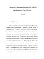
apparent in the morphology of the biopsy specimen (Fig. 102-1) and MRI of the
spine. Cells bearing the CD34 antigen, a marker of early hematopoietic cells, are
greatly diminished, and in functional studies, committed and primitive progenitor
cells are virtually absent; in vitro assays have suggested that the stem cell pool is
reduced to ≤1% of normal in severe disease at the time of presentation.
Figure 102-1
A. Normal bone marrow biopsy. B.
Normal bone marrow aspirate smear.
The marrow is normally 30–
70% cellular, and there is a heterogeneous mix of
myeloid, erythroid, and lymphoid cells. C. Aplastic anemia biopsy. D.
Marrow
smear in a
plastic anemia. The marrow shows replacement of hematopoietic tissue
by fat and only residual stromal and lymphoid cells.
An intrinsic stem cell defect exists for the constitutional aplastic anemias:
cells from patients with Fanconi's anemia exhibit chromosome damage and death
on exposure to certain chemical agents. Telomeres are short in a large proportion
of patients with aplastic anemia, and mutations in genes of the telomere repair
complex (TERC and TERT) can be identified in some adults with apparently
acquired marrow failure and without physical anomalies or typical family history.
Aplastic anemia does not appear to result from defective stroma or growth
factor production.
Drug Injury
Extrinsic damage to the marrow follows massive physical or chemical
insults such as high doses of radiation and toxic chemicals. For the more common
idiosyncratic reaction to modest doses of medical drugs, altered drug metabolism
has been invoked as a likely mechanism. The metabolic pathways of many drugs
and chemicals, especially if they are polar and have limited water solubility,
involve enzymatic degradation to highly reactive electrophilic compounds; these
intermediates are toxic because of their propensity to bind to cellular
macromolecules. For example, derivative hydroquinones and quinolones are
responsible for benzene-induced tissue injury. Excessive generation of toxic
intermediates or failure to detoxify the intermediates may be genetically
determined and apparent only on specific drug challenge; the complexity and
specificity of the pathways imply multiple susceptibility loci and would provide an
explanation for the rarity of idiosyncratic drug reactions.