Chapter 109. Disorders of Platelets and Vessel Wall (Part 6) potx
Bạn đang xem bản rút gọn của tài liệu. Xem và tải ngay bản đầy đủ của tài liệu tại đây (13.74 KB, 5 trang )
Chapter 109. Disorders of Platelets
and Vessel Wall
(Part 6)
Immune Thrombocytopenic Purpura: Treatment
The treatment of ITP utilizes drugs that decrease reticuloendothelial uptake
of the antibody-bound platelet and/or decrease antibody production. However, the
diagnosis of ITP does not necessarily mean that treatment must be instituted.
Patients with platelet counts >30,000/µL appear not to have increased mortality
related to the thrombocytopenia.
Initial treatment in patients without significant bleeding symptoms, severe
thrombocytopenia (<5000/µL), or signs of impending bleeding (such as retinal
hemorrhage or large oral mucosal hemorrhages) can be instituted as an outpatient
using single agents. Traditionally this has been prednisone at 1 mg/kg, although
Rh
0
(D) immune globulin therapy (WinRho SDF) at 50–75 µg/kg is also being
used in this setting. Rh
0
(D) immune globulin must be used only in Rh+ patients as
the mechanism of action is production of limited hemolysis, with antibody-coated
cells "saturating" the Fc receptors, inhibiting Fc receptor function. Hemoglobin
levels usually decrease (mean 1.7 g/dL), although severe intravascular hemolysis
is a rare complication. Doses are reduced if given to anemic patients. Intravenous
gamma globulin (IVIgG), which is pooled, primarily IgG antibodies, also blocks
the Fc receptor system, but appears to work primarily through different
mechanism(s). IVIgG has more efficacy than anti-Rh
0
(D) in post-splenectomized
patients. IVIgG is dosed at 2 g/kg total, given in divided doses over 2–5 days. Side
effects are usually related to the volume of infusion and infrequently include
aseptic meningitis and renal failure. All immunoglobulin preparations are derived
from human plasma and undergo treatment for viral inactivation.
For patients with severe ITP and/or symptoms of bleeding, hospital
admission and combined modality therapy are given using high-dose
glucocorticoids with IVIgG or anti-Rh
0
D therapy, and, as needed, additional
immunosuppressive agents. Rituximab, an anti-CD20 (B cell) antibody, has shown
efficacy in the treatment of refractory ITP.
Splenectomy has been used for treatment of patients who relapse after
glucocorticoids are tapered. Splenectomy remains an important treatment option;
however, more patients than previously thought will go into a remission over time.
Observation, if the platelet count is high enough, or intermittent treatment with
anti-Rh
0
(D) or IVIgG may be a reasonable approach to see if the ITP will resolve.
Vaccination against encapsulated organisms (especially pneumococcus, but also
menningococcus and Haemophilus influenzae, depending on patient age and
potential exposure) is recommended before splenectomy. Accessory spleen(s) are
a very rare cause of relapse.
New drugs for ITP include TPO receptor agonists. This approach to
treatment of ITP stems from the finding that many patients with ITP do not have
increased TPO levels, as was previously hypothesized, nor do they all have
increased platelet destruction. Two agents, one administered subcutaneously and
another orally, have shown response in many patients with refractory ITP. Roles
for these agents in ITP treatment are not fully defined.
Inherited Thrombocytopenia
Thrombocytopenia is rarely inherited, either as an isolated finding or as part
of a syndrome, and may be inherited in an autosomal dominant, autosomal
recessive, or X-linked pattern. Many forms of autosomal dominant
thrombocytopenia are now known to be associated with mutations in the
nonmuscle myosin heavy chain MYH9 gene. Interestingly, these include the May-
Hegglin anomaly and Sebastian, Epstein's, and Fechtner syndromes, all of which
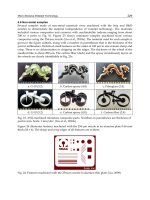
have distinct distinguishing features. A common feature of these disorders is large
platelets (Fig. 109-1C). Autosomal recessive disorders include congenital
amegakaryocytic thrombocytopenia, thrombocytopenia with absent radii, and
Bernard Soulier syndrome. The latter is primarily a functional platelet disorder due
to absence of GPIb-IX-V, the vWF adhesion receptor. X-linked disorders include
Wiskott-Aldrich syndrome and a dyshematopoietic syndrome resulting from a
mutation in GATA-1, an important transcriptional regulator of hematopoiesis.
Thrombotic Thrombocytopenic Purpura and Hemolytic Uremic
Syndrome
Thrombotic thrombocytopenic microangiopathies are a group of disorders
characterized by thrombocytopenia, a microangiopathic hemolytic anemia evident
by fragmented RBCs (Fig. 109-1D) and laboratory evidence of hemolysis, and
microvascular thrombosis. This includes thrombotic thrombocytopenic purpura
(TTP) and hemolytic uremic syndrome (HUS), as well as syndromes complicating
bone marrow transplantation, certain medications and infections, pregnancy, and
vasculitis. In DIC, while thrombocytopenia and microangiopathy are seen, a
coagulopathy predominates, with consumption of clotting factors and fibrinogen
resulting in an elevated prothrombin time (PT) and often activated partial
thromboplastin time (aPTT). The PT and aPTT are characteristically normal in
TTP or HUS.