Chapter 118. Infective Endocarditis (Part 4) pot
Bạn đang xem bản rút gọn của tài liệu. Xem và tải ngay bản đầy đủ của tài liệu tại đây (30.19 KB, 5 trang )
Chapter 118. Infective Endocarditis
(Part 4)
Cardiac Manifestations
Although heart murmurs are usually indicative of the predisposing cardiac
pathology rather than of endocarditis, valvular damage and ruptured chordae may
result in new regurgitant murmurs. In acute endocarditis involving a normal valve,
murmurs are heard on presentation in only 30–45% of patients but ultimately are
detected in 85%. Congestive heart failure develops in 30–40% of patients; it is
usually a consequence of valvular dysfunction but occasionally is due to
endocarditis-associated myocarditis or an intracardiac fistula. Heart failure due to
aortic valve dysfunction progresses more rapidly than does that due to mitral valve
dysfunction. Extension of infection beyond valve leaflets into adjacent annular or
myocardial tissue results in perivalvular abscesses, which in turn may cause
fistulae (from the root of the aorta into cardiac chambers or between cardiac
chambers) with new murmurs. Abscesses may burrow from the aortic valve
annulus through the epicardium, causing pericarditis. Extension of infection into
paravalvular tissue adjacent to either the right or the noncoronary cusp of the
aortic valve may interrupt the conduction system in the upper interventricular
septum, leading to varying degrees of heart block. Although perivalvular abscesses
arising from the mitral valve may potentially interrupt conduction pathways near
the atrioventricular node or in the proximal bundle of His, such interruption occurs
infrequently. Emboli to a coronary artery may result in myocardial infarction;
nevertheless, embolic transmural infarcts are rare.
Noncardiac Manifestations
The classic nonsuppurative peripheral manifestations of subacute
endocarditis are related to the duration of infection and, with early diagnosis and
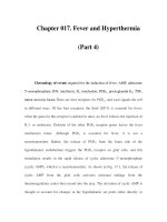
treatment, have become infrequent. In contrast, septic embolization mimicking
some of these lesions (subungual hemorrhage, Osler's nodes) is common in
patients with acute S. aureus endocarditis (Fig. 118-2). Musculoskeletal
symptoms, including nonspecific inflammatory arthritis and back pain, usually
remit promptly with treatment but must be distinguished from focal metastatic
infection. Hematogenously seeded focal infection may involve any organ but most
often is clinically evident in the skin, spleen, kidneys, skeletal system, and
meninges. Arterial emboli are clinically apparent in up to 50% of patients.
Vegetations >10 mm in diameter (as measured by echocardiography) and those
located on the mitral valve are more likely to embolize than are smaller or
nonmitral vegetations. Embolic events—often with infarction—involving the
extremities, spleen, kidneys, bowel, or brain are often noted at presentation. With
effective antibiotic treatment, the frequency of embolic events decreases from 13
per 1000 patient-days during the initial week to 1.2 per 1000 patient-days after the
third week. Emboli occurring late during or after effective therapy do not in
themselves constitute evidence of failed antimicrobial treatment. Neurologic
symptoms, most often resulting from embolic strokes, occur in up to 40% of
patients. Other neurologic complications include aseptic or purulent meningitis,
intracranial hemorrhage due to hemorrhagic infarcts or ruptured mycotic
aneurysms, seizures, and encephalopathy. (Mycotic aneurysms are focal dilations
of arteries occurring at points in the artery wall that have been weakened by
infection in the vasa vasorum or where septic emboli have lodged.)
Microabscesses in brain and meninges occur commonly in S. aureus endocarditis;
surgically drainable intracerebral abscesses are infrequent.
Figure 118-2
Septic emboli with hemorrhage and infarction due to acute
Staphylococcus
aureus endocarditis. (Used with permission of L. Baden.)
Immune complex deposition on the glomerular basement membrane causes
diffuse hypocomplementemic glomerulonephritis and renal dysfunction, which
typically improve with effective antimicrobial therapy. Embolic renal infarcts
cause flank pain and hematuria but rarely cause renal dysfunction.
Manifestations of Specific Predisposing Conditions
In almost 50% of patients who have endocarditis associated with injection
drug use, infection is limited to the tricuspid valve. These patients present with
fever, faint or no murmur, and (in 75% of cases) prominent pulmonary findings
related to septic emboli, including cough, pleuritic chest pain, nodular pulmonary
infiltrates, and occasionally pyopneumothorax. Infection involving valves on the
left side of the heart presents with the typical clinical features of endocarditis.
Health care–associated endocarditis (defined as that which is nosocomial,
arises after recent hospitalization, or is a direct consequence of long-term
indwelling devices) has typical manifestations if it is not associated with a retained
intracardiac device. Endocarditis associated with flow-directed pulmonary artery
catheters is often cryptic, with symptoms masked by comorbid critical illness, and
is commonly diagnosed at autopsy. Transvenous pacemaker lead– and/or
implanted defibrillator–associated endocarditis may be associated with obvious or
cryptic generator pocket infection and results in fever, minimal murmur, and
pulmonary symptoms due to septic emboli.
Late-onset prosthetic valve endocarditis presents with typical clinical
features. Cases arising within 60 days of valve surgery (early onset) lack
peripheral vascular manifestations, and typical symptoms may be obscured by
comorbidity associated with recent surgery. In both early-onset and more delayed
presentations, paravalvular infection is common and often results in partial valve
dehiscence, regurgitant murmurs, congestive heart failure, or disruption of the
conduction system.