DISORDERS OF THE VULVA AND VAGINA pps
Bạn đang xem bản rút gọn của tài liệu. Xem và tải ngay bản đầy đủ của tài liệu tại đây (304.84 KB, 28 trang )
20
DISORDERS OF THE VULVA
AND VAGINA
CHAPTER
571
COMMON VULVOVAGINAL
INFECTIONS ASSOCIATED
WITH LEUKORRHEA
Leukorrhea is a usually whitish vaginal discharge that may occur
at any age and affects virtually all women at some time. Although
some vaginal discharge (mucus) is physiologic and nearly always
present, when it becomes greater or abnormal (bloody or soils cloth-
ing), is irritating, or has an offensive odor, it is considered patho-
logic. Pathologic discharge is often coupled with vulvar irritation.
Commonly, the pathologic conditions are due to infection of the
vagina or cervix. Other causes may include uterine tumors, estro-
genic or psychic stimulation, trauma, foreign bodies (retained tam-
pon), excessive douching (especially with irritating medications),
and vulvovaginal atrophy (hypoestrogenism).
Vulvovaginal disorders constitute the major reason for office gy-
necology visits. These disorders are heavily influenced by the phys-
iologic alterations summarized in Table 20-1. Estrogen and pro-
gesterone influence the nonkeratinized squamous epithelium of the
vagina and vulva. Without hormonal influence, the epithelium is
thin and atrophic and contains little glycogen, and the vaginal fluid
has a high pH. By contrast, with adequate estrogen and proges-
terone, cellular glycogen content increases and the pH decreases
(partially due to breakdown of glycogen to lactic acid). During their
reproductive lives, most women harbor three to eight major types
of pathogenic bacteria at any given time (Table 20-1).
Physiologic vaginal secretions consist mainly of cervical mu-
cus (a transudate from the vaginal squamous epithelium) and exfo-
liated squamous cells. Lesser amounts are contributed by the meta-
bolic products of the microflora, exudates from sebaceous sweat
glands, Bartholin glands, and Skene glands, and small amounts of
Copyright 2001 The McGraw-Hill Companies. Click Here for Terms of Use.
TABLE 20-1
SUMMARY OF THE HORMONAL INFLUENCE, VAGINAL pH, AND USUAL (PREDOMINANT)
VAGINAL ORGANISMS AT DIFFERENT TIMES OF A FEMALE’S LIFE
Time of Life Hormonal Influence Vaginal pH Usual Predominant Vaginal Organisms
Birth Estrogen Progesterone 3.7–6.3 Anaerobic and aerobic
Infant None 6.0–8.0 Gram-positive cocci and bacilli
Puberty–Reproductive Estrogen Progesterone 3.5–4.5 Aerobes (%)
Lactobacillus (70–90)
Staphylococcus epidermidis (30–60)
Diphtheroids (30–60)
Alpha-hemolytic Streptococcus (15–50)
Group D Streptococcus (10–40)
Nonhemolytic Streptococcus (5–30)
Escherichia coli (20–25)
Beta-hemolytic Streptococcus (10–20)
Anaerobes (%)
Bacteroides fragilis (5–40)
Bacteroides species (1–40)
Peptococcus (5–60)
Peptostreptococcus (5–40)
Clostridium (5–15)
Veillonella (10–15)
Menopause Little or none 6.0–8.0 Gram-positive cocci and bacilli
572
endometrial and oviductal fluid. When there is little hormonal stim-
ulation (e.g., prior to puberty and postmenopausally), vaginal se-
cretions are scant and the genital tract is less resistant to infection.
Physiologic events enhancing the amount of cervical mucus and
vaginal discharge occur as a result of sexual or other emotional
stimulation, ovulation, pregnancy, and with the excessive estrogen
produced by feminizing ovarian tumors.
The normal vaginal flora is most likely to be interrupted during
nonphysiologic conditions with the symptomatology noted. The
most common organisms causing leukorrhea include Trichomonas
vaginalis (protozoon), Candida (yeast), Gardnerella (or a combina-
tion of organisms collectively known as Bacterial Vaginosis)
and Chlamydia (bacterial). Helminths (e.g., Oxyuris) may cause
leukorrhea in children. Leukorrhea is unusual in genital gonorrhea
or tuberculosis.
Investigation of vaginal discharge involves collection of histor-
ical information (what, when, where, why, and to what degree); ex-
amination of the vulva, vagina, and cervix; assessment of the dis-
charge (texture, color, odor); and preparation of a saline wet mount
(see p. 523). In the majority of infections, it is not necessary to per-
form a culture for confirmation of diagnosis.
TRICHOMONAS VAGINALIS
Trichomonas infection generally is manifest as a diffuse vaginitis
with varying vulvar involvement. T. vaginalis infections result in
marked pruritus with variable edema and erythema. Numerous red
points (strawberry patches), which rarely bleed, may be scattered
over the vaginal surface and cervical portio. The cervix, urethra,
and bladder may be secondarily infected. The leukorrhea is char-
acterized as thin, yellow-green, and occasionally frothy, with a fetid
odor. The discharge has a pH of 5–6.5. On saline wet mount, the
unicellular flagellate may be observed moving about in a field of
many leukocytes. The trichomonads are pear shaped and smaller
than epithelial cells but larger than white cells.
T. vaginalis is almost always a sexually transmitted infection.
It causes 20%–25% of infectious vaginitis and is responsible for up
to 3 million cases a year (United States). The source often can be
traced to the male partner, who may harbor the flagellate beneath
the prepuce or in the urethra or urethral prostate, yet remain asymp-
tomatic. Moreover, ϳ25% of females harboring T. vaginalis are
also asymptomatic, although some may have urinary frequency and
dyspareunia. T. vaginalis vaginitis is frequently followed by chronic
bacterial cervicitis.
CHAPTER 20
DISORDERS OF THE VULVA AND VAGINA
573
BENSON & PERNOLL’S
574 HANDBOOK OF OBSTETRICS AND GYNECOLOGY
The treatment for trichomoniasis is oral metronidazole (a sin-
gle 2 g dose, 1 g q12h ϫ 2, or 250 mg tid for 5–7 days). The side
effects of metronidazole include nausea, occasional vomiting, a
metallic taste, and intolerance to alcohol. It should not be taken dur-
ing the first trimester of pregnancy. It is necessary to treat both part-
ners. Men usually are treated with metronidazole 2 g PO or 1 g
q12h ϫ 2. In cases of sensitivity to metronidazole, topical clotri-
mazole is used.
CANDIDA ALBICANS
Candida albicans and related pathogens, Candida glabrata and
Candida tropicalis, are natural fungal inhabitants of the bowel and
are also found on the perineal skin. Thus, vaginal contamination
from these sources is common. C. albicans is also found in the vagi-
nal flora of ϳ25% of asymptomatic women. Candidal infections
occur when vaginal flora abnormalities take place (e.g., a decrease
in lactobacilli), and 80%–95% are caused by C. albicans. With Can-
dida infections, there is generally more vulvar pruritus than with
Trichomonas infections but less burning. The usual symptomatol-
ogy includes vaginal discharge, vulvar pruritus, burning, and dys-
pareunia. Candida vaginitis commonly leads to dermatitis of the
vulva and thighs. Symptomatology generally begins in the pre-
menstrual phase of the cycle, but ϳ20% of women with Candida
are asymptomatic. Unlike bacterial or protozoal vaginitis, Candida
infections are not considered a sexually transmitted disease and are
not commonly associated with mixed infections or sexually trans-
mitted diseases. At particular risk for developing candidiasis are
diabetics, oral contraceptive users, those who have recently taken
antibiotics, and pregnant women.
Vaginal discharge due to Candida infection has a cottage
cheese appearance, usually without odor. White, curdlike collec-
tions of exudate often are present, and some are lightly attached
to the cervical and vaginal mucosa. When these are removed,
slight oozing occurs. There may be both erythema and edema of
the vulva and vagina. The discharge with Candida infection has
a pH of 4–5. Mixing the secretions with a drop of 10%–20% KOH
microscopically reveals the characteristic mycelia and hyphae,
with only a moderate leukocyte response. Should culture be nec-
essary, it may be accomplished using Nickerson’s or Sabouraud’s
medium.
The treatment for C. albicans infection is topical 2% mi-
conazole nitrate, 1 applicator or vaginal suppository at bedtime
for 3–7 days. Alternatively, clotimatzole or butoconazole vaginal
suppositories or cream may be used nightly for 7–14 days. If C.
albicans recurs (a frequent occurrence), the patient should have a
glucose screening examination for carbohydrate intolerance. It is
also worthwhile to inquire about the possibility of a sexual partner
with Candida infection about the prepuce. Finally, it is crucial to
recognize that C. glabrata and C. tropicalis are resistant to the
imidazoles and may be the cause of recurrent infections. The dis-
charge must be cultured, and treatment is topical gentian violet
q3–4d ϫ 2–3. Boric acid (600 mg in gelatin caps) inserted high
in the vagina bid and douching every other night (to a total of
three times) with dilute povidone-iodine may be useful therapeutic
adjuncts.
BACTERIAL VAGINOSIS
Bacterial vaginosis (BV) is the clinical diagnosis describing an
overgrowth (100–1000-fold) of certain facultative and obligate
anaerobic bacteria derived from the patient’s endogeneous vaginal
flora. It is also known as Bacterial vaginitis, Nonspecific vaginitis,
Haemophilus vaginalis, and Gardnerella vaginalis. The usual bac-
terial species involved are: Bacteriodes species, Petostreptococcus
species, G. vaginalis, Mycoplasma hominis, and members of the
Enterobacteriaceae. Although asymptomatic in approximately one
half of patients, BV occurs in 10%–25% of general obstetrics and
gynecology patients. The incidence of BV is higher (ϳ2/3) in pa-
tients being seen for STDs.
The primary symptom of BV is a relatively alkaline, malodor-
ous (fishy), gray (dark or dull), watery, homogeneous discharge that
is worse during menses and after intercourse. Vulvar pruritis is a less
frequent symptom. In addition to history and physical examination,
the investigation of BV includes a vaginal pH, a “whiff” (smell) test,
and a microscopic wet-mount. The wet-mount is usually character-
ized by: clue cells, an abundance of bacteria of various morpholo-
gies, the absence of homogeneous bacilli (lactobacilli), and an ab-
sence or paucity of inflammatory cells. Pap tests are not effective
in the diagnosis of BV and cultures are necessary only when the
discharge does not respond to treatment or overgrowth of a specific
organism is suspected. The diagnosis of BV (false-positives Ͻ10%)
is confirmed by 3 of the 4 following criteria:
●
pH Ͼ4.5,
●
Clue cells,
●
Positive KOH,
●
Homogeneous discharge.
CHAPTER 20
DISORDERS OF THE VULVA AND VAGINA
575
BENSON & PERNOLL’S
576 HANDBOOK OF OBSTETRICS AND GYNECOLOGY
Treatment may be local (intravaginal) or systemic (oral). The lo-
cal regimens include: 0.75% metronidazole gel bid for 5 d, and 2%
clindamycin cream once a d for 7 d. Oral metronidazole (500 mg bid,
250 mg tid) for 7 d is Ͼ90% effective, whereas a single 2 g dose is
less effective (ϳ70%) and has a greater incidence of gastrointestinal
upset. Recurrences occur with vexing frequency. Although treatment
of partners is not recommended unless BV is recalcitrant to therapy,
this remains a controversial area. The higher association of BV and
STDs should heighten the practitioner’s suspicion concerning gon-
orrhea, chlamydia, syphilis, hepatitis and HIV.
BV may be associated with furthering the incidence of a num-
ber of gynecological complications, including: PID, postabortal
infections, and posthysterectomy vaginal cuff cellulitis. Although
not completely proven, treatment of the BV appears to decrease
the incidence of these complications and provides at least part of
the rationale for prophylactic antibiotic therapy in these circum-
stances.
Additionally, BV has been incriminated in increasing the inci-
dence of preterm delivery, premature rupture of membranes, am-
nionitis, chorioamnionitis, and postpartum endometritis. Thus, it is
currently recommended that BV screening be considered during
pregnancy in risk patients, but data supporting low-risk screening
has not emerged. There is also no common agreement on therapy
or rescreening. During pregnancy, 2% clindamycin intravaginal
cream may be used once a d for 7 d, but may be less effective. Al-
ternatively, clindamycin 300 mg bid for 7 d may be used. Finally,
metronidazole oral therapy may be used after the first trimester.
CHLAMYDIA TRACHOMATIS
Chlamydial infections are caused by the obligate intracellular bac-
terium, Chlamydia trachomatis. Other closely related infections
are lymphogranuloma venereum, inclusion conjunctivitis, urethritis,
cervicitis, salpingitis, proctitis, epididymitis, and pneumonia of the
newborn. C. trachomatis infection may be the most prevalent sex-
ually transmitted disease in the United States, affecting .3 million
persons annually. It is often asymptomatic (ϳ60%–80% of infected
women and ϳ10% of infected men). The organism is best detected
by enzyme-linked amino acids in a fluorescein-conjugate mono-
clonal antibody test. The infections usually begin as mucopurulent,
often odorous or pruritic discharges, and the principal site of in-
fection is the cervix. Chlamydia can be eradicated from the vagina
and cervix by tetracycline or erythromycin 500 mg PO qid for
7 days.
COMMON VULVOVAGINAL
VIRAL INFECTIONS
HERPES SIMPLEX VIRUS (HSV)
HSV infections of the genital tract are a sexually transmitted dis-
ease. Type 2 HSV accounts for ϳ90% of infections, and 10% are
type 1. This DNA virus has an incubation period of 3–22 days, and
even primary attacks may be asymptomatic, although most patients
complain of fever, malaise, anorexia, local genital pain, leukorrhea,
dysuria, or even vaginal bleeding. Typical genital lesions are mul-
tiple vesicles that progress to shallow ulceration often surrounded
by redness or erythematous patches. Painful bilateral inguinal
adenopathy is usually present during the primary infection. If the
urethra or bladder is affected, dysuria or urinary retention may re-
sult. The lesions gradually heal without scarring (7–10 days) unless
bacterial superinfection occurs.
The diagnosis is usually made on the typical appearance of vesi-
cles and ulcers. Cytologic smear of the ulcers or vesicles demon-
strates classic multinucleated giant cells with acidophilic intranu-
clear inclusion bodies. Definitive culture may be obtained from the
fluid of unruptured vesicles using Hanks medium. However, false-
negative cultures are frequent. Serologic diagnosis is possible, and
use of the gamma globulin or macroglobulin response may deter-
mine if the attack is recurrent or primary.
Affected individuals harbor the virus indefinitely. Recurrent le-
sions may be triggered by emotional distress, exposure to the sun,
or a variety of other stimuli. After the primary lesion, the patient
frequently develops paresthesias in the affected region before a re-
currence (the virus resides in specialized nerve endings during la-
tent intervals). Recurrent lesions account for much of the morbid-
ity but are not as painful as the primary lesions.
Genital herpes during pregnancy is hazardous to the fetus. Ser-
ial cultures for the detection of asymptomatic viral shedding have
been very disappointing as a diagnostic technique during pregnancy.
It is recommended that an infant not be delivered through the birth
canal with active lesions. Although cesarean section does not guar-
antee that the infant will not be infected, it may be undertaken if it
is Ͻ4 h after rupture of the membranes. Delivery through an infected
birth canal with active lesions poses ϳ50% chance of the neonate
developing neonatal herpes. Of those infected, ϳ50% die and ϳ25%
have permanent neurologic sequelae. Additionally, HSV type 2 has
been suggested (but not proven) as etiologic in cervical dysplasia.
CHAPTER 20
DISORDERS OF THE VULVA AND VAGINA
577
BENSON & PERNOLL’S
578 HANDBOOK OF OBSTETRICS AND GYNECOLOGY
Currently, there is no cure for herpes simplex viral infections.
Symptomatic measures include hot sitz baths, douching with Bur-
row’s solution, and oral or parenteral acyclovir. Local or oral acy-
clovir may shorten the course of an initial attack but has little
effect on recurrences. Valacyclovir may also be used for treat-
ment of an initial infection (1 g bid PO for 10 d, started Ͻ72 h af-
ter onset of symptoms), treatment of recurrances (500 mg bid PO
for 5 d, started Ͻ24 h after onset of symptoms) or for suppres-
sion (1 g PO a day, limited to Ͻ1 yr of use). Another suppressive
agent is famciclovir.
General rules for prevention of dissemination include covering
small lesions situated away from the oral or vaginal orifices with
occlusive dressing during sexual contact, the use of condoms, and
the application of contraceptive cream or foam. A partner may be-
come infected despite these precautions. If a regular partner has had
genital herpes or has not been infected despite prolonged exposure,
precautions are probably not necessary.
HUMAN PAPILLOMAVIRUS (HPV)
A member of the Papovavirus group, human papillomavirus causes
condylomata acuminata. The virus is sexually transmitted, com-
monly affects both partners, and affects the same age group as other
venereal diseases. This DNA virus causes easily discernible, raised,
papillomatous lesions of the vulva as well as less discernible le-
sions of the vagina and cervix. The lesions are much more florid in
patients who are diabetic, pregnant, taking oral contraceptives, or
immunosuppressed. The most common complaints concern the le-
sions themselves, but vaginal discharge or pruritus may be present.
The vaginal or cervical lesions are occasionally exophytic or
papillomatous (wartlike) but may also be flat, spiked, or inverted.
The flat condylomata are white lesions with a somewhat granular
surface and a mosaic pattern (some with punctuation) on col-
poscopy. The papillomatous condylomata is a raised white lesion
with fingerlike projections, often containing capillaries. The spiked
condyloma is a hyperkeratotic lesion with surface projection and
prominent capillary tips. Inverted condylomata grow into cervical
glands and, thus, do not occur in the vagina.
Subtypes 6 and 11 are primarily responsible for genital warts.
Cytologic smear or biopsy of vaginal or cervical lesions reveals
koilocytes, which are superficial or intermediate cells character-
ized by an enlarged perinuclear cavity that stains only faintly.
Biopsy often is necessary to distinguish cervical condylomata from
dysplasia.
Treatment in nonpregnant patients generally consists of weekly
applications of podophyllin (25% in tincture of benzoin). If after
4–6 weeks this is not successful, cryosurgery, electrocautery, or
laser therapy may be necessary. Podophyllin should not be used
during pregnancy, and if it is used within 6 weeks of biopsy, the
pathologist must be notified because bizarre changes occur that
could alter the diagnosis. During pregnancy, cryosurgery is most
commonly used for therapy of condylomata. If vaginal or introital
lesions are present, consider cesarean section because of the possi-
bility of bleeding from the very friable lesions as well as the pos-
sibility of the fetus acquiring laryngeal papillomatosis (infection of
the vocal cords by papillomavirus) during the birth process.
MOLLUSCUM CONTAGIOSUM
Molluscum contagiosum is an autoinoculable virus with an incu-
bation period of 1–4 weeks. Asymptomatic pink to gray, discrete,
umbilicated epithelial skin tumors Ͻ1 cm in diameter develop gen-
erally on the vulva. The histologic picture is that of numerous in-
clusion bodies in the cell cytoplasm. Each lesion must be treated
by desiccation, freezing or curettage, and chemical cauterization of
the base.
OTHER VULVOVAGINAL
INFECTIONS
BARTHOLIN DUCT CYST AND ABSCESS
The Bartholin duct is susceptible to infectious occlusion because of
its length and narrowness. Infectious organisms (often Neisseria
gonorrhoeae with secondary streptococci, staphylococci, or Escherichia
coli) become pocketed within the passage to form an abscess. The
inflammation usually resolves, but permanent occlusion of the dis-
tal duct causes retention of mucus produced by the gland, and a cyst
develops. The process is usually unilateral and occurs in up to 2%
of women. The gland is almost never seriously involved with the
ductal infection, but in older women acquiring a mass in the
Bartholin area, carcinoma (see p. 592) must be excluded.
Clinical manifestations include acute pain, tenderness, and dys-
pareunia. Surrounding tissues (at the junction of the mid and lower
thirds of the labia minora) become inflamed and edematous. The
introitus may be distorted, and a fluctuant mass usually is palpable.
CHAPTER 20
DISORDERS OF THE VULVA AND VAGINA
579
BENSON & PERNOLL’S
580 HANDBOOK OF OBSTETRICS AND GYNECOLOGY
Rarely are systemic symptoms reported or signs of infection noted.
Smears and cultures may reveal a specific bacteriologic diagnosis.
By the time the process is seen, however, the culture usually will
not be reliable.
The differential diagnosis includes inclusion cysts, large seba-
ceous cysts, hidradenoma, congenital anomalies, primary malig-
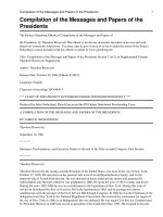
nancy, and metastatic cancers. Treatment consists of drainage of the
infected cyst or abscess, preferably by marsupialization (Fig. 20-1).
This procedure best affords permanent fistula formation. Other pro-
cedures (e.g., simple incision and drainage) frequently lead to re-
currence. Marsupialization is feasible under local anesthesia, and
fine interrupted chromic catgut or polyglycolic acid sutures are gen-
erally employed. If considerable surrounding inflammation is pres-
ent, broad-spectrum antibiotics should be given until appropriate
antibiotics for organisms in the abscess pus (determined by culture
at the time of surgery) can be determined. Bedrest, local dry or
moist heat or both, and analgesics should be used as indicated. Prog-
nosis is good with marsupialization. With other treatment, recurrent
infection and cystic dilation are likely. Rarely, it is necessary to sur-
gically excise the entire gland. Although in all cases it is desirable
to biopsy an area for pathologic section, this becomes crucial in the
perimenopausal or postmenopausal woman because of the risk of
Bartholin carcinoma.
HIDRADENITIS
Hidradenitis is a refractory infection of the apocrine sweat glands
usually caused by staphylococci or streptococci. It is analogous to
cystic acne, and symptoms are soreness and local swelling, edema,
FIGURE 20-1. Marsupialization of Bartholin cyst.
cellulitis, and suppuration of the groin. Involvement of apocrine
glands establishes the diagnosis. Treatment consists of hot, wet
packs, drainage, and specific antibiotics chosen on the basis of cul-
ture and sensitivity testing. Excision may be necessary, but the
wound must be allowed to heal by secondary intention.
TOXIC SHOCK SYNDROME (TSS)
Toxic shock syndrome generally occurs in previously healthy
women of childbearing age (usually 12–24 years). The incidence is
currently ϳ5/100,000 menstruating women per year. TSS is char-
acterized by abrupt onset of high fever (1028F); a diffuse macular
erythematous rash (sunburnlike) over the face, trunk, and proximal
extremities; and hypotension (systolic ,90 mm Hg). Additionally,
there is involvement of three or more of the following systems: gas-
trointestinal (vomiting and watery diarrhea), muscular (tenderness),
mucous membranes (nonpurulent conjunctivitis, sore throat), renal
(failure), hepatic (failure), hematologic (thrombocytopenia), and
central nervous system (nuchal rigidity, headaches, confusion). Re-
nal failure and cardiac failure are manifestations in severe cases and
generally occur within Ͻ48 h of onset.
Coagulase-positive Staphylococcus aureus has been isolated
from the vagina of victims, but blood cultures are negative. The
cause is most likely an exotoxin (exfoliatin) produced by some
strains of staphylococci. TSS begins (95% of cases) within 5 days
of the onset of menses in which tampons are used, and superab-
sorbent tampons appear to be linked to causation. Other potential
sources of TSS include delivery, diaphragm usage, surgery, soft-
tissue abscess, pyelonephritis, and osteomyelitis.
The laboratory workup must include a CBC with differential
count, electrolytes, UA, BUN, creatinine, liver function studies,
blood culture, throat culture, and vaginal culture. A lumbar punc-
ture should be performed if signs of meningitis are present, and
the CSF should be analyzed and cultured. The differential diag-
nosis includes Kawasaki disease (in children), scarlet fever, Rocky
Mountain spotted fever, leptospirosis, gram-negative sepsis, and
measles.
Treatment includes removal of a tampon if present (as well as
culture for penicillinase-producing S. aureus), admission to a crit-
ical care unit for intensive (often invasive) monitoring, correction
of fluid and electrolyte deficiencies (sizable deficits occur from third
spacing), corticosteroid therapy (methylprednisolone 30 mg/kg or
dexamethasone 3 mg/kg as a bolus, repeated q4h prn), antistaphylo-
coccal antibiotics (beta-lactamase-resistant antibiotics, e.g., nafcillin,
CHAPTER 20
DISORDERS OF THE VULVA AND VAGINA
581
BENSON & PERNOLL’S
582 HANDBOOK OF OBSTETRICS AND GYNECOLOGY
oxycillin, or methicillin 1 g IV q4h or vancomycin 500 mg IV q6h
if penicillin allergy exists), and management of renal and cardiac
insufficiency. It may be necessary to give blood and blood prod-
ucts (packed RBCs, fresh frozen plasma, platelets). Corticosteroids
shorten the fever duration and reduce the severity of illness.
Dopamine infusion may be necessary (2–5 m g/kg/min) if fluids
do not correct hypotension. Naloxone may be used in persistent
hypotension for its antiendorphin activity. Since gram-negative
sepsis is in the differential diagnosis, an aminoglycoside should
be given until gram-negative sepsis is ruled out. For both van-
comycin and the aminoglycoside, drug levels must be carefully
monitored.
Complications include adult respiratory distress syndrome
(ARDS), intractable hypotension, and hemorrhage from dissemi-
nated intravascular coagulation, any of which can be fatal. Mor-
tality from TSS is 3%–6%. Desquamation, especially of palms and
soles, occurs 1–2 weeks after onset of TSS. There is a 30% recur-
rence rate, especially in the first 3 months after the attack. The re-
currences are reduced to ϳ5% by administration of antistaphylo-
coccal antibiotics in the initial episode. If a woman recovers from
TSS, she should forego the use of tampons until cervicovaginal and
nasal cultures for S. aureus are negative twice at 4-week intervals
and then avoid tampon use at night.
FURUNCULOSIS
Furuncular abscesses caused by staphylococcal infections are
termed furunculosis or boils. Symptoms usually include throbbing
pain and regional tenderness. Pustular areas require incision and
drainage, with culture of the pus. Treatment includes segregation,
topical moist heat periodically, and systemic antibiotics (e.g.,
cephalosporin).
TUBERCULOSIS
Vulvovaginal tuberculosis, rare even in developing countries, is
manifest by chronic, minimally painful, exudative sores that are red-
dish, raised, moderately firm and nodular, with central apple jelly-
like contents. Later, ulcerative, undermined, necrotic, discharging
lesions develop. There is some tendency toward healing with heavy
scarring, but induration and sinus formation are common in the
scrofulous type of infection. The differential diagnosis includes
cancer and sexually transmitted diseases. Demonstration of My-
cobacterium tuberculosis is necessary for diagnosis. Treatment con-
sists of antituberculosis chemotherapy.
VULVAR INFESTATIONS
PEDICULOSIS PUBIS
Pthirus pubis (crab louse) is transmitted by sexual contact or from
bedding or clothing. The eggs are laid at the base of the pubic,
axillary, or scalp hair. When the eggs hatch, the lice attach to the
skin and cause intense itching. Close observation reveals minute,
pale brown insects and their ova attached to hair shafts near
the skin. The treatment consist of 1% gamma benzene hexachlo-
ride cream/lotion or shampoo (not recommended for pregnant or
lactating women) or pyritherans applied to the infestation and
adjacent hair areas. Retreatment may be required in 1 week. It
is necessary to treat all contacts and sterilize infected bedding
and clothing.
SCABIES
Sarcoptes scabei causes intractable itching and excoriation of the
surface in the vicinity of minute skin burrows where the parasites
have deposited ova. The mite is transmitted directly from person to
person. Treatment is 1% gamma benzene hexachloride cream/lotion
from the neck down overnight, washing off thoroughly after 8 h, or
10% crotamiton cream or lotion applied from the neck down twice
nightly and washed off thoroughly after the second application. With
this infestation, contacts must be treated, and all infected clothing
and bedding must be sterilized.
ENTEROBIASIS (PINWORMS)
Enterobius vermicularis is a short, spindle-shaped roundworm that
commonly infects children. The usual symptomatology is noctur-
nal perianal itching, which leads to excoriation. The usual diag-
nostic technique is a short strip of cellophane pressure-sensitive tape
applied to the perianal region and then spread on a slide. This re-
veals the adult worms or ova in Ͼ90% of cases. Therapy is a sin-
gle oral dose of mebendazole 100 mg.
CHAPTER 20
DISORDERS OF THE VULVA AND VAGINA
583
BENSON & PERNOLL’S
584 HANDBOOK OF OBSTETRICS AND GYNECOLOGY
BENIGN VULVAR LESIONS
ECZEMA
Eczema is a nonspecific, common, pruritic, moist dermatitis char-
acterized by excoriation and crusting with later lichenification.
Eczema is often a contact dermatitis caused by irritants in soap, bath
oils, or deodorant medications, dyes in clothing, or allergy to wool
or silk. Sensitivity tests and the exclusion of other dermatitis aid in
diagnosis. General treatment depends on elimination of the irritant.
Therapy is Burrow’s solution followed by steroid creams (e.g., 0.5%
hydrocortisone bid).
PSORIASIS
Pruritic, reddened, slightly elevated lesions (without the typical sil-
very scale seen on elbows and knees) are seen in body folds. The
elbows and knees are frequently affected by the scaly lesions, how-
ever. Psoriasis is a chronic, often familial, disorder of unknown eti-
ology. Exacerbations often occur in winter, and treatment includes
improving hygiene and 0.5% hydrocortisone cream applied bid.
More extensive lesions require dermatologic consultation.
BENIGN NEOPLASIA
A number of benign tumors may involve the vulvovaginal
area. These are generally characterized as either cystic or solid.
The cysts include epidermal cysts, sebaceous cysts, and apocrine
sweat gland cysts. A cyst of epidermal origin may arise from trauma
or occlusion of pilosebaceous ducts. These tend to be small, soli-
tary, lined with squamous epithelium, and filled with sebaceous
material as well as desquamated epithelial cells. Most are asymp-
tomatic.
Cysts of the sebaceous or sweat glands are frequently multiple
and almost always involve the labia majora. They are asymptomatic
unless infection develops. Apocrine sweat glands become functional
after puberty. Then, occlusion of the ducts results in an extremely
pruritic, microcytic disorder, Fox-Fordyce disease. Should the apo-
crine glands become infected by streptococci or staphylococci, the
process termed hidradenitis supprativa occurs.
Less common cysts or pseudocysts include Skene duct cysts,
urethral diverticula, inguinal hernia, occlusion of a persistently
patent vaginalis (canal of Nuck), dilation of mullerian duct vestiges,
and supernumerary mammary tissue.
The most worrisome benign vulvar solid tumors are pigmented
nevi. Because nearly all vulvar nevi are of the junctional type, they
may give rise to malignant melanomas. Thus, vulvar pigmented nevi
should be viewed more cautiously than elsewhere on the body. All
small pigmented lesions of the vulva are suspect and should be re-
moved with a 0.5–1 cm margin. Other benign solid tumors usually
are incidental findings and, like the cystic tumors, usually are pro-
visionally diagnosed by clinical examination. If therapy is required,
excisional biopsy is usually sufficient.
An acrochordon (or skin tag) is a small, flesh-colored, polypoid
tumor composed of fibrous epithelial elements and is never malig-
nant. Mesodermal vulvar tumors are infrequent, although leiomy-
omas arise from the round ligament and fibromas and lipomas also
occur. Neurofibromas are usually small lesions that arise from the
neural sheath and are of little consequence unless associated with
general neurofibromatosis (von Recklinghausen disease).
VULVAR DYSTROPHIES
Disorders of vulvar epithelial growth and nutrition produce nu-
merous nonspecific gross changes collectively termed vulvar dys-
trophies. These abnormalities are divided into hypertrophic, at-
rophic, and mixed types. Generally, the lesions are circumscribed
or diffuse white lesions of the vulva and do not have a uniform
microscopic appearance throughout. Therefore, multiple biopsies
are necessary. The toluidine blue test and colposcopy may assist
in detailing areas most suitable for biopsy. The malignant poten-
tial of vulvar dystrophies is ,5%. Table 20-2 is the International So-
ciety for the Study of Vulvar Disease classification of vulvar
dystrophies.
Treatment of atrophic dystrophies is topical 2% testosterone
proprionate in petrolatum bid for 1 week, then daily, gradually
decreasing to one to two applications per week. Androgenic side
effects may occur—thus the amount used should be minimal. Con-
trol of itching is accomplished by removal of any source of irrita-
tion (e.g., nylon panties, use of strong soaps), intermittent Bur-
row’s solution wet dressings (bid or tid), and topical fluorinated
corticosteroid (e.g., 0.025%–0.1% triamcinolone acetonide) bid for
1–2 weeks. Because these latter compounds may cause vulvar at-
rophy and contracture, the dose must be decreased as symptoms
subside. Surgical repair is indicated in cases of lichen sclerosis with
severe constriction of the vulva at the posterior fourchette.
CHAPTER 20
DISORDERS OF THE VULVA AND VAGINA
585
TABLE 20-2
CLASSIFICATION OF VULVAR DYSTROPHIES ADOPTED BY THE INTERNATIONAL SOCIETY
FOR THE STUDY OF VULVAR DISEASE
Clinical Features Histologic Features
Lichen sclerosis Pruritic, thin, parchment-like atrophic Thin, loss of rete homogenization;
area; introital stenosis inflammatory infiltrate
Hyperplastic
a
Pruritic, thick, gray or white plaques Acanthosis, hyperkeratosis,
on skin or mucosa inflammatory infiltrate
Mixed
a
Areas compatible with both forms may (See above)
be present at the same time
a
Atypia may accompany hyperplastic dystrophy and is graded as mild, moderate, or severe.
586
HYPERTROPHIC DYSTROPHIES
Chronic vaginal infection or other chronic irritation may cause
benign epithelial thickening and hyperkeratosis. In the acute
phase, this lesion may be red and moist, often with evidence of
secondary infection. Following subsequent epithelial thickening
and maceration, a raised white lesion (lichen simplex chronicus
or neurodermatitis) often develops, which may involve any of the
external genital area. Diagnosis is afforded by multiple biopsy
assessment. Characteristically, hyperkeratosis and acanthosis
with thickening of the epithelium and elongation of the rete pegs
occur. If advancement to atypical hyperplasia or carcinoma in situ
occurs, expect pleomorphism and loss of epithelial cellular po-
larity. The patient must be reexamined periodically to rule out
advancement to frank cancer. Surgical excision of more advanced
lesions is indicated.
ATROPHIC DYSTROPHIES
Lichen sclerosis et atrophicus (LSA) is a cutaneous degenerative
disorder of unknown cause. The vulva is most frequently affected,
but the skin of the back, axillas, beneath the breast, neck, and arms
also may be affected. The topical disease can occur in most age
groups but is most common in white women .65 years. In the per-
ineal area, LSA classically involves the vulvar, perineal, and peri-
anal areas in an hourglass pattern. The skin is white, thin, and wrin-
kled, and there may be surface atrophy of the labia minora and
majora. The chief symptom is pruritus.
Microscopically, LSA is distinguished by hyperkeratosis, ep-
ithelial atrophy, and flattening of the rete pegs. Beneath the epi-
dermis is a homogeneous, collagenous, acellular, pink-staining
zone. Below this lies a concentration of plasma cells. Cellular pleo-
morphism and loss of epithelial cell polarity are typical. Although
the lesion appears atrophic, the rate of cellular turnover is higher
than in normal skin or many hypertrophic lesions. Thus, there is an
enhanced rate of malignancy.
Treatment of hypertrophic and atrophic lesions involves elimi-
nating infection and the cautious use of estrogenic creams or topi-
cal corticosteroids and testosterone (e.g., 1% hydrocortisone and
2%–3% testosterone tid to qid). Treat carcinoma in situ or invasive
cancer in a dystrophic area definitively. Lesser vulvar intraepithe-
lial neoplasias (VIN I or mild dysplasia, VIN II or moderate dys-
plasia) should be treated conservatively to relieve symptoms. How-
ever, arrange close follow-up for signs of progression.
CHAPTER 20
DISORDERS OF THE VULVA AND VAGINA
587
BENSON & PERNOLL’S
588 HANDBOOK OF OBSTETRICS AND GYNECOLOGY
VULVAR CARCINOMA IN SITU
SQUAMOUS CELL CARCINOMA
A vulvar carcinoma in situ (CIS) is diagnosed when the full ep-
ithelial thickness is replaced by hyperchromatic cells with poorly
defined cellular boundaries. Increased cellular density, abnormal
mitoses, multinucleated cells, and increased nuclear/cytoplasmic ra-
tios may also be noted. Chronic infections, granulomatous disease,
and the vulvar dystrophies have long been associated with enhanced
susceptibility to vulvar CIS. There is increasing evidence that pa-
pillomavirus infections may play a major role in the etiology of
vulvar intraepithelial neoplasia (VIN). VIN is most often found
in women Ͼ40 years of age. However, with evidence of papillo-
mavirus, the median age falls to ϳ31 years. The progression rate
from VIN to carcinoma appears to be low.
CIS of the vulva is likely to be located posterior to the vaginal
orifice in the vulvar and perineal areas. VIN III (severe dysplasia
and CIS), like the vulvar dystrophies and VIN I and II, is most fre-
quently multifocal, and contiguous areas may be affected. For ex-
ample, with vulvar CIS the following may be affected: anus (22%),
clitoral glans (18%), vagina (10%), and urethral meatus (2%).
The symptoms of vulvar CIS usually are nonspecific (e.g., mild
irritation or itching). The gross appearance of the vulva with CIS is
variable (white patches, reddish nodules, dystrophic areas, pigmented
nevi). Biopsy is mandatory to establish the diagnosis. Because the le-
sions are usually discrete and multifocal, the toluidine blue test or
colposcopy or both are helpful to identify the correct area(s) for sam-
pling. Colposcopic examination will not reveal the characteristic tis-
sue and vascular patterns often found on the cervix, but it is useful
in identifying white or pigmented lesions for biopsy.
The toluidine blue test, which stains nuclei in the superficial
epithelium, is not diagnostic of CIS, but the dye is a useful adjunct.
Aqueous 1% toluidine blue is applied to the vulva, and after drying
for Ͼ1 min, the excess is gently removed with a cotton swab mois-
tened with 1% acetic acid. The areas retaining a blue color are the
ones to be biopsied. Although exfoliative cytology may be useful in
ulcerated lesions, it is of much less value for vulvar than for cervical
lesions because the thick keratinized skin does not shed cells readily.
Biopsy is easily accomplished using a 4–5 mm Keyes dermal
punch after local anesthesia has been administered. The dermal
thickness is penetrated, the specimen is elevated, and the underly-
ing stroma is incised. Bleeding may be controlled using pressure or
Monsell’s solution (ferrous subsulfate) or silver nitrate.
Therapy for CIS requires the removal of all vulvar VIN together
with any condylomata acuminata. Currently, the therapeutic modali-
ties include laser vaporization, topical 5-fluorouracil (5-FU), and sur-
gery. Carbon dioxide laser treatment allows healing in 2–3 weeks
without scarring. Ablation is usually to a depth of 3–4 mm under lo-
cal or general anesthesia. More than one therapy session may be nec-
essary for very extensive lesions. The use of 5-FU will successfully
eliminate CIS in ϳ75% of patients, but it causes vulvar edema, and
severe pain may be reported for ϳ6 weeks. Wide local excision has
all but been replaced by laser therapy. Should surgical therapy be nec-
essary, wide local excision, a skinning vulvectomy (i.e., removal of
the superficial vulvar skin and replacement with a split-thickness graft
while preserving the clitoris) and simple vulvectomy are options.
Prognosis for patients with CIS is good with all modes of therapy.
PAGET’S DISEASE
This rare vulvar intraepithelial lesion, which most often affects
postmenopausal Caucasian women, is associated with other vulvar
disorders (31%) or more distant carcinoma or CIS. The latter group
approaches 30% of cases and includes the breast, cervix, rectum,
urethra, and skin. Therefore, identification of vulvar Paget’s disease
mandates a thorough search for other cancers.
Vulvar Paget’s disease may be confused with other chronic pru-
ritic vulvar lesions. Paget’s disease is typically a velvety, red skin dis-
coloration that comes to resemble eczema with secondary maceration
and the development of white plaques. It is slowly growing but may
spread to the perineum, perianal area, or thighs. The primary symp-
tom is pruritus. Biopsy is mandatory, and Paget cells on microscopy
are pathognomonic (it is equivalent to Paget’s disease of the breast).
Extramammary Paget’s disease is an in situ lesion that warrants
simple vulvectomy with careful pathologic examination, including
the surgical margins. Local recurrence is a major problem, and
repeated local surgical excisions may be necessary. However, pro-
gression to adenocarcinoma is rare. Women with vulvar Paget’s
disease posttherapy should have an annual breast evaluation (Chap-
ter 17), vulvar evaluation, cervical cytologic study, and screening
for malignant gastrointestinal disease.
CANCER OF THE VULVA
Cancer of the vulva occurs primarily in postmenopausal women.
There is usually a long history of vulvar irritation, with itching,
CHAPTER 20
DISORDERS OF THE VULVA AND VAGINA
589
BENSON & PERNOLL’S
590 HANDBOOK OF OBSTETRICS AND GYNECOLOGY
local discomfort, and possibly bloody discharge. Whereas early
lesions may appear as chronic vulvar dermatitis, the late lesions
appear as nodules, exophytic lesions, or hard ulcerated areas. Di-
agnosis requires biopsy.
Vulvar tumors are 85%–90% epidermoid in origin. Nonethe-
less, cancer of the vulva may arise also from the urethra, glandular
elements of the vulva, or mucosa of the lower third of the vagina.
Vulvar cancers are intraepithelial or invasive. Vulvar cancer is the
fourth most common female genital cancer (after endometrial, cer-
vical, and ovarian cancer) and accounts for ϳ5% of gynecologic
malignancies. The patient with vulvar malignancy is predisposed
also to other malignancies; 22% will have another primary tumor
(most commonly of the cervix). The average age of patients with
vulvar cancer is 65, and .50% of afflicted women are .50 years.
The cause of vulvar cancer is unknown, although HSV type 2 and
HPV are possible etiologic agents. Preexisting genital condylomata
are the sites of ϳ5% of vulvar cancers. Although most patients with
vulvar cancer give no history of predisposing conditions, many other
local disorders may be present [e.g., hypertrophic and atrophic vul-
var dystrophies, chronic granulomatous disorders (especially lym-
phogranuloma venereum, syphilis, and granuloma inguinale), chronic
irritation, extramammary Paget’s disease, pigmented moles, irradia-
tion, and intraepithelial carcinoma]. Associated etiologic factors in-
clude poor hygiene and lack of proper medical care. The mean age
of patients with vulvar CIS is ϳ10 years less than patients with in-
vasive cancer.
Cancers of the vulva are diagnosed most often (in order of fre-
quency) in the labia majora, the prepuce of the clitoris, the labia
minora, Bartholin gland, and the vaginal vestibule. Vulvar cancer
usually begins as a surface growth, with ulceration and extension
downward and laterally. Slow growth is typical, and although
metastases are unpredictable, the malignant cells may remain in the
regional lymph nodes for some time before further dissemination.
Eventual metastases occur via lymphatic channels of the vulva to
the superficial and deep inguinal or femoral nodes and the external
iliac and obturator nodes. Since the lymphatics of the vulva cross,
tumor cells may spread from one side to the other.
TYPES OF VULVAR CANCER
EPIDERMOID VULVAR CANCER
Epidermoid cancer most frequently involves the anterior half of the
vulva and arises in the labia (major and minor) in 65% of patients
and in the clitoris in 25%. Over one third of tumors are midline or bi-
lateral. There is no positive correlation as to frequency of metastases
between the gross appearance, exophytic (cauliflower-like), ulcerative
lesions, or red velvety tumors. The primary determinant of metastases
and subsequent outcome is tumor size. However, histologic grading
is pertinent to potential metastasis if the tumor is Ͻ2 cm.
Typical grade I epidermoid carcinomas of the vulva are com-
posed of well-differentiated spicule or prickle cells, many form-
ing keratin pearls. Occasional mitoses are seen. Malignant cells
invade the subepithelial tissues, and leukocytes and lymphocytes
infiltrate the stroma and tissues adjacent to the tumor. Grades II
and III epidermoid cancers are composed of increasingly poorly
differentiated cells. Verrucous carcinoma, a variant of epidermoid
cancer, grossly resembles condylomata acuminata. Local spread
is common, but lymphatic metastasis in elderly patients is un-
common.
MALIGNANT MELANOMA
Malignant melanoma, comprising 6%–11% of all vulvar carcino-
mas, is the second most common type of vulvar cancer. Melanomas,
extremely aggressive malignancies, usually arise from pigmented
nevi of the vulva. Melanomas predominantly affect postmenopausal
white women. Malignant melanomas most frequently involve the
labia minora or the clitoris. Generally, malignant melanomas are
single, hyperpigmented, raised, nontender, ulcerated lesions that
bleed easily. All malignant melanomas spread early by the venous
system. Also, local recurrences are frequent. Treatment is similar to
that for squamous cell carcinomas.
BASAL CELL CARCINOMA
Basal cell carcinomas are ulcerative lesions composed of small,
rounded, basophilic malignant cells derived from the innermost
layer of the epidermis. The cells are arranged in irregular groups
and often penetrate the underlying connective tissue. Occasional mi-
toses are observed, but there is no keratinization. Unlike keratiniz-
ing squamous cell carcinoma, basal cell carcinomas metastasize
infrequently and late; however, local recurrence is common. Basal
cell carcinomas account for 2%–3% of vulvar cancers, and they
almost always arise in the skin of the labia majora. The usual treat-
ment, is wide local excision because the tumor does not metasta-
size readily. However, ϳ20% recur. One exception to therapy is the
basal–squamous cell type tumor, which requires treatment similar
to that for invasive squamous cell carcinoma.
CHAPTER 20
DISORDERS OF THE VULVA AND VAGINA
591
BENSON & PERNOLL’S
592 HANDBOOK OF OBSTETRICS AND GYNECOLOGY
BARTHOLIN GLAND CARCINOMA
Although the cure rates are the same, stage by stage, for Bartholin
gland carcinoma and squamous cell carcinoma, two factors make
Bartholin gland carcinomas more dangerous. Generally, the diagnosis
of cancer of the Bartholin gland is delayed because it is slightly less
accessible than cervical cancer and may be interpreted as a Bartholin
cyst. Additionally, because the tumors have access to the lymphatic
channels draining the rectum, they may metastasize directly to the
deep pelvic lymph nodes. Nonetheless, therapy for Bartholin gland
carcinoma is similar to that for squamous cell carcinoma.
VULVAR SARCOMAS
Sarcomas of the vulva represent Ͻ2% of vulvar cancers. The most
common of these stromal cell cancers are leiomyosarcoma and fibrous
histiocytoma. Adenocarcinomas of the vulva (except those of Bartholin
origin) are extremely rare. Metastatic cancers to the vulva may come
from other genital tract tumors or from the kidney or urethra.
CLINICAL FINDINGS
Pruritus is the most common symptom of ulcerated vulvar cancer.
A lump may be present for months or years before the patient con-
sults a physician. A sore (ulceration), odorous discharge, and bleed-
ing usually occur later, but in postgranulomatous cases, these signs
often occur early. Lymphadenopathy is always suggestive of metas-
tasis. Pain, a late symptom, depends on the tumor’s size and loca-
tion as well as the presence or absence of infection. On physical
examination, nodular, ulcerative lesions, especially those occurring
in postmenopausal women and those containing granulomatous or
leukoplakia changes, are particularly suggestive of vulvar cancer.
STAGING
Staging is summarized in Table 20-3.
DIAGNOSTIC PROCEDURES
In the workup of vulvar carcinoma, obtain CBC (with differential
and HCT), BUN, AST, lactic dehydrogenase, and electrolytes. It is
useful also to have a UA, chest x-ray, and IVP. An ECG should help
identify patients at risk from anesthesia or operative procedures. A
repeat biopsy should be obtained if the first is inadequate. Toluidine
blue dye may be used to determine better sites for biopsy. Colposcopy
CHAPTER 20
DISORDERS OF THE VULVA AND VAGINA
593
TABLE 20-3
STAGING OF CARCINOMA OF THE VULVA
a
Cases should be classified as carcinoma of the vulva
when the primary site of the growth is in the vulva. Tumors
present in the vulva as secondary growths from either a
genital or extragenital site should be excluded from reg-
istration, as should cases of malignant melanoma.
FIGO Nomenclature
Stage 0 Carcinoma in situ.
Stage I Tumor confined to vulva—2 cm or less in
diameter. Nodes are not palpable or are
palpable in either groin, not enlarged,
mobile (not clinically suspicious of
cancer).
Stage II Tumor confined to the vulva—more than
2 cm in diameter. Nodes are not palpable
or are palpable in either groin, not
enlarged, mobile (not clinically
suspicious of cancer).
Stage III Tumor of any size with (1) adjacent spread
to the urethra and any or all of the
vagina, the perineum, and the anus, and/
or (2) nodes palpable in either or both
groins (enlarged, firm, and mobile, not
fixed but clinically suspicious of cancer).
Stage IV Tumor of any size (1) infiltrating the
bladder mucosa or the rectal mucosa or
both, including the upper part of the
urethral mucosa, and/or (2) fixed to the
bone or other distant metastases. Fixed or
ulcerated nodes in either or both groins.
TNM Nomenclature
1.1 Primary tumor (T)
TIS, T1, T2, T3, T4
See corresponding FIGO stages.
1.2 Nodal involvement (N)
NX Not possible to assess the regional nodes.
N0 No involvement of regional nodes.
N1 Evidence of regional node involvement.
N3 Fixed or ulcerated nodes.
(Continued)
BENSON & PERNOLL’S
594 HANDBOOK OF OBSTETRICS AND GYNECOLOGY
TABLE 20-3
(Continued)
N4 Juxtaregional node involvement.
1.3 Distant metastasis (M)
MX Not assessed.
M0 No (known) distant metastasis.
M1 Distant metastasis present.
Specify
a
American Joint Committee for Cancer Staging and End-Results
Reporting; Task Force on Gynecologic Sites: Staging System for
Cancer at Gynecologic Sites, 1979.
may demonstrate the need for multiple biopsies and detail suspicious
areas. Lymphangiography is indicated for cancer in stages II-IV
(Table 20-3). Cystoscopy, colposcopy, proctoscopy, or barium en-
ema is required if the symptoms suggest involvement of pelvic or-
gans by the tumor or injury to pelvic organs during therapy. A liver
scan is required for malignant melanoma.
TREATMENT
The primary treatment of vulvar cancer (except in those previously
noted instances requiring local excision) is radical vulvectomy and
regional lymphadenectomy. The operative extent may be modified
according to the medical condition of the patient or by the site or
extent of the cancer.
Lymphadenectomy may involve unilateral or bilateral deep or
superficial inguinal lymph node areas. Cloquet’s node is the high-
est deep inguinal lymph node beneath the inguinal ligament, and
it must be submitted for a frozen section examination. If metasta-
tic disease is present, an ipsilateral extraperitoneal deep pelvic
lymphadenectomy should be performed eliminating the common
iliac, external iliac, hypogastric, presacral, and obturator lymph
nodes. Contralateral lymph node dissections are usually done if the
ipsilateral lymph nodes are positive.
Anemia and metabolic and cardiovascular diseases should be
treated intensively before surgery. Preoperatively, broad-spectrum
antibiotic therapy for several days may be beneficial if local infec-
tion is present. Additionally, minidose heparin prophylaxis (5000 U
SC bid or tid) started preoperatively and continued postoperatively
is useful to prevent deep venous thrombophlebitis.
CHAPTER 20
DISORDERS OF THE VULVA AND VAGINA
595
Topical fluorouracil (2%–5% bid) has been used for treatment
of vulvar CIS. Radiotherapy is not a primary treatment but may be
of great value in the treatment of a cancer recurrence, particularly
basal cell carcinoma. Radiation also is useful in instances of known
incomplete surgery or for palliation of inoperable cancer. Paget’s
disease and malignant melanoma do not respond well to radiation.
Routine follow-up involves examination every 3 months for
2 years and every 6 months thereafter. Five-year survival is ϳ60%
after surgical treatment of invasive squamous cell carcinoma. With
tumors 2 cm diameter, the incidence of nodal metastasis is
10%–15%, and when nodal metastasis occurs, the 5-year survival
rate is 15%–30%. Operative mortality rate is ϳ5%, and death may
be due to cardiovascular complications, primary or secondary hem-
orrhage, infection, or venous thrombosis.
CANCER OF THE VAGINA
Cancer of the vagina is usually asymptomatic and is most often re-
vealed by abnormal vaginal cytology. Early in its course, there may
be painless bleeding from an ulcerated tumor. In late cases, expect
pain, bleeding, weight loss, or local swelling. Squamous cell carci-
noma represents ϳ85% of primary vaginal cancers. The rest (in de-
creasing frequency) includes adenocarcinomas, sarcomas, and
melanomas. Primary cancer of the vagina represents ϳ1%–2% of
gynecologic malignancies and usually develops about 10 years af-
ter the menopause.
Two special vaginal carcinomas are noteworthy: clear cell car-
cinoma and sarcoma botryoides. Clear cell carcinoma of the cervix
or vagina occurs in females age 10–30 years whose mother received
diethylstilbestrol during early pregnancy. The tumor is multicentric
but is most commonly found in the upper third of the vagina. Sar-
coma botryoides occurs most frequently in young children. In all
cases, however, loose connective tissue and rich vascular lymphatic
circulation favor rapid growth and early cancer dissemination. Tu-
mors of the lower vagina metastasize in the same manner as vul-
var cancer, whereas those in the upper vagina spread like cervical
cancer.
Painless bleeding is the initial manifestation in ϳ50% of cases
of carcinoma of the vagina. Primary vaginal carcinoma must be dis-
tinguished from extensions of the vulvar or cervical cancer and can-
cer metastasis from the urinary tract, gastrointestinal tract, or ovary.
Staging is summarized in Table 20-4.
The preferred treatment is radiation. Radical surgery (exenter-
ation) should be reserved for vaginal cancers near the introitus, for
sarcomas, and for definitely localized cancers involving the urethra