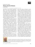
SEXUALLY TRANSMITTED DISEASES docx
Bạn đang xem bản rút gọn của tài liệu. Xem và tải ngay bản đầy đủ của tài liệu tại đây (290.78 KB, 26 trang )
681
A sexually transmitted disease (STD) is any infection acquired pri-
marily through sexual contact. STD is a general term, and the
causative organisms, which are harbored in the blood or body se-
cretions, include viruses, mycoplasmas, bacteria, fungi, spirochetes,
and minute parasites (e.g., crab lice, scabies). Some of the organ-
isms involved are found exclusively in the genital (reproductive)
tract, but others exist simultaneously in other systems. Additionally,
various STDs often coexist, and when one is found, others should
be suspected. There is a range of intimate bodily contact that may
transmit STDs, including kissing, sexual intercourse, anal inter-
course, cunnilingus, anilingus, fellatio, and mouth or genital to
breast contact. Physicians are required to report most STDs to lo-
cal public health departments.
The vast majority of female genital tract infections are acquired
sexually. Female genital tract infections are divided into lower geni-
tal tract and upper genital tract (or pelvic) infections. The lower gen-
ital tract infections (including a number of STDs and their sequelae)
are discussed in Chapters 20 and 21, and they include viral infections
(herpes simplex, human papillomavirus, and molluscum contagio-
sum) and vulvar infestations (pedicularis pubis and scabies). Com-
mon types of vulvovaginitis (e.g., Trichomonas, bacterial vaginosis,
and Candida) and some of the sequelae of STDs (e.g., infections of
Bartholin glands and cervicitis) also are discussed in Chapter 20. This
chapter deals with upper genital tract infections, the most serious,
most directly sexually transmitted diseases and their sequelae.
HUMAN IMMUNODEFICIENCY
VIRUS (HIV) INFECTIONS
The human immunodeficiency virus (HIV) was first reported to
cause disease in 1981. In the United States, AIDS is now the fifth
24
SEXUALLY TRANSMITTED
DISEASES
CHAPTER
Copyright 2001 The McGraw-Hill Companies. Click Here for Terms of Use.
BENSON & PERNOLL’S
682 HANDBOOK OF OBSTETRICS AND GYNECOLOGY
leading cause of death among women of childbearing age. More-
over, it is the leading cause of death in this age group in New
York City. This is now a worldwide crisis, with millions affected,
especially in developing countries. One of the problems in recog-
nition of HIV infection is a long, asymptomatic latency of
2 months to 5 years. The mean age at diagnosis of HIV infection
is 35 years.
The virus is present in blood and all body fluids and is trans-
mitted by sexual contact (.70%), by parenteral exposure to infected
blood or body fluids, or by transplacental passage of the virus from
mother to fetus. The highest-risk groups for HIV infection are ho-
mosexuals, bisexual men, intravenous drug abusers, and hemo-
philiacs receiving blood transfusions. Others at high risk are pros-
titutes and heterosexual partners of men in the high-risk groups. All
blood must be screened for HIV before transfusion to minimize
transfusion risk. Women acquire the virus more easily from men
rather than the reverse because the concentration of HIV in semen
is high and mucosal breaks at the introitus or vagina with inter-
course occur more commonly than do breaks in penile skin.
Although anti-HIV antibodies develop within 12 weeks of ex-
posure, 45%–90% of persons infected with HIV will develop symp-
toms of an acute infection similar to mononucleosis within a few
months. They experience weight loss, fever, night sweats, pharyn-
gitis, lymphadenopathy, and an erythematous maculopapular rash.
Most of these symptoms resolve within a few weeks, although the
patients remain infectious despite being asymptomatic. Some will
progress to develop symptoms of AIDS-related complex (ARC),
with early immunosuppression (decreased CD4ϩ lymphocytes).
ARC is usually marked by generalized lymphadenopathy, weight
loss, diarrhea, malabsorption, and wasting. Some patients experi-
ence further immunosuppression and develop AIDS (any of the
symptoms of acute sepsis, opportunistic infections, Kaposi’s sar-
coma, cognitive difficulties, or depression). Once AIDS has been
diagnosed, mortality is Ͼ90%. Immunologic abnormalities associ-
ated with AIDS include (but are not limited to) lymphopenia, de-
creased T helper cells, decreased T lymphocytes, hypergamma-
globulinemia, and an inverted T4/T8 ratio.
Because there is no cure for HIV, current therapy only slows the
progression of the disease. Hence, there is every reason to stress
prevention. Other than abstinence or having a monogamous rela-
tionship with a known noninfected partner, using latex condoms lu-
bricated with nonoxynol 9 is the most effective method of limiting
the risk of infection. If a woman is HIV positive, she should be
counseled (1) not to donate blood, plasma, tissue, or organs; (2) to
avoid pregnancy; (3) to maintain a monogamous relationship;
and (4) to assiduously use condoms lubricated with nonoxynol 9
during any sexual contact.
HIV antibody testing begins with the enzyme-linked im-
munosorbent assay (ELISA), which has a Ͼ95% sensitivity and a
Ͼ99% specificity if repeatedly positive. If the ELISA is positive, a
Western blot assay must be performed to confirm the diagnosis.
False-negative results are rare unless the patient is too early in the
disease to have formed antibodies. HIV screening (after informed
consent has been obtained and assurances of confidentiality pro-
vided) should be encouraged for women in the following cate-
gories: intravenous drug users, prostitutes, sex partner(s) of men
who are HIV positive or at risk for HIV, those with other sexually
transmitted disease, those who received blood transfusions between
1978 and 1985, those with clinical signs and symptoms of HIV in-
fection, inhabitants of a country with high endemic heterosexual
HIV infection, prison inmates, and one who considers herself at
risk.
Pregnancy does not appear to alter the progression of HIV in-
fection, but the chance of the fetus acquiring the virus is 20%–50%.
The neonate may be infected during labor and delivery by mater-
nal blood or body fluids or may be infected during breastfeeding.
The mode of delivery does not influence the development of pedi-
atric AIDS. The acute illness associated with HIV in pregnancy may
be misdiagnosed if HIV serologic testing is not performed. When
HIV infection is diagnosed during pregnancy, treatment should be
delayed because of the teratogenic potential of the medications
used. The pregnant HIV-infected woman should be screened for
other STDs, along with evaluation for opportunistic infection. A
baseline serologic study for CMV and toxoplasmosis, TB skin test-
ing, and chest radiograph are recommended. Recently AZT and other
chemotherapeutic agents have been found to decrease maternal–fetal
and neonatal transmission of HIV. When caring for HIV positive
mothers, health care providers should obtain the very latest infor-
mation in this important and rapidly evolving area.
Care of the HIV-positive woman and her infant in the peri-
partum and postpartum interval includes protection of health care
workers by using universal infection control guidelines (e.g.,
water-repellent gowns, gloves, masks, goggles for potential splash
situations, wall or bulb suctioning). Scalp electrodes and fetal
scalp blood samples should be avoided (potential entry site for
HIV if fetus is not already infected). Circumcision should not be
done if the neonate is HIV positive. Because anti-HIV IgG anti-
body passes through the placenta, the infant may be seropositive
without being infected. Abnormal facial features have been de-
scribed in some HIV-positive newborns, but this is not common.
CHAPTER 24
SEXUALLY TRANSMITTED DISEASES
683
BENSON & PERNOLL’S
684 HANDBOOK OF OBSTETRICS AND GYNECOLOGY
If neonatal/pediatric AIDS develops, the course of the disease is
much more rapid than in adults, with death in months rather than
years.
GONORRHEA
Neisseria gonorrhoeae (one of the most common causes of STD)
is a gram-negative diplococcus that usually resides in the female in
the urethra, cervix, pharynx, or anal canal. The infection primary
involves the columnar and transitional epithelium of the genitouri-
nary tract. The organism is very fastidious and sensitive to drying,
sunlight, heat, and most disinfectants. Special media (e.g., Thayer-
Martin) are required to achieve optimal recovery. Culture of the
lower genital tract is usually obtained by rotating a cotton swab for
15–20 sec deep in the endocervical canal. If a rectal swab is taken,
the incidence of recovery increases from 85% to Ͼ90%. In upper
genital tract infections (salpingitis, peritonitis) proven by laparo-
scopically obtained culture, only ϳ50% of the lower genital tract
cultures will reveal N. gonorrhoeae.
After exposure to an infected partner, 60%–90% of women and
20%–50% of men will become infected. Untreated, 10%–17% of
women will develop pelvic inflammatory disease (PID). If a woman
is positive for N. gonorrhoeae, she has a 20%–40% chance of also
having chlamydial infection, syphilis, or hepatitis.
Early symptoms typically include vaginal discharge, urinary
frequency, and rectal irritation. Some report burning, itching, or in-
flammation of the vulva, vagina, cervix, or urethra, although most
women are asymptomatic. Bartholin duct(s) and gland(s) may be
involved, as evidenced by swelling or abscess formation (Chapter
20). Acute pharyngitis and tonsillitis may occur, but this is uncom-
mon. Rarely, the asymptomatic carrier will develop a disseminated
infection with polyarthralgia, tenosynovitis, and dermatitis or
meningitis or endocarditis. Although ophthalmic infection most
commonly occurs in neonates born to an infected mother, adult oph-
thalmitis may result from autoinoculation.
The diagnosis may be presumed when a stained smear from the
involved sites reveals intracellular gram-negative diplococci. How-
ever, confirmation after growth on selective medium is essential.
The culture for gonorrhea must include penicillin resistance testing
because 2%–3% of strains in the United States are penicillin re-
sistant. Gonorrhea must be reported to the state public health au-
thorities.
The patient and all sexual partners must be treated. Other
concomitant diseases must be ruled out and treated if present. The
preferred adult regimen for uncomplicated disease is ceftriaxone
125 mg IM (single dose), cefixime 400 mg PO (single dose), or
spectinomycin 2 g IM (single dose for patients with cephalosporin
intolerance). Although spectinomycin is not reliable therapy for pha-
ryngeal infection, ceftriaxone and cefixime are effective in all sites.
Given the high rate of coinfection, treatment for chlamydial infec-
tion (see the following section) is necessary. Because of the emer-
gence of resistant organisms, repeat cultures should be performed
within 7 d of completion of therapy to ensure cure.
Disseminated disease requires hospitalization. Meningitis and
endocarditis must be confirmed or ruled out. Recommended therapy
is ceftriaxone 1 g IM or IV qd or cefotaxime or ceftrizoxime 1 g IV
q8h. Patients with allergy to beta-lactamase drugs may be treated
with spectromycin 2 g IM q12h. If sensitivity testing confirms that
the organism is penicillin-sensitive, ampicillin 1 g q6h may be
given. Whichever regimen is chosen, therapy should be continued
for 7 days. The prognosis for properly treated gonorrhea is good,
but future fertility may be compromised.
CHLAMYDIAL INFECTIONS
Chlamydia trachomatis is an obligate intracellular microorganism
with a cell wall similar to that of gram-negative bacteria. Although
they are classified as bacteria, contain both DNA and RNA, and di-
vide by binary fission. Chlamydia grow only intracellularly, as do
viruses. Since most of the C. trachomatis serotypes attack only
columnar epithelial cells (except the aggressive L serotypes), signs
and symptoms tend to be localized to the infected area (e.g., eye or
genital tract) without deep tissue invasion.
CERVICITIS
C. trachomatis cervical and tubal infections occur in women of
young age (2–3 times higher in women Ͻ20 years), with numerous
sexual partners, of low socioeconomic status, with other STDs, and
with oral contraceptive use. Barrier contraception tends to decrease
the infection rate. Pregnant women have an incidence of 8%–12%.
SIGNS AND SYMPTOMS
Typically, a mucopurulent discharge develops with cervical chlamy-
dial infection, and the cervix shows hypertrophic inflammation (mu-
copurulent cervicitis). The infection may be asymptomatic in 15%
of nonpregnant, sexually active women.
CHAPTER 24
SEXUALLY TRANSMITTED DISEASES
685
BENSON & PERNOLL’S
686 HANDBOOK OF OBSTETRICS AND GYNECOLOGY
LABORATORY FINDINGS
The most frequently used method of detection is a direct fluores-
cein-conjugated monoclonal antibody test (available in kit form).
This is rapid, sensitive (85%–93%), and specific (ϳ99%). Speci-
mens are generally obtained as described for gonorrhea (p. 684).
Tissue culture is required for culture of C. trachomatis, and because
of the high cost, limited availability, and 2–6 day delay, it is used
infrequently. Although Giemsa staining of conjunctival specimens
in neonates is fairly successful in identifying chlamydial inclusions,
this technique is only 40% accurate in genital infections.
DIFFERENTIAL DIAGNOSIS
N. gonorrhoeae is the only other predominant organism causing a
mucopurulent cervicitis. Thus, fluorescent antibody tests or cultures
on selective medium are mandatory for differentiation. Both or-
ganisms may be present simultaneously.
TREATMENT
Cure rates of .95% can be achieved with the use of one of several
regimens. The preferred regimen is tetracycline 500 mg PO qid for
7 days or doxycycline 100 mg bid for 7 days. If tetracyclines can-
not be taken or are contraindicated, erythromycin base 500 mg qid
for 7 days or erythromycin ethylsuccinate 800 mg qid for 7 days
may be prescribed.
COMPLICATIONS
The primary complication of C. trachomatis cervical infection is
salpingitis. Unfortunately, if the patient is pregnant and untreated,
the vaginally delivered neonate will develop chlamydial conjunc-
tivitis in 50% of cases and late onset pneumonitis in 10%. Prema-
ture delivery and early postpartum endometritis are also associated
problems.
SALPINGITIS
C. trachomatis salpingitis may be as prevalent as that caused by
N. gonorrhoeae. However, there are marked differences in the
pathophysiology and symptomatology. C. trachomatis salpingitis
(which is also an ascending infection) has an insidious onset, it
usually causes minimal symptoms, and the organism remains in
the tube (primarily in the epithelium) for months. In contrast,
N. gonorrhoeae infections have an acute onset, cause more acute
symptoms, and remain in the tubes only 24–48 h. Gonorrheal in-
fections appear to have a much greater cytotoxic effect on the tubal
epithelium.
Although C. trachomatis salpingitis usually causes fewer symp-
toms, the gross appearance of the tubes suggests even more severe
involvement. Salpingitis is a consequence of C. trachomatis cer-
vicitis. Treatment of C. trachomatis salpingitis may be accom-
plished with tetracyclines or erythromycin. The sequelae of C. tra-
chomatis salpingitis include ectopic pregnancy and infertility,
although the exact incidence of these complications is unknown.
LYMPHOGRANULOMA VENEREUM
The L serotypes of C. trachomatis cause lymphogranuloma venereum,
which usually occurs in tropical or subtropical areas (including the
southern United States). The incubation period is 7–21 days, and
men are affected 6 times more often than women. In the United
States, Ͻ500 cases/year are reported, and most occur in men.
Lymphogranuloma venereum begins with a vesicopustular erup-
tion that progresses to very painful inguinal and vulvar ulceration,
lymphedema, and secondary bacterial invasion. Clinically, a de-
pression between the groups of inguinal nodes and the genitocrural
fold produces the appearance of a double genital crural fold (the
groove sign). There is a reddish to purplish blue, hard induration
that occurs 10–30 days after exposure. Anorectal lymphedema
causes painful defecation and blood-streaked stools. Later in the
disease, progressive rectal strictures form, which may even prevent
defecation. Vaginal strictures may cause distortion and narrowing,
with resultant dyspareunia. Headache, arthralgia, chills, and ab-
dominal cramps may occur late in this disease. Late complications
include vulvar elephantiasis.
The diagnosis is confirmed by tissue culture and serotype de-
termination, but complement fixation for Chlamydia with titer
Ն1:16 is presumptive, as is a rising titer (Ͼ1:64 is diagnostic). Im-
munofluorescent testing is available. The differential diagnosis for
the cutaneous lesions includes granuloma inguinale, tuberculosis,
syphilis, chancroid, vulvar cancer, genital herpes, and Hodgkin’s
disease. With systemic symptoms, meningitis, arthritis, peritonitis,
and pleurisy must be considered.
Treatment for lymphogranuloma venereum includes doxycy-
cline 100 mg PO bid for 21 days. Persistent disease requires a sec-
ond course. Alternative drugs include tetracycline, erythromycin, or
sulfisoxizole, each at 500 mg PO qid for 21 days. After the disease
CHAPTER 24
SEXUALLY TRANSMITTED DISEASES
687
BENSON & PERNOLL’S
688 HANDBOOK OF OBSTETRICS AND GYNECOLOGY
is under control, surgery may be necessary (e.g., partial vulvec-
tomy). Abscesses should not be excised but treated by aspiration.
Anal strictures should be dilated weekly. A diversionary colostomy
may be required for severe anal stricture.
SYPHILIS
Syphilis is a disease caused by the spirochete Treponema pallidum,
which is transmitted by direct contact with an infectious moist le-
sion. These organisms can pass through intact mucous membranes
or abraded skin or may be acquired transplacentally. A single sex-
ual encounter with an infected partner carries ϳ10% chance of ac-
quiring syphilis. Untreated, the disease progresses from primary to
secondary to latent and, finally, to tertiary syphilis. Congenital
syphilis has its own course and symptoms. There are .280,000 new
cases of syphilis in the United States each year.
The primary lesion of syphilis is the hard chancre, an indurated,
firm, painless papule or ulcer with raised borders, which appears
10 days to 3 months (average is 3 weeks) after the treponemes have
entered the body. The chancre may be located on the external gen-
italia, cervix, or vagina or any area of skin or mucous membrane
of the body but is often not noted in women. The primary lesion
persists for 1–5 weeks and is followed in most by spontaneous heal-
ing. Any lesion suspected of being a chancre should be subjected
to darkfield examination, seeking treponemes, because culture is
not available. Serologic tests for syphilis should be performed
weekly for 6 weeks or until positive (usually reactive 1–4 weeks
after the chancre appears).
The generalized cutaneous eruption (macular, maculopapular,
papular, or pustular) of secondary syphilis appears 2 weeks to 6
months after the primary lesion. The rash is a diffuse, bilateral, sym-
metric papulosquamous eruption that may involve the palms and
soles. Perineal lesions (moist papules, condyloma latum) are pres-
ent and positive for treponemes on darkfield examination or im-
munofluorescent studies. Other mucous patches may be present, as
well as patchy alopecia, hepatitis, or nephritis. Generalized lym-
phadenopathy is typical. The secondary lesions last 2–6 weeks and
heal spontaneously. Serologic tests are almost always positive at
this stage.
Latent syphilis is untreated syphilis after secondary symptoms
have subsided. These patients remain infectious for 1–2 years and
may have relapses resembling the secondary stage. Latency may be
lifelong or end with the development of tertiary syphilis, which oc-
curs in one third of patients.
Tertiary syphilis is marked by the presence of destructive le-
sions of skin, bone (gummas), cardiovascular system (e.g., aortic
aneurysm or insufficiency), or nervous system disorders (e.g.,
meningitis, tabes dorsalis, paresis). Tertiary syphilis is fatal in 25%
of those affected.
Although the maternal course of syphilis is unaltered by preg-
nancy, it is frequently not recognized unless detected by serologic
screening. The treponemes may pass transplacentally throughout
pregnancy, but if the disease is discovered and treated Ͻ18 weeks
gestation, the fetus appears to suffer few sequelae. After 18 weeks,
the classic signs of congenital syphilis occur in the fetus. The risk of
fetal infection is greater during the secondary stage than during the
primary or latent stages. The incidence of stillbirth and premature
delivery is increased with syphilis. Hydramnios may be present. The
placenta is involved; it has a waxy, hydropic appearance. Infection
late in pregnancy results in fetal or neonatal infection in 40%–50%.
Congenital syphilis occurs in the fetus or newborn whose mother
has untreated syphilis. Depending on time of acquisition of
infection, there may be signs of intrauterine infection (e.g., he-
patosplenomegaly, radiographic changes in bone, anemia, jaundice,
lymphadenitis, and meningitis) or the baby may appear unaffected,
only to develop signs and symptoms equivalent to secondary
syphilis sometime after birth.
Classically, the newborn with congenital syphilis may be un-
dergrown, with wrinkled facies because of reduced subcutaneous
fat. The skin may have a brownish (café-au-lait) tint. The most
common lesion of early congenital syphilis in the newborn is a
bullous rash, so-called syphilitic pemphigus. Large blebs may
appear over the palms and soles and, occasionally, in all other areas.
Seropurulent fluid from the lesions swarms with treponemes.
Mucositis identical with that of secondary syphilis in older patients
may be noted in the mouth and upper respiratory passages of the
newborn. The nasal discharge (syphilitic snuffles) is very infectious
because it contains large numbers of T. pallidum.
The bones usually show signs of osteochronditis, and on x-ray,
an irregular epiphyseal juncture (Guerin’s line) is characteristic.
Abnormalities of the eyes and other organs or the central nervous
system may be apparent at birth, or defects may develop later in
untreated cases. Any infant with the stigmata of syphilis should be
placed in isolation until a definitive diagnosis can be made and ap-
propriate treatment given.
Because serologic testing evaluates IgG antibodies that are
transplacentally acquired, the baby will be positive if the mother is
positive. Effective neonatal treatment is shown by progressively
falling titers over weeks to months.
CHAPTER 24
SEXUALLY TRANSMITTED DISEASES
689
BENSON & PERNOLL’S
690 HANDBOOK OF OBSTETRICS AND GYNECOLOGY
LABORATORY FINDINGS
Visualization of the treponemal organisms requires the presence of
a moist cutaneous lesion for darkfield examination (fresh smear),
immunofluorescent staining (dried smear), or silver staining for the
treponemes in a biopsy specimen. Because the organisms are
demonstrable for only a short time, diagnosis usually relies on his-
tory and serologic testing.
Screening for syphilis is accomplished primarily by nonspecific
nontreponemal antibody testing (e.g., VDRL, RPR). All pregnant
women should be tested at the first visit. High-risk patients should
be screened at 28–32 weeks gestation and at delivery. These tests
become positive 3–6 weeks after infection. The titers are high in
secondary syphilis and fall to low titers or even become negative
in late syphilis. Titers that have a 4-fold drop or are falling in early
syphilis indicate adequate treatment.
False-positive tests may be associated with collagen disease, in-
fectious mononucleosis, malaria, leprosy, febrile illnesses, vaccina-
tion, drug addiction, old age, and pregnancy itself. The titer seen
with false-positive tests usually is low. However, any positive test
should be investigated by an antitreponemal antibody test. The most
widely performed antitreponemal antibody test is the fluorescent
treponemal antibody absorption (FTA-ABS) test. The test remains
positive regardless of therapy. Thus, titers are not determined.
DIFFERENTIAL DIAGNOSIS
The differential diagnosis for primary syphilis includes chancroid,
granuloma inguinale, lymphogranuloma venereum, herpes, carci-
noma, scabies, trauma, lichen planus, psoriasis, drug eruption,
aphthosis, mycotic infection, Reiter’s syndrome, and Bowen’s
disease.
The differential diagnosis for secondary syphilis includes pityr-
iasis rosea, psoriasis, lichen planus, tinea versicolor, drug eruption,
“id” eruptions, perleche, parasitic infection, iritis, neuroretinitis,
condylomata accuminata, acute exanthems, infectious mononucle-
osis, alopecia, and sarcoidosis.
TREATMENT
Treatment should be initiated if exposure has occurred even if evi-
dence of disease is not present. During pregnancy, it is better to
treat any suspicion of disease rather than risk congenital syphilis.
Contacts and patients with early syphilis (primary, secondary,
and latent Ͻ1 year) should be treated with one of the following
regimens: (1) benzathine penicillin G 2.4 million units IM, (2)
tetracycline hydrochloride 500 mg PO qid or doxycycline 100 mg
bid for 14 days (for penicillin allergy but not during pregnancy),
or (3) erythromycin (stearate, ethylsuccinate, or base) 500 mg PO
qid for 15 days (30 g total) for penicillin allergy and if unable to
take tetracycline. A short-lived (Ͻ24 h) febrile reaction occurs in
50%–75% of those receiving penicillin therapy, presumably due
to a release of toxic treponemal products. The fever, which occurs
4–12 h after injection, is a Jarisch-Herxheimer reaction.
Congenital syphilis is treated with benzathine penicillin G
50,000 units/kg IM if the infant is asymptomatic and there is no
evidence of neurosyphilis. Symptomatic congenital syphilis or
neurosyphilis is treated with aqueous crystalline penicillin G
50,000 units/kg/day IV, divided in two doses for 10 days or aque-
ous procaine penicillin G 50,000 units/kg daily for 10 days.
CHANCROID
Chancroid (soft chancre) is caused by the gram-negative rod
Haemophilus ducreyi and is uncommon in the United States (Ͻ1500
cases/year). This infection begins in females as a papule or vesico-
pustular lesion on the perineum, cervix, or vagina 3–5 days after
exposure. The lesion progresses over 48–72 h to a very tender
saucer-shaped ragged ulcer. Several ulcers may develop in a clus-
ter. The heavy discharge produced by the ulcer(s) is foul-smelling
and infectious. Over 50% of patients develop painful inguinal
lymphadenitis that may become necrotic and drain spontaneously.
Aspiration of pus from a bubo may yield the organism. Syphilis must
be ruled out, although the differential diagnosis also includes herpes
simplex, lymphogranuloma venereum, and granuloma inguinale.
Treatment includes sitz baths, soap and water plus antibiotics.
The therapeutic regimen will vary depending on sensitivity of the
pathogen. Ceftriaxone 250 mg IM qd, erythromycin 500 mg PO
qid, and trimethoprim (160 mg)/sulfamethoxazole (800 mg) PO bid
have been effective. Treatment should continue for a minimum of
10 days until the ulcer(s) and lymph nodes are healed. Abscessed
nodes should be aspirated rather than incised and drained.
GRANULOMA INGUINALE
Granuloma inguinale is caused by Calymmatobacterium granulo-
matis. A characteristic finding in the lesions is the Donovan body
CHAPTER 24
SEXUALLY TRANSMITTED DISEASES
691
BENSON & PERNOLL’S
692 HANDBOOK OF OBSTETRICS AND GYNECOLOGY
(bacteria encapsulated in mononuclear leukocytes). It is almost
never seen in the United States (ϳ100 cases/year) but is common
in India, Brazil, and the West Indies. The incubation period is 1–12
weeks. Granuloma inguinale may be spread by repeated sexual or
nonsexual contact.
The disease usually is localized to the vulva and inguinal area
but may involve the cervix, uterus, ovary, or mouth. It begins as an
asymptomatic papule or nodule that ulcerates to form a red, gran-
ular area with sharp borders. The ulcer drains a foul-smelling dis-
charge. Healing is extremely slow, but there are few local or sys-
temic manifestations. Satellite ulcers may coalesce into one large
ulcer. Buboes may occur late in the disease. Pain may be present if
the urethra or anus is involved.
Late complications include dyspareunia if the introitus con-
stricts from chronic disease. The differential diagnosis includes car-
cinoma, chancroid, lymphogranuloma venereum, and syphilis. The
diagnosis is confirmed by finding Donovan bodies in a biopsy spec-
imen or smear using Wright, Giemsa, or a silver stain.
The drug of choice for treatment of granuloma inguinale is tetra-
cycline 500 mg qid for a minimum 21 days. Other choices include
erythromycin 500 mg qid for 14–21 days, doxycycline 100 mg bid
for 21 days, or sulfamethoxazole 1 g bid for 21 days.
PELVIC INFECTIONS
Infections may occur in any or all portions of the upper genital
tract: endometrium (endometritis), uterine wall (myositis), oviducts
(salpingitis), ovary (oophoritis), uterine serosa and broad ligaments
(parametritis), and pelvic peritoneum (peritonitis). Although the ex-
act incidence of upper genital tract infection is unclear, over 10%
of U.S. women of reproductive age have received treatment for an
upper genital tract infection. A functional classification of pelvic in-
fections is shown in Table 24-1.
Organisms may disseminate to and throughout the pelvis in any
of five ways.
●
Intraluminal. Nonpuerperal acute pelvic inflammatory dis-
ease nearly always (ϳ99%) follows a progression of en-
trance of pathogens through the cervix into the uterine
cavity. Infection then spreads to the uterine tubes, with pus
eventually entering the peritoneal cavity from the ostia.
Organisms known to spread by this mechanism include
N. gonorrhoeae, C. trachomatis, Streptococcus agalactiae,
cytomegalovirus, and herpes simplex virus. Three fourths of
women with acute PID have concomitant endometritis,
whereas ϳ40% of those with mucopurulent cervicitis and
50% of those with positive C. trachomatis or N. gonorrhoeae
endocervical cultures have concomitant endometritis. The
endometritis phase is generally asymptomatic, often brief,
and occurs at the end of a menses.
●
Lymphatic. Puerperal infections (including abortion) and
IUD-related infections are disseminated through the lym-
phatic system, as are nonpuerperal Mycoplasma infections.
●
Hematogenous. Hematogenous dissemination of pelvic dis-
ease is limited to certain diseases (e.g., tuberculosis) and is
uncommon in the United States.
●
Intraperitoneal. Intraabdominal infections (e.g., appendici-
tis, diverticulitis) as well as intraabdominal accidents (e.g.,
perforated viscus or ulcer) may lead to an infectious process
involving the internal genital system.
CHAPTER 24
SEXUALLY TRANSMITTED DISEASES
693
TABLE 24-1
FUNCTIONAL CLASSIFICATION OF
PELVIC INFECTIONS
Pelvic inflammatory disease (PID)
Limited (salpingitis)
Pelvic abscess (cul-de-sac or tuboovarian)
Puerperal infections
Cesarean section (common)
Vaginal (less common)
Postgynecologic procedure infection
Acute PID after diagnostic instrumentation
Abortion-related infections
Postabortal cellulitis
Incomplete septic abortion
Cuff cellulitis and parametritis
Vaginal cuff abscess
Tuboovarian abscess
Pelvic infection secondary to other infections or
intraabdominal accidents
Appendicitis
Diverticulitis
Tuberculosis
Traumatic viscus rupture
BENSON & PERNOLL’S
694 HANDBOOK OF OBSTETRICS AND GYNECOLOGY
●
Contiguous. The postgynecologic surgical infections are the
result of local spread of infection from areas of tissue necro-
sis and infection.
PELVIC INFLAMMATORY DISEASE (PID)
Given the anatomic intimacy and functional proximity of the re-
productive system, it is infrequent that infection is confined to just
a single anatomic site. Thus, PID is a general clinical term for up-
per genital tract infections. PID is an extraordinary health problem.
There are about 1 million cases of acute PID a year in the United
States, and the total cost is estimated to exceed $7 billion per year.
Over a quarter of PID cases require hospitalization. PID affects
1%–2% of sexually active females yearly and is more frequent in
young women (75% of those affected are Ͻ25 years). In the United
States, PID annually results in 2.5 million physician visits, nearly
270,000 inpatient admissions, about 120,000 operative procedures,
and 0.29 deaths/100,000 women age 15–44.
There are a number of risk factors for PID, but the greatest risk
centers about sexual activity. PID, which usually arises after a men-
strual period, in sexually active females accounts for 85% of cases,
but 15% occur after procedures in which the mucosal surface is in-
jured (e.g., IUD insertion, endometrial biopsy, curettage, hystero-
salpingogram). In Ͻ1% of PID, transperitoneal spread to the upper
genital tract occurs from appendicitis, diverticulitis, or traumatic
rupture of a viscus.
The risk of acquiring any STD is directly related to the number
of sex partners. A woman with Ն10 lifetime sexual partners is Ͼ3
times more likely to have PID than a woman with one partner. Young
age (adolescence) is a risk factor because of the less stable sexual
relationships marking this time of life and possibly because of less
immunity. Other risk factors for PID include: contraceptive prac-
tice (barrier methods markedly reduce the risk), ethnicity (in United
States, African-American women report 17%, Caucasians 10%),
postmarital status (3 times that of never married), bacterial vagi-
nosis, vaginal douching and cigarette smoking.
Approximately two thirds of acute pelvic infections are polymi-
crobial. N. gonorrhoeae is responsible for one third of acute PID,
N. gonorrhoeae with a mixed endogenous anaerobic and aerobic flora
is responsible for another one third, and mixed anaerobes and aerobes
are responsible for the remaining third. In combination with other or-
ganisms, C. trachomatis is found in up to 30% of cases. The aerobes
and anaerobes found in PID usually are normal vaginal and gastroin-
testinal flora. The anaerobes (e.g., Bacteroides, Peptostreptococcus,
Peptococcus) predominate in abscesses. Common aerobes include
Escherichia coli, group B Streptococcus, Streptococcus faecalis,
and coagulase-negative Staphlococcus. Mycoplasma hominis and
Ureaplasma urealyticum do not appear to be pathogenic in PID.
The signs and symptoms of acute PID are generally nonspecific
but are related to both the extent of the infection and the organisms
involved. Lower abdominal pain of Ͻ7 days duration occurs in
Ͼ90% of patients with acute PID. The pain is usually characterized
as constant and dull (but may be aching, burning, cramping, or stab-
bing) and is enhanced by movement or sexual activity. Endocervi-
cal infection is present in 75% of patients with PID. Abnormal vagi-
nal bleeding, increased discharge or abnormal vaginal odor occurs
in 40% of patients. Other symptoms are nonspecific, including fever
(30%), malaise, gastrointestinal (vomiting, diarrhea, constipation,
and tenesmus), urinary (dysuria, frequency, and urgency), and
headache.
The white blood cell count may be normal, increased, or
decreased and should not be relied on to rule out PID. Abdominal
x-ray films (KUB and upright) may demonstrate adynamic ileus or
free peritoneal gas or both. Transvaginal sonography may be the
most useful imaging and will reveal peritoneal fluid, thicknened ede-
matous structures, hydrosalpingx, and so forth. Sonography may
also be useful for monitoring therapy. Culdocentesis is simple, rel-
atively painless, and may be diagnostic, but is not currently used a
great deal because of sonography and laparoscopy.
Laparoscopy is the “gold standard” for definitive diagnosis.
Evaluating the fluid within the abdomen is of assistance in inter-
preting the visualized alterations. Pus suggests a ruptured tu-
boovarian abscess, ruptured appendix, ruptured viscus, ruptured di-
verticular abscess, or a uterine abscess involving a myoma. Cloudy
fluid suggests pelvic peritonitis (as seen with acute gonococcal sal-
pingitis), adnexal torsion, or other causes of peritonitis (e.g., ap-
pendicitis, pancreatitis, cholecystitis, perforated ulcer, carcino-
matosis, or echinococcosis). Blood may be found with ruptured
ectopic pregnancy, hemorrhage from corpus luteum cyst, retrograde
menstruation, ruptured spleen or liver, gastrointestinal bleeding, or
acute salpingitis.
If laparoscopy is performed the sterile collection of specimens
is mandatory. Acute PID may be polymicrobial, but in two thirds
N. gonorrhoeae, C. trachomatis, or both, are recovered. The other
one third have mixed anaerobic organisms (Prevotella species and
peptostreptococci) and facultative bacteria (Gardnerella vaginalis,
streptococci, Escherichia coli, and Haemophilus influenzae).
Reports are beginning to accumulate linking bacterial vaginosis
and PID.
CHAPTER 24
SEXUALLY TRANSMITTED DISEASES
695
BENSON & PERNOLL’S
696 HANDBOOK OF OBSTETRICS AND GYNECOLOGY
The infection may be so widespread that in 5%–10% of acute
PID, perihepatic inflammation develops. Here, the symptoms are
right upper quadrant distress, pleuritic pain, and right upper quad-
rant tenderness. This condition is called the Curtis-Fitzhugh syn-
drome and often results in perihepatic adhesions.
Early diagnosis and prompt effective treatment may decrease
the sequelae (e.g., pelvic adhesions, tissue necrosis, abscess for-
mation, intestinal obstruction, infertility, and tubal pregnancy), but
25% of women with acute PID develop significant complications.
There is a 6–10 times increase in ectopic pregnancy and a 4 times
increase in chronic pelvic pain, dyspareunia, and pelvic adhesions.
Infertility is enhanced (20% of patients with just one attack—a
7 times increase), depending on the number and severity of attacks.
Tuboovarian abscess occurs in 5%–10% of hospitalized acute PID
patients.
PID may be decreased or prevented by limiting the number of
sexual contacts; determining if sexual contacts have STDs; using
condoms, spermicides, diaphragms and spermicides; and employ-
ing postcoital toilet (urination, washing, or douching with antisep-
tic solution).
ACUTE SALPINGITIS
As noted previously, the most common organisms initiating acute
salpingitis-peritonitis is Neisseria gonorrhoeae and Chlamydia tra-
chomatis. Approximately 15% of asymptomatic infections will re-
sult in acute salpingitis.
Symptoms and Signs
Typically, salpingitis occurs in young (often teenage) women who
have multiple sexual partners and are not using vaginal contracep-
tion. Symptoms typically begin shortly after cessation of menses or
following instrumentation. The onset of lower abdominal and pelvic
pain (frequently bilateral) is usually acute but may be insidious.
Pain may radiate from the back down the leg(s). There may be a
purulent vaginal discharge. Systemic symptoms include fever
(30%), headache, malaise, nausea, and vomiting.
Physical examination reveals abdominal tenderness, usually of
the lower quadrants. Rebound tenderness is noted in the presence
of peritonitis. Bowel sounds may be decreased or absent. The
paraurethral and Bartholin glands may be inflamed and discharging
purulent material. The cervix often exudes a purulent discharge.
Movement of the cervix or uterus is exquisitely painful. The adnexa
are tender to palpation. The criteria for diagnosis of salpingitis are
summarized in Table 24-2.
Laboratory Findings
A smear of the cervical discharge nearly always reveals infection
and may suggest the etiology (e.g., gram-negative diplococci of
N. gonorrhoeae), but confirmation is essential, using selective me-
dia for N. gonorrhoeae and LCR or PCR for C. trachomatis. The
WBC may be elevated or normal. Women with suspected PID should
have a quantitative hCG test because 3%–4% of them will have ec-
topic gestation. Culdocentesis usually produces cloudy fluid, which
should be sent for cell count, (Ͼ30,000 WBC/mL is associated with
PID), gram stain, culture, and sensitivity.
If abdominal x-ray films show evidence of free air under the di-
aphragm, laparotomy is mandatory. Ultrasonic scanning is useful
in the patient who is too tender to examine properly to rule out ec-
topic gestation or to reveal abscesses.
CHAPTER 24
SEXUALLY TRANSMITTED DISEASES
697
TABLE 24-2
CRITERIA FOR DIAGNOSIS OF SALPINGITIS
Criteria
Abdominal direct tenderness,
with or without rebound
tenderness
Tenderness with motion All 3 necessary for
of cervix and uterus diagnosis
Adnexal tenderness
plus
Gram stain of endocervix
positive for gram-negative,
intracellular diplococci
Fever (Ͼ38ЊC)
Leukocytosis (Ͼ10,000) One or more necessary
Purulent material for diagnosis
(WBCs present)
from peritoneal cavity by
culdocentesis or laparoscopy
Pelvic abscess or inflammatory
complex on bimanual
examination or on sonography
Modified from W.E. Hager, D.A. Eschenbach, M.R. Spence, et al.
Criteria for diagnosis and grading of salpingitis. Obstet Gynecol
61:114, 1983.
144244314444424444443
BENSON & PERNOLL’S
698 HANDBOOK OF OBSTETRICS AND GYNECOLOGY
Differential Diagnosis
Included in the differential diagnosis are appendicitis, ectopic preg-
nancy, ruptured corpus luteum cyst with hemorrhage, diverticulitis,
infected septic abortion, degeneration of a uterine leiomyoma, tor-
sion of an adnexal mass, endometriosis, acute urinary tract infec-
tion, ulcerative colitis, and regional enteritis.
Treatment
Free peritoneal air is a surgical emergency, but the decision to hos-
pitalize for treatment is most frequently based on the following find-
ings: peritonitis in upper quadrants (nonpelvic peritonitis), gas-
trointestinal symptoms (including ileus), tuboovarian abscess,
pregnancy, uncertain diagnosis, presence of an intrauterine device,
history of instrumentation, inadequate response to outpatient ther-
apy, or nulliparity.
Patients requiring hospitalization should be put at bedrest, ini-
tially kept NPO, given IV fluids, and placed on nasogastric suction
for abdominal distention or ileus. Antibiotic therapy should be IV
until the patient has shown clinical improvement for 48 h. Recom-
mended
*
drug regimens include: cefotetan 2 g IV every 12 h, or
cefeoxitin 2 g IV every 6 h, plus doxycycline 100 mg IV or PO
every 12 h. An alternative
*
regimen is clindamycin 900 mg IV every
8 h, plus gentamicin, IV loading of 2 mg/kg followed by mainte-
nance of 1.5 mg/kg every 8 h. For both regimens, 24 h after clinical
improvement, parenteral therapy may be discontinued; however oral
doxycycline 100 mg bid should be administered for 14 d. Alterna-
tive parenteral regimens recommended by various authorities in-
clude: ofloxacin 400 mg IV every 12 h plus metronidazole 500 mg
IV every 8 h, ampicillin-sulbactam 3 g IV every 6 h, plus doxycy-
cline 100 mg IV or PO every 12 h, Ciprofloxacin 200 mg IV every
12 h plus doxycycline 100 mg IV or PO every 12 h plus metron-
idazole 500 mg IV or PO every 8 h.
An IUD should be removed once therapy has been begun. Anal-
gesics with or without codeine may provide relief.
Surgical exploration is reserved for those in whom life is threat-
ened, the condition deteriorates, a tuboovarian abscess ruptures, a
pelvic abscess is pointing into the cul-de-sac, the abdominal symp-
toms persist despite intensive therapy, and there are persistent
masses. In women not desiring future childbearing, the tuboovar-
ian masses may be removed. In the majority, however, every effort
should be made to preserve ovarian function, especially in those
who may want in vitro fertilization, when the uterus should remain.
*
Centers for Disease Control and Prevention: 1998 guidelines for treatment of sexually
transmitted diseases. MMWR 1998;47(RR-1):82–84.
Treatment is reasonable on an outpatient basis for the majority
of mild PID (ϳ75% of cases). The patient must not be pregnant and
must not appear acutely ill, and the diagnosis must be certain. The
recommended
*
outpatient oral regimens is ofloxacin, 400 mg PO
bid for 14 d, plus metronidazole 500 mg PO bid for 14 d. The rec-
ommended
*
outpatient parenteral regimens include: ceftriaxone
250 mg IM once, or cefoxitin 2 g IM plus probenecid 1 g PO. Other
possibilities
*
include other parenteral third-generation cephalos-
porins (e.g., ceftizoxime or cefotaxime) plus doxycycline 100 mg
PO bid for 14 d. Additionally, many authorities advocate adding
metronidazole 500 mg bid PO to the previously noted IM regimens
to cover the anaerobes associated with bacterial vaginosis. Alter-
natively amoxicillin-clavulanic acid plus doxycycline 100 mg PO
ever 12 h for 14 d may be used. The patient should be reevaluated
in 48–72 h after starting therapy. If she fails to improve, she should
be hospitalized for treatment. Other indications for hospitalization
include: inability to follow or tolerate an oral regimen, severe ill-
ness (high fever, nausea, vomiting), tuboovarian abscess, and an
immunodeficiency state. Follow-up examination, including cervi-
cal cultures, should be performed 2 weeks after therapy to assure a
cure.
Prognosis
The prognosis depends on prompt therapy with broad-spectrum an-
tibiotics and rest. Complications include hydrosalpinx, pyosalpinx,
tuboovarian abscess, infertility, ectopic pregnancy (at least 2 times
increased), and chronic pelvic pain (20% after just one episode).
Probably between 1%–2% of U.S. women are infertile as a result
of PID. Approximately 6% of aggressively treated gonococcal sal-
pingitis will result in infertility, whereas 17% of nongonoccal case
will suffer infertility. Laparoscopy is useful in relating the extent of
the disease to future infertility. About 10% with observable mild
disease will be infertile, compared to Ͼ40% of those with severe
disease. The prognosis for fertility decreases substantially with each
infection. Infertility is present in 12%–18% with the first (or only)
episode of acute salpingitis, 25% with two episodes, and 60% with
three episodes of salpingitis.
Recurrent infections are so common (25%) that in the past a
designation of chronic infection was given to cases in which ther-
apy was appropriate but the patient returned with another infection.
Thus, effective treatment of sexual partners is mandatory. The part-
ners of women with N. gonorrhoeae and C. trachomatis are usu-
ally teated with ceftriaxone 125 mg IM plus 100 mg of doxycycline
PO bid for 7 d. An alternative is azithromycin 1 g PO (a single
dose).
CHAPTER 24
SEXUALLY TRANSMITTED DISEASES
699
BENSON & PERNOLL’S
700 HANDBOOK OF OBSTETRICS AND GYNECOLOGY
PELVIC ABSCESS
Cul-De-Sac Abscess
Pelvic abscesses may be tuboovarian or in the cul-de-sac. Most
often, pelvic abscesses follow acute pelvic infection or septic
abortion, although they may follow appendicitis, perforated vis-
cus, or recurrent pelvic infection. Bacteroides (either fragilis or
bovis) is the most commonly cultured organism. The symptoms
may be those of acute pelvic infection plus a fluctuant mass in the
adnexa or cul-de-sac. Pain usually is more severe than with acute
PID or salpingitis, especially to the rectum and back and during
defecation.
The differential diagnosis includes tuboovarian abscess, peri-
appendiceal abscess, ectopic pregnancy, retroflexed and incarcer-
ated uterus, endometriosis, diverticulitis with perforation, and car-
cinomatosis.
Antibiotic treatment must be effective against both aerobic and
anaerobic bacteria. Recommended regimens include (1) penicillin
G 20–30 million units IV per day, plus chloramphenicol 4–6 g IV
per day (monitor for idiosyncratic aplastic anemia); (2) penicillin
G 20–30 million units IV per day, clindamycin 600–1200 mg IV
qid, and gentamicin 5 mg/kg per day; (3) cefoxitin 8–12 g IV per
day and gentamicin or tobramycin 5 mg/kg/day; and (4) cefotaxime
6–8 g IV per day.
The patient should be reevaluated frequently (and gently) for
signs of peritoneal involvement or dissection into the rectovaginal
septum with fixation. If any of these occur, surgical intervention is
necessary. If extension downward occurs, vaginal drainage with a
large Pezar-type catheter should be accomplished. This should be
followed by low-pressure irrigation with sterile saline q6h until the
space is obliterated. If fever persists despite antibiotic therapy,
percutaneous drainage and irrigation of the cul-de-sac may be
performed.
If peritoneal signs develop or if the patient’s condition deterio-
rates despite therapy, an exploratory laparotomy is indicated. When
the patient has no further desire for children, total abdominal hys-
terectomy (TAH), bilateral salpingo-oophorectomy (BSO), and ly-
sis of adhesions should be performed. More conservative surgery
may be dictated by age, parity, condition of the tubes and ovaries,
and desire for childbearing. In some cases, salvage of even an ovary
may be desirable.
Although the prognosis for fertility is guarded, the overall
prognosis is good if the abscess is localized and is treated
early. If rupture into the peritoneum occurs, the prognosis is
serious.
Tuboovarian Abscess (TOA)
Although tuboovarian abscess may occur after an initial episode of
acute salpingitis, it most often is associated with recurrent adnexal
infections (Fig. 24-1). The ovarian site of ovulation is thought to be
a portal of infection for abscess formation. TOA is bilateral in
60%–80%.
Fulminant peritonitis with a 5%–10% mortality rate results from
sudden rupture of an abscess, whereas a slow leak may result in
signs and symptoms of a cul-de-sac abscess. Granulomatous dis-
ease (e.g., tuberculosis, actinomycosis) and use of IUDs are asso-
ciated with TOA.
Patients may be asymptomatic or clearly be in septic shock. The
typical patient has a history of previous pelvic infection, is young,
with low parity, and has had symptoms for ,1 week. The onset is
usually 2 weeks after menses, with pelvic and abdominal pain (of
varying degrees), nausea, vomiting, fever, and tachycardia. The en-
tire abdomen is tender, and guarding may be present. Because of
extreme tenderness of the adnexa, pelvic examination may be dif-
ficult. Culdocentesis may rupture the abscess, thus ultrasonography
is preferred for diagnosis. The WBC count may be low, normal, or
greatly elevated. The abdominal x-ray may show adynamic ileus or
free air under the diaphragm if rupture has occurred.
The differential diagnosis in the asymptomatic patient may
be ovarian cyst, ovarian neoplasm, unruptured ectopic pregnancy,
CHAPTER 24
SEXUALLY TRANSMITTED DISEASES
701
FIGURE 24-1. Tuboovarian abscess.
BENSON & PERNOLL’S
702 HANDBOOK OF OBSTETRICS AND GYNECOLOGY
uterine leiomyoma, hydrosalpinx, or periappendiceal abscess. In the
symptomatic patient whose tuboovarian abscess remains unrup-
tured, the differential diagnosis includes appendiceal abscess, ap-
pendiceal rupture, diverticular abscess, perforated diverticulum,
perforated peptic ulcer, porphyria, and diabetes mellitus. Compli-
cations include septic shock, septic emboli, peritonitis, bowel ob-
struction, recurrent infection, ectopic pregnancy, and infertility.
Treatment depends on whether or not the abscess is sympto-
matic and whether or not it has ruptured. If the tuboovarian abscess
is unruptured and asymptomatic, it may be treated by antibiotics
(see below). If the mass does not shrink within 2–3 weeks of an-
tibiotic therapy or increases in size, surgery is indicated. Usually
TAH and BSO are performed, although a more conservative ap-
proach (unilateral adnexectomy) is reasonable in select patients. If
the abscess is unruptured but symptomatic, the patient should be
hospitalized, placed at bedrest in semi-Fowler position, and main-
tained NPO with nasogastric suction applied, and she should re-
ceive IV fluids with close monitoring of electrolytes. Antibiotics
should be administered IV in one of the following combinations.
Penicillin G (or ampicillin) and chloramphenicol
Penicillin G (or ampicillin) plus metronidazole
Clindamycin plus an aminoglycoside (gentamicin, amikacin, to-
bramycin)
Cefoxitin or cefamandole plus either clindamycin, metronida-
zole, or choramphenicol
Moxalactam or cefotaxime with or without clindamycin,
metronidazole, chloramphenicol, or an aminoglycoside
If the initial therapy results in improvement, continue oral an-
tibiotics (tetracycline, doxycycline) for 10–14 days. If rupture or
leakage is suspected or if the patient does not respond to antibiotic
therapy, perform an exploratory laparotomy. About 50% of patients
with unruptured symptomatic tuboovarian abscess will require sur-
gery.
A ruptured tuboovarian abscess is a life-threatening emergency
and must be treated as such. Admit the patient to an intensive care
unit (or its equivalent) in preparation for surgery. Monitor urine out-
put hourly, and monitor the central venous pressure, administer oxy-
gen, and correct hypovolemia. Corticosteroids (e.g., methylpred-
nisolone succinate 15–30 mg/kg IV q4–6h for 4 doses) may be
advisable. Surgery must be performed as soon as the patient has
stabilized sufficiently to tolerate a major operation.
The abdomen should be entered through a midline incision. The
pus encountered should be sent for aerobic and anaerobic culture.
The entire abdomen should be explored, and all abscesses should
be drained. Thorough irrigation with suction is necessary. Normally,
TAH and BSO are performed, but therapy must be individualized.
Supracervical hysterectomy may be necessary to shorten operative
time in the unstable patient. Drains should be inserted through the
open vaginal cuff or cul-de-sac. Leave drains in place as long as
there is purulent discharge. Close the fascia but leave the subcuta-
neous space open.
The prognosis for survival with an unruptured tuboovarian ab-
scess is excellent, although fertility is markedly diminished. Mor-
tality from ruptured tuboovarian abscess is 5%–10%, with sterility
a consequence of hysterectomy and oophorectomy.
POSTOPERATIVE PELVIC INFECTIONS
Hysterectomy carries a high rate of infection presumably because
the vaginal flora cannot be eliminated from the operative site. The
common infectious sequelae of hysterectomy includes cuff indura-
tion (cellulitis), infected cuff hematoma (cuff abscess), salpingitis,
pelvic cellulitis, suppurative pelvic thrombophlebitis, and tu-
boovarian abscess. These infections are usually polymicrobial. The
incidence of posthysterectomy infection may be lowered to 5% by
a single dose of prophylactic antibiotic therapy.
Postoperative infection usually begins as cuff cellulitis that
spreads from the vaginal apex to the parametrial tissues. If there is
a hematoma present, an abscess forms. Antibiotic therapy and ad-
equate drainage may halt further progression to salpingitis, diffuse
pelvic cellulitis, and tuboovarian abscess. If the pelvic veins be-
come involved (as is common with anaerobic infection), suppura-
tive pelvic thrombophlebitis results, and septic emboli may occur
to lungs, brain, spleen, and other sites.
CLINICAL FINDINGS
Postoperative pelvic infection causes fever (Ͼ38ЊC or Ͼ100ЊF)
within 24–36 h in 50% of patients. Alternatively, temperature ele-
vation may be due to atelectasis, urinary tract infection, or phlebitis.
Early onset of fever without symptoms may resolve spontaneously,
whereas fever by 72 h may require antibiotic therapy.
Examination of the vaginal cuff after hysterectomy may disclose
hyperemia, edema, and purulent or seropurulent exudate whether
the wound is infected or not. If the host immune system cannot
control the inoculum of vaginal flora, the parametrium will usually
become indurated, causing pain and tenderness. Infection may
then spread to the tubes and ovaries, followed by paralytic ileus.
CHAPTER 24
SEXUALLY TRANSMITTED DISEASES
703
BENSON & PERNOLL’S
704 HANDBOOK OF OBSTETRICS AND GYNECOLOGY
Spiking diurnal fever after the sixth postoperative day is suggestive
of suppurative pelvic thrombophlebitis. The patient may not appear
toxic (despite a high temperature) unless septic embolization has
occurred.
Palpation is difficult, but ultrasonography may detect an in-
fected pelvic hematoma. Typically, there may be no abnormality
other than fever. An unexpectedly low HCT and fever are sugges-
tive of an infected hematoma.
Because laboratory and x-ray findings are not often helpful in
making the diagnosis of postoperative pelvic infection, the diagno-
sis must be made clinically. If the next step is a workup for post-
operative fever, it is useful to recall the clinical mnemonic 3 Ws
(wind, water and wound). Include in the differential diagnosis at-
electasis, which causes fever within 12–36 h and is suggested by
auscultation and demonstrated by chest x-ray films. Dehydration
may cause mild fever within 24–48 h of surgery and usually can be
suspected by the patient’s clinical appearance, fluid losses, urine
output, and HCT. The abdominal incision may become infected and
should be probed carefully if the pelvic examination is unremark-
able. Except for extremely virulent infections (e.g., clostridia), it is
uncommon for the abdominal wound to cause marked fever before
the third postoperative day.
Other causes of postoperative fever to be ruled out include
phlebitis and urinary tract infection. Phlebitis of a superficial vein
at an IV site may cause fever, especially if antibiotics are being in-
fused. Urinary tract infection is common after gynecologic surgery
because of the use of indwelling catheters. However, fever usually
occurs principally when pyelonephritis is present.
COMPLICATIONS
Complications of postoperative infections include wound abscess,
tuboovarian abscess, pelvic or intraabdominal abscess, metastatic
septic emboli, and septicemia.
PREVENTION
Conscientious attempts should be made to prevent or decrease sep-
sis because of the severity of postoperative pelvic infection. Help-
ful measures reportedly include:
●
Preoperative vaginal douches with topical antibacterial
agents (e.g., povidone-iodine, hexochlorophene) for several
days
●
Preoperative insertion of antibacterial vaginal creams or sup-
positories (especially if vaginitis or cervicitis is present)
CHAPTER 24
SEXUALLY TRANSMITTED DISEASES
705
●
Meticulous operative hemostasis without strangulation of
tissues
●
Use of nonreactive suture material
●
Suction drainage at sites of suboptimal hemostasis (with
vaginal surgical margin left open or closed)
●
Prophylactic antibiotics preoperatively with two optional ad-
ditional doses 6 and 12 h postoperatively (note: usual drugs
for single-dose prophylaxis are cefonicid, ceforanide, cefo-
taxime, cefotetan, ceftriaxone, and cefuroxime)
●
Treatment of a mild infection before it becomes severe
TREATMENT
Treatment depends on diagnosis. A cuff hematoma or abscess
must be adequately drained and antibiotics must be initiated.
Single-agent therapy with one of the newer cephalosporins usu-
ally eliminates fever within 48–72 h. Large hematomas will
require suction drainage and more prolonged antibiotic therapy.
Tuboovarian abscess therapy has been described previously.
Diagnosis of suppurative pelvic thrombophlebitis is made by
exclusion when fever persists after 7–10 days of antibiotic
administration. Heparin to a therapeutic dose, e.g., 5000 units
q6h IV should be administered. If fever persists despite heparin
and antibiotic therapy, addition of another antibiotic effective
against anaerobic organisms (e.g., clindamycin, chloramphenicol,
metronidazole) is recommended. Surgical intervention is usually
necessary only if heparin therapy is contraindicated, if em-
bolization continues despite adequate therapy, or if the patient
fails to respond to therapy.
PELVIC TUBERCULOSIS
Pelvic tuberculosis (TB), often resulting from lymphatic spread of
pulmonary TB, is rare in the United States and complicates pul-
monary tuberculosis in about 5%. Direct extension to other
abdominal organs from the pelvic organs is common. The pelvic
organs primarily involved are oviducts and the endometrium.
Clinical findings may be minimal. Presentation may be for in-
fertility, although pelvic pain, dysmenorrhea, or signs of tubercu-
lous peritonitis may be present. Patients with abdominal symptoms
may complain of low-grade fever and weight loss. Tuberculous peri-
tonitis is usually accompanied by ascites. If the pelvic TB is found
incidentally during pelvic surgery, it may be mistaken for chronic
pelvic inflammation. However, the adhesions are much more dense,