Converging Technologies for Improving Human Performance Episode 2 Part 1 docx
Bạn đang xem bản rút gọn của tài liệu. Xem và tải ngay bản đầy đủ của tài liệu tại đây (269.9 KB, 20 trang )
Converging Technologies for Improving Human Performance (pre-publication on-line version)
ï Cells
x,y; #; Mean±SD
ï Molecules
x,y; #; Mean±SD
ï Layers
x,y; #; Mean±SD
ï Structures
Digital
Imaging
x,y; #; Mean±SD
ï Matrix
Tissue
x,y; #; Mean±SD
Automated Digital
Tissue Analysis
Pathologist Knowledge
Incorporated Here
187
Computation
Data Available For Rich
Digital Correlation With
Other Datasets, Including
Genomic, Proteomic, Etc.
Figure C.6. Capture of tissue information in hyperquantitative fashion. All components of the tissue
that can be made visible are located simultaneously after robotic capture of slide-based
images. This step automates the analysis of tissue, putting it immediately into a form that
enables sharing of images and derived data.
Preparation of tissue information in this way requires two steps:
a. automated imaging that enables location of tissue on a microscope slide and the capture of a
composite image of the entire tissue — or tissues — on the slide
b. the application of image analytic software that has been designed to automatically segregate and
co-localize in Cartesian space the visible components of tissue (including molecular probes, if
applied)
Tissue information captured in this way enables very precise mathematical comparison of tissues to
detect change (as in toxicology testing or, ultimately, clinical diagnostics). In each case, substantial
work must first be done to collect normative reference data from tissue populations of interest.
More importantly, when tissue information is reduced to this level of scale, the data is made available
for more precise correlation with other data sets in the continuum of bioinformatics in the following
applications:
• Backward correlation: “Sorter” of genomic and proteomic data
Rationale: When gene or protein expression data are culled from a tissue that has undergone
hyperquantitative analysis, tighter correlations are possible between molecular expression patterns
and tissue features whose known biological roles help to explain the mechanisms of disease —
and therefore may help to identify drug targets more sharply.
• Forward correlation: Stratifier of diagnosis with respect to prognosis
Rationale: When tissue information is collected along with highly detailed clinical descriptions
and outcome data, subtle changes in tissue feature patterns within a diagnostic group may help to
further stratify prognoses associated with a diagnosis and may prompt more refined diagnostic
classifications.
• Pan Correlation: Tighten linkage of prognosis with molecular diagnostics
C. Improving Human Health and Physical Capabilities
188
Rationale: Since tissue is the classical “site of diagnosis,” the use of tissue information to correlate
with molecular expression data and clinical outcome data validates those molecular expression
patterns with reference to their associated diseases, enabling their confident application as
molecular diagnostics.
Nanotechnology developments applicable to imaging and computational science will aid and abet
these discoveries.
Information Management
The physical management of the large volumes of information needed to represent the COB is
essentially an information storage and retrieval problem. Although only several years ago the amount
of information that required management would have been a daunting problem, this is far less so
today. Extremely large storage capacities in secure and fast computer systems are now commercially
available. While excellent database systems are also available, none has yet been developed that
completely meets the needs of the COB as envisioned. Database system development will continue to
be required in order for the COB to be applied maximally. Several centers are now attempting the
development of representative databases of this type.
Extracting Value From the Continuum of Bioinformatics
Once the COB is constructed and its anonymized data becomes available, it can be utilized by
academia, industry, and government for multiple critical purposes. Table C.4 shows a short list of
applications.
Table C.4
Applications of the COB in multiple sectors
Research
•
Drug Development
Medical Device Development
•
Tissue Engineering
•
Marketing
•
Population Epidemiology
•
Disease Tracking
•
Government Applications
Education
•
Industrial Applications
•
•
Academic Applications
Healthcare Cost Management
In order for COB data to be put to best use, considerable work will be needed to incorporate statistical
methodology and robust graphical user interfaces into the COB. In some cases, the information
gleaned will be so complex that new methods of visualization of data will need to be incorporated.
The human mind is a powerful interpreter of graphical patterns. This may be the reason why tissue
data — classically having its patterns interpreted visually by a pathologist — was the last in the
continuum to be reduced to discrete digital form.
As the COB develops, we are likely to see novel data visualization methods applied in ways that
cannot be envisioned at all today. In each instance, the robustness of these tools will ultimately
depend on the validity of the data that was entered into the COB and on the mode of application of
statistical tools to the data being analyzed.
Converging Technologies for Improving Human Performance (pre-publication on-line version)
189
Impact on Human Health
The COB will significantly enhance our ability to put individual patterns of health and disease in
context with that of the entire population. It will also enable us to better understand the mechanisms
of disease, how disease extends throughout the population, and how it may be better treated. The
availability of the COB will resect time and randomness from the process of scientific hypothesis
testing, since data will be available in a preformed state to answer a limitless number of questions.
Finally, the COB will enable the prediction of healthcare costs more accurately. All of these
beneficial reesults will be accelerated through the application of nanotechnology principles and
techniques to the creation and refinement of imaging, computational, and sensing technologies.
Reference
D’Trends, Inc. />West, J.L., and N.J. Halas. 2000. Applications of nanotechnology to biotechnology commentary, Curr. Opin.
Biotechnol. 11(2):215-7 (Apr.).
SENSORY REPLACEMENT AND SENSORY SUBSTITUTION: OVERVIEW AND
PROSPECTS FOR THE FUTURE
Jack M. Loomis, University of California, Santa Barbara
The traditional way of dealing with blindness and deafness has been some form of sensory substitution
— allowing a remaining sense to take over the functions lost as the result of the sensory impairment.
With visual loss, hearing and touch naturally take over as much as they can, vision and touch do the
same for hearing, and in the rare cases where both vision and hearing are absent (e.g., Keller 1908),
touch provides the primary contact with the external world. However, because unaided sensory
substitution is only partially effective, humans have long improvised with artifices to facilitate the
substitution of one sense with another. For blind people, braille has served in the place of visible
print, and the long cane has supplemented spatial hearing in the sensing of obstacles and local features
of the environment. For deaf people, lip reading and sign language have substituted for the loss of
speech reception. Finally, for people who are both deaf and blind, fingerspelling by the sender in the
palm of the receiver (Jaffe 1994; Reed et al. 1990) and the Tadoma method of speech reception
(involving placement of the receiver’s hand over the speaker’s face) have provided a means by which
they can receive messages from others (Reed et al. 1992).
Assistive Technology and Sensory Substitution
Over the last several decades, a number of new assistive technologies, many based on electronics and
computers, have been adopted as more effective ways of promoting sensory substitution. This is
especially true for ameliorating blindness. For example, access to print and other forms of text has
been improved with these technologies: electronic braille displays, vibtrotactile display of optically
sensed print (Bliss et al. 1970), and speech display of text sensed by video camera (Kurzweil 1989).
For obstacle avoidance and sensing of the local environment, a number of ultrasonic sensors have been
developed that use either auditory or tactile displays (Brabyn 1985; Collins 1985; Kay 1985). For help
with large-scale wayfinding, assistive technologies now include electronic signage, like the system of
Talking Signs (Crandall et al. 1993; Loughborough 1979; see also and
navigation systems relying on the Global Positioning System (Loomis et al. 2001), both of which
make use of auditory displays. For deaf people, improved access to spoken language has been made
possible by automatic speech recognition coupled with visible display of text; in addition, research has
C. Improving Human Health and Physical Capabilities
190
been conducted on vibrotactile speech displays (Weisenberger et al. 1989) and synthetic visual
displays of sign language (Pavel et al. 1987). Finally, for deaf-blind people, exploratory research has
been conducted with electromechanical Tadoma displays (Tan et al. 1989) and finger spelling displays
(Jaffe 1994).
Interdisciplinary Nature of Research on Sensory Replacement / Sensory Substitution
This paper is concerned with compensating for the loss of vision and hearing by way of sensory
replacement and sensory substitution, with a primary focus on the latter. Figure C.7 shows the stages
of processing from stimulus to perception for vision, hearing, and touch (which often plays a role in
substitution) and indicates the associated basic sciences involved in understanding these stages of
processing. (The sense of touch, or haptic sense, actually comprises two submodalities: kinesthesis
and the cutaneous sense [Loomis and Lederman 1986]; here we focus on mechanical stimulation).
What is clear is the extremely interdisciplinary nature of research to understand the human senses.
Not surprisingly, the various attempts to use high technology to remedy visual and auditory
impairments over the years have reflected the current scientific understanding of these senses at the
time. Thus, there has been a general progression of technological solutions starting at the distal stages
(front ends) of the two modalities, which were initially better understood, to solutions demanding an
understanding of the brain and its functional characteristics, as provided by neuroscience and cognitive
science.
Scientific discipline(s)
Vision
Hearing
Touch
Cognitive
processing
Cognitive Science/
Neuroscience
Multiple brain
areas
Multiple brain
areas
Multiple brain
areas
Sensory
processing
Psychophysics/
Neuroscience
Visual pathway
Auditory pathway
Somatosensory
pathway
Transduction
Biophysics/Biology
Retina
Cochlea
Mechanoreception
Conduction
Physics/Biology
Optics of eye
Outer/middle ears
Skin
Stimulus
Physics
Light
Sound
Force
Figure C.7. Sensory modalities and related disciplines.
Sensory Correction and Replacement
In certain cases of sensory loss, sensory correction and replacement are alternatives to sensory
substitution. Sensory correction is a way to remedy sensory loss prior to transduction, the stage at
which light or sound is converted into neural activity (Figure C.7). Optical correction, such as
eyeglasses and contact lenses, and surgical correction, such as radial keratotomy (RK) and laser in situ
keratomileusis (LASIK), have been employed over the years to correct for refractive errors in the
Converging Technologies for Improving Human Performance (pre-publication on-line version)
191
optical media prior to the retina. For more serious deformations of the optical media, surgery has been
used to restore vision (Valvo 1971). Likewise, hearing aids have long been used to correct for
conductive inefficiencies prior to the cochlea. Because our interest is in more serious forms of sensory
loss that cannot be overcome with such corrective measures, the remainder of this section will focus
on sensory replacement using bionic devices.
In the case of deafness, tremendous progress has already been made with the cochlear implant, which
involves replacing much of the function of the cochlea with direct electrical stimulation of the auditory
nerve (Niparko 2000; Waltzman and Cohen 2000). In the case of blindness, there are two primary
approaches to remedying blindness due to sensorineural loss: retinal and cortical prostheses. A retinal
prosthesis involves electrically stimulating retinal neurons beyond the receptor layer with signals from
a video camera (e.g., Humayun and de Juan 1998); it is feasible when the visual pathway beyond the
receptors is intact. A cortical prosthesis involves direct stimulation of visual cortex with input driven
by a video camera (e.g., Normann 1995). Both types of prosthesis present enormous technical
challenges in terms of implanting the stimulator array, power delivery, avoidance of infection, and
maintaining long-term effectiveness of the stimulator array.
There are two primary advantages of retinal implants over cortical implants. The first is that in retinal
implants, the sensor array will move about within the mobile eye, thus maintaining the normal
relationship between visual sensing and eye movements, as regulated by the eye muscle control
system. The second is that in retinal implants, connectivity with the multiple projection centers of the
brain, like primary visual cortex and superior colliculus, is maintained without the need for implants at
multiple sites. Cortical implants, on the other hand, are technically more feasible (like the delivery of
electrical power), and are the only form of treatment for blindness due to functional losses distal to
visual cortex. For a discussion of other pros and cons of retinal and cortical prostheses, visit the Web
site ( of Professor Richard Normann of
the University of Utah.
Interplay of Science and Technology
Besides benefiting the lives of blind and deaf people, information technology in the service of sensory
replacement and sensory substitution will continue to play another very important role — contributing
to our understanding of sensory and perceptual function. Because sensory replacement and sensory
substitution involve modified delivery of visual and auditory information to the perceptual processes
in the brain, the way in which perception is affected or unaffected by such modifications in delivery is
informative about the sensory and brain processes involved in perception. For example, the success or
lack thereof of using visual displays to convey the information in the acoustic speech signal provides
important clues about which stages of processing are most critical to effective speech reception. Of
course, the benefits flow in the opposite direction as well: as scientists learn more about the sensory
and brain processes involved in perception, they can then use the knowledge gained to develop more
effective forms of sensory replacement and substitution.
Sensory Replacement and the Need for Understanding Sensory Function
To the layperson, sensory replacement might seem conceptually straightforward — just take an
electronic sensor (e.g., microphone or video camera) and then use its amplified signal to drive an array
of neurons somewhere within the appropriate sensory pathway. This simplistic conception of “sensory
organ replacement” fails to recognize the complexity of processing that takes place at the many stages
of processing in the sensory pathway. Take the case of hearing. Replacing an inoperative cochlea
involves a lot more than taking the amplified signal from a microphone and using it to stimulate a
collection of auditory nerve fibers. The cochlea is a complex transducer that plays sound out in terms
of frequency along the length of the cochlea. Thus, the electronic device that replaces the inoperative
C. Improving Human Health and Physical Capabilities
192
cochlea must duplicate its sensory function. In particular, the device needs to perform a running
spectral analysis of the incoming acoustic signal and then use the intensity and phase in the various
frequency channels to drive the appropriate auditory nerve fibers. This one example shows how
designing an effective sensory replacement begs detailed knowledge about the underlying sensory
processes. The same goes for cortical implants for blind people. Simply driving a large collection of
neurons in primary visual cortex by signals from a video camera after a simple spatial sorting to
preserve retinotopy overlooks the preprocessing of the photoreceptor signals being performed by the
intervening synaptic levels in the visual pathway. The most effective cortical implant will be one that
stimulates the visual cortex in ways that reflect the normal preprocessing performed up to that level,
such as adaptation to the prevailing illumination level.
Sensory Substitution: An Analytic Approach
If sensory replacement seems conceptually daunting, it pales in comparison with sensory substitution.
With sensory substitution, the goal is to substitute one sensory modality that is impaired or
nonfunctioning with another intact modality (Bach-y-Rita 1972). It offers several advantages over
sensory replacement: (1) Sensory substitution is suitable even for patients suffering sensory loss
because of cortical damage and (2) because the interface with the substituting modality involves
normal sensory stimulation, there are no problems associated with implanting electrodes. However,
because the three spatial modalities of vision, hearing, and touch differ greatly in terms of their
processing characteristics, the hope that one modality, aided by some single device, can simply
assume all of the functions of another is untenable. Instead, a more reasonable expectation is that one
modality can only substitute for another in performance of certain limited functions (e.g., reading of
print, obstacle avoidance, speech reception). Indeed, research and development in the field of sensory
substitution has largely proceeded with the idea of restoring specific functions rather than attempting
to achieve wholesale substitution. A partial listing follows of the functions performed by vision and
hearing, which are potential goals for sensory substitution:
•
Some functions of vision = potential goals for sensory substitution
− access to text (e.g., books, recipes, assembly instructions, etc.)
− access to static graphs/pictures
− access to dynamic graphs/pictures (e.g., animations, scientific visualization)
−
−
−
−
access to environmental information (e.g., business establishments and their locations)
obstacle avoidance
navigation to remote locations
controlling dynamic events in 3-D (e.g., driving, sports)
− access to social signals (e.g., facial expressions, eye gaze, body gestures)
− visual aesthetics (e.g., sunset, beauty of a face, visual art)
•
Some functions of audition = potential goals for sensory substitution
− access to signals and alarms (e.g., ringing phone, fire alarm)
− access to natural sounds of the environment
− access to denotative content of speech
− access to expressive content of speech
− aesthetic response to music
Converging Technologies for Improving Human Performance (pre-publication on-line version)
193
An analytic approach to using one sensory modality (henceforth, the “receiving modality”) to take
over a function normally performed by another is to (1) identify what optical, acoustic, or other
information (henceforth, the “source information”) is most effective in enabling that function and (2)
to determine how to transform the source information into sensory signals that are effectively coupled
to the receiving modality.
The first step requires research to identify what source information is necessary to perform a function
or range of functions. Take, for example, the function of obstacle avoidance. A person walking
through a cluttered environment is able to avoid bumping into obstacles, usually by using vision under
sufficient lighting. Precisely what visual information or other form of information (e.g., ultrasonic,
radar) best affords obstacle avoidance? Once one has identified the best information to use, one is
then in a position to address the second step.
Sensory Substitution: Coupling the Required Information to the Receiving Modality
Coupling the source information to the receiving modality actually involves two different issues:
sensory bandwidth and the specificity of higher-level representation. After research has determined
the information needed to perform a task, it must be determined whether the sensory bandwidth of the
receiving modality is adequate to receive this information. Consider the idea of using the tactile sense
to substitute for vision in the control of locomotion, such as driving. Physiological and
psychophysical research reveals that the sensory bandwidth of vision is much greater than the
bandwidth of the tactile sense for any circumscribed region of the skin (Loomis and Lederman 1986).
Thus, regardless of how optical information is transformed for display onto the skin, it seems unlikely
that the bandwidth of tactile processing is adequate to allow touch to substitute for this particular
function. In contrast, other simpler functions, such as detecting the presence of a bright flashing alarm
signal, can be feasibly accomplished using tactile substitution of vision.
Even if the receiving modality has adequate sensory bandwidth to accommodate the source
information, this is no guarantee that sensory substitution will be successful, because the higher-level
processes of vision, hearing, and touch are highly specialized for the information that typically comes
through those modalities. A nice example of this is the difficulty of using vision to substitute for
hearing in deaf people. Even though vision has greater sensory bandwidth than hearing, there is yet no
successful way of using vision to substitute for hearing in the reception of the raw acoustic signal (in
contrast to sign language, which involves the production of visual symbols by the speaker). Evidence
of this is the enormous challenge in deciphering an utterance represented by a speech spectrogram.
There is the celebrated case of Victor Zue, an engineering professor who is able to translate visual
speech spectrograms into their linguistic descriptions. Although his skill is an impressive
accomplishment, the important point here is that enormous effort is required to learn this skill, and
decoding a spectrogram of a short utterance is very time-consuming. Thus, the difficulty of visually
interpreting the acoustic speech signal suggests that presenting an isomorphic representation of the
acoustic speech signal does not engage the visual system in a way that facilitates speech processing.
Presumably there are specialized mechanisms in the brain for extracting the invariant aspects of the
acoustic signal; these invariant aspects are probably articulatory features, which bear a closer
correspondence with the intended message. Evidence for this view is the relative success of the
Tadoma method of speech reception (Reed et al. 1992). Some deaf-blind individuals are able to
receive spoken utterances at nearly normal speech rates by placing a hand on the speaker’s face. This
direct contact with articulatory features is presumably what allows the sense of touch to substitute
more effectively than visual reception of an isomorphic representation of the speech signal, despite the
fact that touch has less sensory bandwidth than vision (Reed et al. 1992).
194
C. Improving Human Health and Physical Capabilities
Although we now understand a great deal about the sensory processing of visual, auditory, and haptic
perception, we still have much to learn about the perceptual/cognitive representations of the external
world created by each of these senses and the cortical mechanisms that underlie these representations.
Research in cognitive science and neuroscience will produce major advances in the understanding of
these topics in the near future. Even now, we can identify some important research themes that are
relevant to the issue of coupling information normally sensed by the impaired modality with the
processing characteristics of the receiving modality.
Achieving Sensory Substitution Through Abstract Meaning
Prior to the widespread availability of digital computers, the primary approach to sensory substitution
using electronic devices was to use analog hardware to map optical or acoustic information into one or
isomorphic dimensions of the receiving modality (e.g., using video to sense print or other high contrast
2-D images and then displaying isomorphic tactile images onto the skin surface). The advent of the
digital computer has changed all this, for it allows a great deal of signal processing of the source
information prior to its display to the receiving modality. There is no longer the requirement that the
displayed information be isomorphic to the information being sensed. Taken to the extreme, the
computer can use artificial intelligence algorithms to extract the “meaning” of the optical, acoustic, or
other information needed for performance of the desired function and then display this meaning by
way of speech or abstract symbols.
One of the great success stories in sensory substitution is the development of text-to-speech devices
for the visually impaired (Kurzweil 1989). Here, printed text is converted by optical character
recognition into electronic text, which is then displayed to the user as synthesized speech. In a similar
vein, automatic speech recognition and the visual display of text may someday provide deaf people
with immediate access to the speech of any desired interactant. One can also imagine that artificial
intelligence may someday provide visually impaired people with detailed verbal descriptions of
objects and their layout in the surrounding environment. However, because inculcating such
intelligence into machines has proven far more challenging than was imagined several decades ago,
exploiting the intelligence of human users in the interpretation of sensory information will continue to
be an important approach to sensory substitution. The remaining research themes deal with this more
common approach.
Amodal Representations
For 3-D space perception (e.g., perception of distance) and spatial cognition (e.g., large-scale
navigation), it is quite likely that vision, hearing, and touch all feed into a common area of the brain,
like the parietal cortex, with the result that the perceptual representations created by these three
modalities give rise to amodal representations. Thus, seeing an object, hearing it, or feeling it with a
stick, may all result in the same abstract spatial representation of its location, provided that its
perceived location is the same for the three senses. Once an amodal representation has been created, it
then might be used to guide action or cognition in a manner that is independent of the sensory
modality that gave rise to it (Loomis et al. 2002). To the extent that two sensory modalities do result
in shared amodal representations, there is immediate potential for one modality substituting for the
other with respect to functions that rely on the amodal representations. Indeed, as mentioned at the
outset of this chapter, natural sensory substitution (using touch to find objects when vision is impaired)
exploits this very fact. Clearly, however, an amodal representation of spatial layout derived from
hearing may lack the detail and precision of one derived from vision because the initial perceptual
representations differ in the same way as they do in natural sensory substitution.
Converging Technologies for Improving Human Performance (pre-publication on-line version)
195
Intermodal Equivalence: Isomorphic Perceptual Representations
Another natural basis for sensory substitution is isomorphism of the perceptual representations created
by two senses. Under a range of conditions, visual and haptic perception result in nearly isomorphic
perceptual representations of 2-D and 3-D shape (Klatzky et al. 1993; Lakatos and Marks 1999;
Loomis 1990; Loomis et al. 1991). The similar perceptual representations are probably the basis both
for cross-modal integration, where two senses cooperate in sensing spatial features of an object (Ernst
et al. 2001; Ernst and Banks 2002; Heller et al. 1999), and for the ease with which subjects can
perform cross-modal matching, that is, feeling an object and then recognizing it visually (Abravanel
1971; Davidson et al. 1974). However, there are interesting differences between the visual and haptic
representations of objects (e.g., Newell et al. 2001), differences that probably limit the degree of crossmodal transfer and integration. Although the literature on cross-modal integration and transfer
involving vision, hearing, and touch goes back years, this is a topic that is receiving renewed attention
(some key references: Ernst and Banks 2002; Driver and Spence 1999; Heller et al. 1999; Martino and
Marks 2000; Massaro and Cohen 2000; Welch and Warren 1980).
Synesthesia
For a few rare individuals, synesthesia is a strong correlation between perceptual dimensions or
features in one sensory modality with perceptual dimensions or features in another (Harrison and
Baron-Cohen 1997; Martino and Marks 2001). For example, such an individual may imagine certain
colors when hearing certain pitches, may see different letters as different colors, or may associate
tactual textures with voices. Strong synesthesia in a few rare individuals cannot be the basis for
sensory substitution; however, much milder forms in the larger population, indicating reliable
associations between intermodal dimensions that may be the basis for cross-modal transfer (Martino
and Marks 2000), might be exploited to produce more compatible mappings between the impaired and
substiting modalities. For example, Meijer (1992) has developed a device that uses hearing to
substitute for vision. Because the natural correspondence between pitch and elevation is space (e.g.,
high-pitched tones are associated with higher elevation), the device uses the pitch of a pure tone to
represent the vertical dimension of a graph or picture. The horizontal dimension of a graph or picture
is represented by time. Thus, a graph portraying a 45º diagonal straight line is experienced as a tone of
increasing pitch as a function of time. Apparently, this device is successful for conveying simple 2-D
patterns and graphs. However, it would seem that images of complex natural scenes would result in a
cacophony of sound that would be difficult to interpret.
Multimodal Sensory Substitution
The discussion of sensory substitution so far has assumed that the source information needed to
perform a function or functions is displayed to a single receiving modality, but clearly there may be
value in using multiple receiving modalities. A nice example is the idea of using speech and audible
signals together with force feedback and vibrotactile stimulation from a haptic mouse to allow visually
impaired people to access information about 2-D graphs, maps, and pictures (Golledge 2002, this
volume). Another aid for visually impaired people is the “Talking Signs” system of electronic signage
(Crandall et al. 1993), which includes transmitters located at points of interest in the environment that
transmit infrared signals carrying speech information about the points of interest. The user holds a
small receiver in the hand that receives the infrared signal when pointed in the direction of the
transmitter; the receiver then displays the speech utterance by means of a speaker or earphone. In
order to localize the transmitter, the user rotates the receiver in the hand until receiving the maximum
signal strength; thus, haptic information is used to orient toward the transmitter, and speech
information conveys the identity of the point of interest.
196
C. Improving Human Health and Physical Capabilities
Rote Learning Through Extensive Exposure
Even when there is neither the possibility of extracting meaning using artificial intelligence algorithms
nor the possibility of mapping the source information in a natural way onto the receiving modality,
effective sensory substitution is not completely ruled out. Because human beings, especially when
they are young, have a large capacity for learning complex skills, there is always the possibility that
they can learn mappings between two sensory modalities that differ greatly in their higher-level
interpretative mechanisms (e.g., use of vision to apprehend complex auditory signals or of hearing to
apprehend complex 2-D spatial images). As mentioned earlier, Meijer (1992) has developed a device
(The vOICe) that converts 2-D spatial images into time-varying auditory signals. While based on the
natural correspondence between pitch and height in a 2-D figure, it seems unlikely that the higherlevel interpretive mechanisms of hearing are suited to handling complex 2-D spatial images usually
associated with vision. Still, it is possible that if such a device were used by a blind person from very
early in life, the person might develop the equivalent of rudimentary vision. On the other hand, the
previously discussed example of the difficulty of visually interpreting speech spectrograms is a good
reason not to base one’s hope too much on this capacity for learning.
Brain Mechanisms Underlying Sensory Substitution and Cross-Modal Transfer
In connection with his seminal work with the Tactile Vision Substitution System, which used a video
camera to drive an electrotactile display, Bach-y-Rita (1967, 1972) speculated that the functional
substitution of vision by touch actually involved a reorganization of the brain, whereby the incoming
somatosensory input came to be linked to and analyzed by visual cortical areas. Though a radical idea
at the time, it has recently received confirmation by a variety of studies involving brain imaging and
transcranial magnetic stimulation (TMS). For example, research has shown that (1) the visual cortex
of skilled blind readers of braille is activated when they are reading braille (Sadata et al. 1996),
(2) TMS delivered to the visual cortex can interfere with the perception of braille in similar subjects
(Cohen et al. 1997), and (3) that the visual signals of American Sign Language activate the speech
areas of deaf subjects (Neville et al. 1998).
Future Prospects for Sensory Replacement and Sensory Substitution
With the enormous increases in computing power, the miniaturization of electronic devices
(nanotechnology), the improvement of techniques for interfacing electronic devices with biological
tissue, and increased understanding of the sensory pathways, the prospects are great for significant
advances in sensory replacement in the coming years. Similarly, there is reason for great optimism in
the area of sensory substitution. As we come to understand the higher level functioning of the brain
through cognitive science and neuroscience research, we will know better how to map source
information into the remaining intact senses. Perhaps even more important will be breakthroughs in
technology and artificial intelligence. For example, the emergence of new sensing technologies, as yet
unknown, just as the Global Positioning System was unknown several decades ago, will undoubtedly
provide blind and deaf people with access to new types of information about the world around them.
Also, the increasing power of computers and increasing sophistication of artificial intelligence
software will mean that computers will be increasingly able to use this sensed information to build
representations of the environment, which in turn can be used to inform and guide visually impaired
people using synthesized speech and spatial displays. Similarly, improved speech recognition and
speech understanding will eventually provide deaf people better communication with others who
speak the same or even different languages. Ultimately, sensory replacement and sensory substitution
may permit people with sensory impairments to perform many activities that are unimaginable today
and to enjoy a wide range of experiences that they are currently denied.
Converging Technologies for Improving Human Performance (pre-publication on-line version)
197
References
Abravanel, E. 1971. Active detection of solid-shape information by touch and vision. Perception &
Psychophysics, 10, 358-360.
Bach-y-Rita, P. 1967. Sensory plasticity: Applications to a vision substitution system. Acta Neurologica
Scandanavica, 43, 417-426.
Bach-y-Rita, P. 1972. Brain mechanisms in sensory substitution. New York: Academic Press.
Bliss, J.C., M.H. Katcher, C.H. Rogers, and R.P. Shepard. 1970. Optical-to-tactile image conversion for the
blind. IEEE Transactions on Man-Machine Systems, MMS-11, 58-65.
Brabyn, J.A. 1985. A review of mobility aids and means of assessment. In Electronic spatial sensing for the
blind, D.H. Warren and E.R. Strelow, eds. Boston: Martinus Nijhoff.
Cohen, L.G., P. Celnik, A. Pascual-Leone, B. Corwell, L. Faiz, J. Dambrosia, M. Honda, N. Sadato, C. Gerloff,
M.D. Catala, and M. Hallett, M. 1997. Functional relevance of cross-modal plasticity in blind humans.
Nature, 389: 180-183.
Collins, C.C. 1985. On mobility aids for the blind. In Electronic spatial sensing for the blind, D.H. Warren and
E.R. Strelow, eds. Boston: Martinus Nijhoff.
Crandall, W., W. Gerrey, and A. Alden. 1993. Remote signage and its implications to print-handicapped
travelers. Proceedings: Rehabilitation Engineering Society of North America RESNA Annual Conference,
Las Vegas, June 12-17, 1993, pp. 251-253.
Davidson, P.W., S. Abbott, and J. Gershenfeld. 1974. Influence of exploration time on haptic and visual
matching of complex shape. Perception and Psychophysics, 15 : 539-543.
Driver, J., and C. Spence. 1999. Cross-modal links in spatial attention. In Attention, space, and action: Studies
in cognitive neuroscience, G.W. Humphreys and J. Duncan, eds. New York: Oxford University Press.
Ernst, M.O. and M.S. Banks. 2002. Humans integrate visual and haptic information in a statistically optimal
fashion. Nature 415: 429 - 433.
Ernst, M.O., M.S. Banks, and H.H. Buelthoff. 2000. Touch can change visual slant perception. Nature
Neuroscience 3: 69-73.
Golledge, R.G. 2002. Spatial cognition and converging technologies. This volume.
Harrison, J., and S. Baron-Cohen. 1997. Synaesthesia: An introduction. In Synaesthesia: Classic and
contemporary readings, S. Baron-Cohen and J.E. Harrison eds. Malden, MA: Blackwell Publishers.
Heller, M.A., J.A. Calcaterra, S.L. Green, and L. Brown. 1999. Intersensory conflict between vision and touch:
The response modality dominates when precise, attention-riveting judgments are required. Perception and
Psychophysics 61: 1384-1398.
Humayun, M.S., and E.T. de Juan, Jr. 1998. Artificial vision. Eye 12: 605-607.
Jaffe, D.L. 1994. Evolution of mechanical fingerspelling hands for people who are deaf-blind. Journal of
Rehabilitation Research and Development 3: 236-244.
Kay, L. 1985. Sensory aids to spatial perception for blind persons: Their design and evaluation. In Electronic
spatial sensing for the blind, D.H. Warren and E.R. Strelow, eds. Boston: Martinus Nijhoff.
Keller, H. 1908. The world I live in. New York: The Century Co.
Klatzky, R.L., J.M. Loomis, S.J. Lederman, H. Wake, and N. Fujita. 1993. Haptic perception of objects and their
depictions. Perception and Psychophysics 54 : 170-178.
Kurzweil, R. 1989. Beyond pattern recognition. Byte 14: 277.
Lakatos, S., and L.E. Marks. 1999. Haptic form perception: Relative salience of local and global features.
Perception and Psychophysics 61: 895-908.
198
C. Improving Human Health and Physical Capabilities
Loomis, J.M. 1990. A model of character recognition and legibility. Journal of Experimental Psychology:
Human Perception and Performance 16: 106-120.
Loomis, J.M., R.G. Golledge, and R.L. Klatzky. 2001. GPS-based navigation systems for the visually impaired.
In Fundamentals of wearable computers and augmented reality, W. Barfield and T. Caudell, eds. Mahwah,
NJ: Lawrence Erlbaum Associates.
Loomis, J.M., R.L. Klatzky, and S.J. Lederman. 1991. Similarity of tactual and visual picture perception with
limited field of view. Perception 20: 167-177.
Loomis, J.M., and S.J. Lederman. 1986. Tactual perception. In K. Boff, L. Kaufman, and J. Thomas (Eds.),
Handbook of perception and human performance: Vol. 2. Cognitive processes and performance (pp. 31.131.41). New York: Wiley.
Loomis, J.M., Y. Lippa, R.L. Klatzky, and R.G. Golledge. 2002. Spatial updating of locations specified by 3-D
sound and spatial language. J. of Experimental Psychology: Learning, Memory, and Cognition 28: 335-345.
Loughborough, W. 1979. Talking lights. Journal of Visual Impairment and Blindness 73: 243.
Martino, G., and L.E. Marks. 2000. Cross-modal interaction between vision and touch: The role of synesthetic
correspondence. Perception 29: 745-754.
_____. 2001. Synesthesia: Strong and weak. Current Directions in Psychological Science 10: 61-65.
Massaro, D.W., and M.M. Cohen. 2000. Tests of auditory-visual integration efficiency within the framework of
the fuzzy logical model of perception. Journal of the Acoustical Society of America 108: 784-789.
Meijer, P.B.L. 1992. An experimental system for auditory image representations. IEEE Transactions on
Biomedical Engineering 39: 112-121.
Neville, H.J., D. Bavelier, D. Corina, J. Rauschecker, A. Karni, A. Lalwani, A. Braun, V. Clark, P. Jezzard, and
R. Turner. 1998. Cerebral organization for language in deaf and hearing subjects: Biological constraints and
effects of experience. Neuroimaging of Human Brain Function, May 29-31, 1997, Irvine, CA. Proceedings
of the National Academy of Sciences 95: 922-929.
Newell, F.N., M.O. Ernst, B.S. Tjan, and H.H. Buelthoff. 2001. Viewpoint dependence in visual and haptic
object recognition. Psychological Science 12: 37-42.
Niparko, J.K. 2000. Cochlear implants: Principles and practices. Philadelphia: Lippincott Williams & Wilkins.
Normann, R.A. 1995. Visual neuroprosthetics: Functional vision for the blind. IEEE Engineering in Medicine
and Biology Magazine 77-83.
Pavel, M., G. Sperling, T. Riedl, and A. Vanderbeek. 1987. Limits of visual communication: The effect of
signal-to-noise ratio on the intelligibility of American Sign Language. Journal of the Optical Society of
America, A 4: 2355-2365.
Reed, C.M., L.A. Delhorne, N.I. Durlach, and S.D. Fischer. 1990. A study of the tactual and visual reception of
fingerspelling. Journal of Speech and Hearing Research 33: 786-797.
Reed, C.M., W.M. Rabinowitz, N.I. Durlach, L.A. Delhorne, L.D. Braida, J.C. Pemberton, B.D. Mulcahey, and
D.L. Washington. 1992. Analytic study of the Tadoma method: Improving performance through the use of
supplementary tactual displays. Journal of Speech and Hearing Research 35: 450-465.
Sadato, N., A. Pascual-Leone, J. Grafman, V. Ibanez, M-P Deiber, G. Dold, and M. Hallett. 1996. Activation of
the primary visual cortex by Braille reading in blind subjects. Nature 380: 526-528.
Tan, H.Z., W.M. Rabinowitz, and N.I. Durlach. 1989. Analysis of a synthetic Tadoma system as a
multidimensional tactile display. Journal of the Acoustical Society of America 86: 981-988.
Valvo, A. 1971. Sight restoration after long-term blindness: the problems and behavior patterns of visual
rehabilitation, L L. Clark and Z.Z. Jastrzembska, eds.. New York, American Foundation for the Blind.
Waltzman, S.B., and N.L. Cohen. 2000. Cochlear implants. New York: Thieme.
Converging Technologies for Improving Human Performance (pre-publication on-line version)
199
Weisenberger, J.M., S.M. Broadstone, and F.A. Saunders. 1989. Evaluation of two multichannel tactile aids for
the hearing impaired. Journal of the Acoustical Society of America 86: 1764-1775.
Welch, R.B., and D.H. Warren. 1980.
Psychological Bulletin 88: 638-667.
Immediate perceptual response to intersensory discrepancy.
VISION STATEMENT: INTERACTING BRAIN
Britton Chance, University of Pennsylvania, and Kyung A. Kang, University of Louisville
Brain functional studies are currently performed by several instruments, most having limitations at this
time. PET and SPECT use labeled glucose as an indicator of metabolic activity; however, they may
not be used within a short time interval and also can be expensive. MRI is a versatile brain imaging
technique, but is highly unlikely to be “wearable.” MEG is an interesting technology to measure axonderived currents with a high accuracy at a reasonable speed; this still requires minimal external
magnetic fields, and a triply shielded micro-metal cage is required for the entire subject. While
thermography has some advantages, the penetration is very small, and the presence of overlying
tissues is a great problem. Many brain responses during cognitive activities may be recognized in
terms of changes in blood volume and oxygen saturation at the brain part responsible. Since
hemoglobin is a natural and strong optical absorber, changes in this molecule can be monitored by
near infrared (NIR) detection method very effectively without applying external contrast agents
(Chance, Kang, and Sevick 1993). NIR can monitor not only the blood volume changes (the variable
that most of the currently used methods are measuring) but also hemoglobin saturation (the variable
that provides the actual energy usage) (Chance, Kang, and Sevick 1993;Hoshe et al. 1994; Chance et
al 1998). Among the several brain imagers, the “NIR Cognoscope” (Figure C.8) is one of a few that
have wearability (Chance et al. 1993; Luo, nioka, and Chance 1996; Chance et al 1998). Also, with
fluorescent-labeled neuroreceptors or metabolites (such as glucose), the optical method will have a
similar capability for metabolic activities as PET and SPECT (Kang et al. 1998).
Nanotechnology and information technology (IT) can be invaluable for the development of future
optical cognitive instruments. Nano-biomarkers targeted for cerebral function representing
biomolecules will enable us to pinpoint the areas responsible for various cognitive activities as well as
to diagnose various brain disorders. Nano-sized sources and detectors operated by very long lasting
nano-sized batteries will be also very useful for unobstructed studies of brain function. It is important
to acknowledge that in the process of taking cognitive function measurements, the instrument itself or
the person who conducts the measurements should not (or should minimally) interfere with or distract
the subject’s cognitive activities. The ultimate optical system for cognitive studies, therefore, requires
wireless instrumentation.
It is envisioned that once nanotech and IT are fully incorporated into the optical instrumentation, the
sensing unit will be very lightweight, disposable Band-aid™ sensor/detector applicators or hats (or
helmets) having no external connection. Stimuli triggering various cognitive activities can be given
through a computer screen or visor with incorporating a virtual reality environment. Signal
acquisition will be accomplished by telemetry and will be analyzed in real time. The needed feedback
stimulus can also be created, depending on the nature of the analysis needed for further tests or
treatments. Some of the important future applications of the kind of “cognoscope” described above
are as follows:
1. Medical diagnosis of brain diseases (Chance, Kang, and Sevick 1993)
2. Identification of children with learning disabilities (Chance et al. 1993; Hoshe et al. 1994; Chance
et al. 1998)
C. Improving Human Health and Physical Capabilities
200
3. Assessment of effectiveness in teaching techniques (Chance et al. 1993; Hoshe et al. 1994;
Heekeren et al. 1997; Chance et al. 1998)
4. Applications for cognitive science — study of the thinking process (Chance et al. 1993; Hoshe et
al. 1994; Chance et al. 1998)
5. Localization of brain sites responding for various stimuli (Gratton et al. 1995; Luo, Nioka, and
Chance 19997; Heekeren et al. 1997; Villringer and Chance 1997)
6. Identification of the emotional state of a human being
7. Communicating with others without going through currently used sensory systems
In Room I
(a)
In Room II
(b)
Figure C.8. A schematic diagram of the future NIR Cognosope. (a) A wireless, hat-like multiple sourcedetector system can be used for brain activities while the stimulus can be given though a
visor-like interactive device. While a subject can be examined (or tested) in a room (room I)
without any disturbance by examiners or other non-cognitive stimuli, the examiner can obtain
the cognitive response through wireless transmission, analyze the data in real-time, and also
may be able to additional stimuli to the subjects for further tests, in another room (room II).
References
Chance, B., Anday, E., Nioka, S., Zhou, S., Hong, L., Worden, K., Li, C., Overtsky, Y., Pidikiti, D., and
Thomas, R., 1998. “A Novel Method for Fast Imaging of Brain Function, Noninvasively, with Light.”
Optical Express, 2(10): 411-423.
Chance, B., Kang, K.A., and Sevick, E., 1993. “Photon Diffusion in Breast and Brain: Spectroscopy and
Imaging,” Optics and Photonics News, 9-13.3.
Chance, B., Zhuang, Z., Chu, U., Alter, C., and Lipton, L., 1993. “Cognition Activated Low Frequency
Modulation of Light Absorption in Human Brain,” PNAS, 90: 2660-2774.
Gratton, G., Corballis, M., Cho, E., Gabiani, M., and Hood, D.C., 1995. “Shades of Gray Matter: NoninvasiveNoninvasive Optical Images of Human Brain Responses during Visual Stimulations,”
Psychophysiology, 32: 505-509.
Converging Technologies for Improving Human Performance (pre-publication on-line version)
201
Heekeren, H.R., Wenzel, R., Obrig, H., Ruben, J., Ndayisaba, J-P., Luo, Q., Dale, A., Nioka, S., Kohl, M.,
Dirnagl, U., Villringer, A., and Chance, B., 1997. “Towards Noninvasive Optical Human Brain Mapping Improvements of the Spectral, Temporal, and Spatial Resolution of Near-infrared Spectroscopy,” in Optical
Tomography and Spectroscopy of Tissue: Theory, Instrumentation, Model, and Human Studies, II, Chance,
B., Alfano, R., eds., Proc. SPIE, 2979: 847-857.
Hoshi, Y., Onoe, H., Watanabe, Y., Andersson, J., Bergstrom, M., Lilja, A., Langstom, B., and Tamura, M.,
1994. “Non-synchronous Behavior of Neuronal Activity, Oxidative Metabolism and Blood Supply during
Mental Tasks in Brain,” Neurosci. Lett., 197: 129-133.
Kang, K.A., Bruley, D.F., Londono, J.M., and Chance, B. 1998. “Localization of a Fluorescent Object in a
Highly Scattering Media via Frequency Response Analysis of NIR-TRS Spectra,” Annals of Biomedical
Engineering, 26:138-145.
Luo, Q., Nioka, S., and Chance, B. 1996, “Imaging on Brain Model by a Novel Optical Probe - Fiber Hairbrush,”
in Adv. Optical Imaging and Photon Migration, Alfano, R.R., and Fumiomoto, J.G., eds., II-183-185.
Luo, Q., Nioka, S., and Chance, B. 1997. “Functional Near-infrared Image,” in Optical Tomography and
Spectroscopy of Tissue: Theory, Instrumentation, Model, and Human Studies, II, Chance, B., Alfano, R.,
eds., Proc. SPIE, 2979: 84-93.
Villringer, A., and Chance, B., 1997. “Noninvasive Optical Spectroscopy and Imaging of Human Brain
Function,” Trends in Neuroscience, 20: 435-442.
FOCUSING THE POSSIBILITIES OF NANOTECHNOLOGY FOR COGNITIVE
EVOLUTION AND HUMAN PERFORMANCE
Edgar Garcia-Rill, PhD, University of Arkansas for Medical Sciences
Two statements are advanced in this paper:
1. Nanotechnology can help drive our cognitive evolution.
2. Nanotechnology applications can help us monitor distractibility and critical judgment, allowing
unprecedented improvements in human performance.
The following will provide supporting arguments for these two positions, one general and one specific,
regarding applications of nanotechnology for human performance. This vision and its transforming
strategy will require the convergence of nanoscience, biotechnology, advanced computing and
principles in cognitive neuroscience.
Our Cognitive Evolution
How did the human brain acquire its incomparable power? Our species emerged less than 200,000
years ago, but it has no “new” modules compared to other primates. Our brains have retained vestiges
from our evolutionary ancestors. The vertebrate (e.g., fish) nervous system is very old, and we have
retained elements of the vertebrate brain, especially in the organization of spinal cord and brainstem
systems. One radical change in evolution occurred in the transition from the aquatic to terrestrial
environment. New “modules” arose to deal with the more complex needs of this environment in the
form of thalamic, basal ganglia, and cortical “modules” evident in the mammalian brain. The changes
in brain structure between lower and higher mammals are related to size rather than to any novel
structures. There was a dramatic growth in the size of the cerebral cortex between higher mammals
and monkeys. But the difference between the monkey brain, the ape brain, and the human brain is
again one of size. In comparing these three brains, we find that the size of the primary cortical areas
202
C. Improving Human Health and Physical Capabilities
(those dealing with sensory and motor functions) are similar in size, but in higher species, secondary
and especially tertiary cortical areas (those dealing with higher-level processing of sensory and motor
information) are the ones undergoing dramatic increases in size, especially in the human. That is, we
have conserved a number of brain structures throughout evolution, but we seem to just have more of
everything, especially cortex (Donald 1991).
As individuals, the factors that determine the anatomy of our cortex are genes, environment, and
enculturation (Donald 1991). For instance, the structure of the basic computational unit of the cortex,
the cortical column, is set genetically. However, the connectivity between cortical columns, which
brings great computational power based on experience, is set by the environment, especially during
critical stages in development. Moreover, the process of enculturation determines the plastic
anatomical changes that allow entire circuits to be engaged in everyday human performance. This can
be demonstrated experimentally. Genetic mutations lead to dramatic deficits in function, but if there is
no genetic problem yet environmental exposure is prevented (such as covering the eyes during a
critical period in development) lifelong deficits (blindness) result. If both genetic and environmental
factors proceed normally, but enculturation is withdrawn, symbolic skills and language fail to develop,
with drastic effects.
The unprecedented growth of the cortex exposed to culture allowed us to develop more complex skills,
language, and unmatched human performance. It is thought that it is our capacity to acquire symbolic
skills that has led to our higher intelligence. Once we added symbols, alphabets, and mathematics,
biological memory became inadequate for storing our collective knowledge. That is, the human mind
became a “hybrid” structure built from vestiges of earlier biological stages, new evolutionarily-driven
modules, and external (cultural “peripherals”) symbolic memory devices (books, computers, etc.),
which, in turn, have altered its organization, the way we “think” (Donald 1991). That is, just as we
use our brain power to continue to develop technology, that technological enculturation has an impact
on the way we process information, on the way our brain is shaped. This implies that we are more
complex than any creatures before, and that we may not have yet reached our final evolutionary form.
Since we are still evolving, the inescapable conclusion is that nanotechnology can help drive our
evolution. This should be the charge to our nanoscientists: Develop nanoscale hybrid technology.
What kind of hybird structures should we develop? It is tempting to focus nanotechnology research on
brain-machine integration, to develop implantable devices (rather than peripheral devices) to
“optimize” detection, perception, and responsiveness, or to increase “computational power” or
memory storage. If we can ever hope to do this, we need to know how the brain processes
information. Recent progress in information processing in the brain sciences, in a sense, parallels that
of advances in computation. According to Moore’s Law, advances in hardware development enables a
doubling of computing and storage power every 18 months, but this has not lead to similar advances in
software development, as faster computers seem to encourage less efficient software (Pollack 2002,
this volume). Similarly, brain research has given us a wealth of information on the hardware of the
brain, its anatomical connectivity and synaptic interactions, but this explosion of information has
revealed little about the software the brain uses to process information and direct voluntary movement.
Moreover, there is reason to believe that we tailor our software, developing more efficient “lines of
code” as we grow and interact with the environment and culture. In neurobiological terms, the
architecture of the brain is determined genetically, the connectivity pattern is set by experience, and
we undergo plastic changes throughout our lives in the process of enculturation. Therefore, we need
to hone our skills on the software of the brain.
What kind of software does the brain use? The brain does not work like a computer; it is not a digital
device; it is an analog device. The majority of computations in the brain are performed in analog
format, in the form of graded receptor and synaptic potentials, not all-or-none action potentials that,
after all, end up inducing other grade potentials. Even groups of neurons, entire modules, and multi-
Converging Technologies for Improving Human Performance (pre-publication on-line version)
203
module systems all generate waveforms of activity, from the 40 Hz rhythm thought to underlie binding
of sensory events to slow potentials that may underlie long-term processes. Before we can ever hope
to implant or drive machines at the macro, micro, or nano scale, the sciences of information
technology and advanced computing need to sharpen our skills at analog computing. This should be
the charge to our information technology colleagues: Develop analog computational software.
However, we do not have to wait until we make breakthroughs in that direction, because we can go
ahead and develop nanoscale peripherals in the meantime.
Improving Human Performance
Sensory Gating
Human performance, being under direct control from the brain, is dependent on a pyramid of
processes. Accurate human performance depends on practice gained from learning and memory,
which in turn depends on selective attention to the performance of the task at hand, which in turn
depends on “preattentional” arousal mechanisms that determine a level of attention (e.g., I need to be
awake in order to pay attention). Human performance can be improved with training, which involves
higher-level processes such as learning and memory. However, the most common factor leading to
poor human performance is a lower-level process, lack of attention, or distractibility. Distractibility
can result from fatigue, stress, and disease, to name a few. Is it possible to decrease the degree of
distractibility, or at least to monitor the level of distractibility? Can nanotechnology provide a critical
service in the crucial area of distractibility?
The National Research Council’s Committee on Space Biology and Medicine (1998) has concluded,
Cumulative stress has certain reliable effects, including psychophysiological changes related
to alterations in the sympathetic-adrenal-medullary system and the hypothalamic-pituitaryadrenal axis (hormonal secretions, muscle tension, heart and respiration rate, gastrointestinal
symptoms), subjective discomfort (anxiety; depression; changes in sleeping, eating and
hygiene), interpersonal friction, and impairment of sustained cognitive functioning. The
person’s appraisal of a feature of the environment as stressful and the extent to which he or
she can cope with it are often more important than the objective characteristics of the threat.
It is therefore critical to develop a method for measuring our susceptibility under stress to respond
inappropriately to features of the environment. “Sensory gating” has been conceptualized as a critical
function of the central nervous system to filter out extraneous background information and to focus
attention on newer, more salient stimuli. By monitoring our sensory gating capability, our ability to
appraise and filter out unwanted stimuli can be assessed, and the chances of successful subsequent task
performance can be determined.
One proposed measure of sensory gating capability is the P50 potential. The P50 potential is a
midlatency auditory evoked potential that is (a) rapidly habituating, (b) sleep state-dependent, and
(c) generated in part by cholinergic elements of the Reticular Activating System (the RAS modulates
sleep-wake states, arousal, and fight versus flight responses). Using a paired stimulus paradigm,
sensory gating of the P50 potential has been found to be reduced in such disorders as anxiety disorder
(especially post-traumatic stress disorder, PTSD), depression, and schizophrenia (Garcia-Rill 1997).
Another “preattentional” measure, the startle response, could be used, however, due to its marked
habituation, measurement time is too prolonged (>20 min), and because compliance using startling,
loud stimuli could also be a problem, the use of the P50 potential is preferable.
Sensory gating deficits can be induced by stress and thus represent a serious impediment to proper
performance under complex operational demands. We propose the development of a nanoscale
module designed for the use of the P50 potential as a measure of sensory gating (Figure C.9).
204
C. Improving Human Health and Physical Capabilities
A method to assess performance readiness could be used as a predictor of performance success,
especially if it were noninvasive, reliable, and not time-consuming. If stress or other factors have
produced decreased sensory gating, then remedial actions could be instituted to restore sensory gating
to acceptable levels, e.g., coping strategies, relaxation techniques, pharmacotherapy. It should be
noted that this technique also may be useful in detecting slowly developing (as a result of cumulative
stress) chronic sensory gating deficits that could arise from clinical depression or anxiety disorder, in
which case remedial actions may require psychopharmacological intervention with, for example,
anxiolytics or antidepressants.
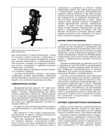
Figure C.9. Nanotechnology application: helmet incorporating P50 midlatency auditory evoked potential
recording and near-infrared detection of frontal lobe blood flow to measure sensory gating
and hypofrontality, respectively. A. Evoked potential module including audio stimulator
(earphones), surface electrodes (vertex, mastoids, forehead), amplifiers, averager with wave
recognition software, and data storage device for downloading. B. Near-infrared detection
module for frontal lobe blood flow measurement. C. Flip-down screen for tracking eye
movements and display of results from sensory gating and frontal blood flow measurements.
Implementation of this methodology would be limited to the ability to record midlatency auditory
evoked responses in varied environments. The foreseen method of implementation would involve the
use of an electronically shielded helmet (Figure C.9) containing the following: (1) P50 potential
recording electrodes at the vertex, mastoids, and ground; (2) eye movement recording using a flipdown transparent screen to monitor the movements of one eye within acceptable limits that do not
interfere with P50 potential acquisition; and (3) electrodes on the forehead to monitor muscle
contractions that could interfere with P50 potential acquisition. The helmet would incorporate an
audio stimulator for delivering click stimuli, operational amplifiers for the three measures, averaging
software, wave detection software (not currently available), and simple computation and display on
the flip-down screen of sensory gating as a percent. A high percentage compared to control conditions
would be indicative of a lack of sensory gating (indicating increased distractibility, uncontrolled
anxiety, etc.). An individual could don the helmet and obtain a measure of sensory gating within 5-7
minutes.
The applications for this nanotechnology would be considerable, including military uses for selfmonitoring human performance in advance of and during critical maneuvers; for self-monitoring by
astronauts on long-duration space missions; for pilots, drivers and operators of sensitive and complex
equipment, etc. It should be noted that this physiological measure can not be “faked” and is applicable
across languages and cultures.
Converging Technologies for Improving Human Performance (pre-publication on-line version)
205
Hypofrontality
In general, the role of the frontal cortex is to control, through inhibition, those old parts of the brain we
inherited from our early ancestors, the emotional brainstem (Damasio 1999). If the frontal cortex
loses some of its inhibitory power, “primordial” behaviors are released. This can occur when the
cortex suffers from decreased blood flow, known as “hypofrontality.” Instinctive behaviors then can
be released, including, in the extreme, exaggerated fight versus flight responses to misperceived
threats, i.e., violent behavior in an attempt to attack or flee. “Hypofrontality” is evident in such
disorders as schizophrenia, PTSD, and depression, as well as in neurodegenerative disorders like
Alzheimer’s and Huntington’s diseases. Decreased frontal lobe blood flow can be induced by alcohol.
Damage, decreased uptake of glucose, reduced blood flow, and reduced function have all been
observed in the frontal cortex of violent individuals and murderers.
The proposed method described below could be used to detect preclinical dysfunction (i.e., could be
used to screen and select crews for military or space travel operations); to determine individual
performance under stress (i.e., could be used to prospectively evaluate individual performance in flight
simulation/virtual emergency conditions); and to monitor the effects of chronic stressors (i.e., monitor
sensory gating during long-duration missions). This nanomethodology would be virtually realtime;
would not require invasive measures (such as sampling blood levels of cortisol, which are difficult to
carry out accurately, are variable and delayed rather than predictive); and would be more reliable than,
for example, urine cortisol levels (which would be delayed, or could be compensated for during
chronic stress). Training in individual and communal coping strategies is crucial for alleviating some
of the sequelae of chronic stress, and the degree of effectiveness of these strategies could be
quantitatively assessed using sensory gating of the P50 potential as well as frontal lobe blood flow.
That is, these measures could be used to determine the efficacy of any therapeutic strategy, i.e., to
measure outcome.
A detecting module located over frontal areas with a display on the flip-down screen could be
incorporated in the helmet to provide a noninvasive measure of frontal lobe blood flow for selfmonitoring in advance of critical maneuvers. The potential nanotechnology involved in such measures
has already been addressed (Chance and Kang n.d.). Briefly, since hemoglobin is a strong absorber,
changes in this molecule could be monitored using near-infrared detection. This promising field has
the potential for monitoring changes in blood flow as well as hemoglobin saturation, a measure of
energy usage.
Peripheral nanotechnology applications such as P50 potential recordings and frontal blood flow
measures are likely to provide proximal, efficient, and useful improvements in human performance.
Nanotechnology, by being transparently integrated into our executive functions, will become part of
the enculturation process, modulating brain structure and driving our evolution.
References
Chance, B., Kang, K. 2002. Optical identification of cognitive state. Converging technology (NBIC) for
improving human performance (this volume).
Damasio, A. 1999. The Feeling of What Happens, Body and Emotion in the Making of Consciousness, Harcourt
Brace & Co., New York, NY.
Donald, M.W. 1991. Origins of the Modern Mind, Three Stages in the Evolution of Culture and Cognition,
Harvard University Press, Cambridge, MA.
Garcia-Rill, E. 1997. Disorders of the Reticular Activating System. Med. Hypoth. 49, 379-387.
C. Improving Human Health and Physical Capabilities
206
National Research Council Committee on Space Biology and Medicine. 1998. Strategy for Research in Space
Biology and Medicine into the Next Century. National Academy Press: Washington, DC.
Pollack, J. 2002. The limits of design complexity. Converging technology (NBIC) for improving human
performance (this volume).
SCIENCE AND TECHNOLOGY AND THE TRIPLE D (DISEASE, DISABILITY,
DEFECT)
Gregor Wolbring, University of Calgary
Science and technology (S&T) have had throughout history — and will have in the future — positive
and negative consequences for humankind. S&T is not developed and used in a value neutral
environment. S&T activity is the result of human activity imbued with intention and purpose and
embodying the perspectives, purposes, prejudice and particular objectives of any given society in
which the research takes place. S&T is developed within the cultural, economical, ethical, and moral
framework of the society in which the research takes place. Furthermore, the results of S&T are used
in many different societies reflecting many different cultural, economical, ethical, moral frameworks. I
will focus on the field of Bio/Gene/Nanomedicine. The development of Bio/Gene/Nanotechnology is
— among other things — justified with the argument that it holds the promises to fix or help to fix
perceived disabilities, impairments, diseases and defects and to diminish suffering. But who decides
what is a disability, disease, an impairment and a ‘defect’ in need of fixing? Who decides what the
mode of fixing (medical or societal) should be, and who decides what is suffering? How will these
developments affect societal structures?
Perception
The right answers to these questions will help ensure that these technologies will enhance human life
creatively, rather than locking us into the prejudices and misconceptions of the past. Consider the
following examples of blatant insensitivity:
Fortunately the Air Dri-Goat features a patented goat-like outer sole for increased traction so
you can taunt mortal injury without actually experiencing it. Right about now you’re
probably asking yourself “How can a trail running shoe with an outer sole designed like a
goat’s hoof help me avoid compressing my spinal cord into a Slinky on the side of some
unsuspecting conifer, thereby rendering me a drooling, misshapen non- extreme-trailrunning husk of my former self, forced to roam the earth in a motorized wheelchair with my
name embossed on one of those cute little license plates you get at carnivals or state fairs,
fastened to the back?” (Nike advertisement, Backpacker Magazine, October 2000).
Is it more likely for such children to fall behind in society or will they through such
afflictions develop the strengths of character and fortitude that lead to the head of their
packs? Here I’m afraid that the word handicap cannot escape its true definition — being
placed at a disadvantage. From this perspective seeing the bright side of being handicapped
is like praising the virtues of extreme poverty. To be sure, there are many individuals who
rise out of its inherently degrading states. But we perhaps most realistically should see it as
the major origin of asocial behavior (Watson 1996).
American bioethicist Arthur Caplan said in regards to human genetic technology, “the understanding
that our society or others have of the concept of health, disease and normality will play a key role in
shaping the application of emerging knowledge about human genetics” (Caplan 1992). I would add
Nanomedicine/Nanotechnology into Caplan’s quote because parts of nanotechnology development are