Guidelines on - Upper Urinary Tract Urothelial Cell Carcinomas pot
Bạn đang xem bản rút gọn của tài liệu. Xem và tải ngay bản đầy đủ của tài liệu tại đây (147.59 KB, 20 trang )
Guidelines on
Upper Urinary
Tract Urothelial
Cell Carcinomas
M. Rouprêt, R. Zigeuner, J. Palou, A. Boehle, E. Kaasinen,
R. Sylvester, M. Babjuk, W. Oosterlinck
© European Association of Urology 2011
2 MARCH 2011
TABLE OF CONTENTS PAGE
1. INTRODUCTION 3
2. METHODOLOGY 3
3. EVIDENCE SYNTHESIS 3
3.1 Epidemiology 3
3.2 Risk factors 3
3.3 Histology and classification 4
3.3.1 Histologic types 4
3.3.2 Classification 4
3.3.2.1 Tumour Node Metastasis (TNM) classification 4
3.3.2.2 Tumour grade 5
3.4 Symptoms 5
3.5 Diagnosis 5
3.5.1 Imaging 5
3.5.1.1 Multidetector computed tomographic urography 5
3.5.1.2 Magnetic resonance imaging 5
3.5.2 Cystoscopy and urinary cytology 6
3.5.3 Diagnostic ureteroscopy 6
3.6 Prognostic factors 6
3.6.1 Tumour stage and grade 6
3.6.2 Age and gender 6
3.6.3 Tumour location 6
3.6.4 Lymphovascular invasion 7
3.6.5 Other factors 7
3.6.6 Molecular markers 7
3.7 Treatment 7
3.7.1 Localised disease 7
3.7.1.1 Radical nephroureterectomy 7
3.7.1.2 Conservative surgery 8
3.7.1.2.1 Ureteroscopy 8
3.7.1.2.2 Segmental resection 8
3.7.1.2.3 Percutaneous access 8
3.7.1.3 Adjuvant topical agents 9
3.7.2 Advanced disease 9
3.7.2.1 Nephroureterectomy 9
3.7.2.2 Chemotherapy 9
3.7.2.3 Radiation therapy 9
3.8 Follow-up 10
4. CONCLUSION 11
5. REFERENCES 11
6. ABBREVIATIONS USED IN THE TEXT 19
MARCH 2011 3
1. INTRODUCTION
The most recent summary of the European Association of Urology (EAU) guidelines on upper urinary tract
urothelial cell carcinomas (UUT-UCCs) was published in 2004 (1). The EAU Guideline Group for UUT-UCCs has
prepared current guidelines to provide evidence-based information for the clinical management of these rare
tumours and to help clinicians incorporate these recommendations into their practice. The current update is
based on a structured literature search.
2. METHODOLOGY
A Medline search was performed on urothelial malignancies and UUT-UCC management using combinations
of the following terms: urinary tract cancer, urothelial carcinomas, upper urinary tract, carcinoma, transitional
cell, renal pelvis, ureter, bladder cancer, chemotherapy, nephroureterectomy, adjuvant treatment, neoadjuvant
treatment, recurrence, risk factors, and survival. The publications concerning UUT-UCCs were mostly
retrospective, including some large multicentre studies. Due to the scarcity of randomised data, articles
were selected for these guidelines based on the following criteria: evolution of concepts, intermediate- and
long-term clinical outcomes, study quality, and relevance. Older studies were included selectively if they
were historically relevant or if data were scarce in recent publications. To facilitate evaluation of the quality
of information provided, levels of evidence (LE) and recommendation grades (GR) were inserted according to
general principles of evidence-based medicine (EBM) (2).
3. EVIDENCE SYNTHESIS
3.1 Epidemiology
Urothelial carcinomas are the fourth most common tumours after prostate (or breast) cancer, lung cancer,
and colorectal cancer (3,4). They can be located in the lower urinary tract (bladder and urethra) or the upper
urinary tract (pyelocaliceal cavities and ureter). Bladder tumours account for 90-95% of urothelial carcinomas
(4) and are the most common malignancy of the urinary tract and the second most common malignancy of the
urogenital tract after prostate cancer (5,6). However, UUT-UCCs are uncommon and account for only 5-10%
of urothelial carcinomas (3,7-9). The estimated annual incidence of UUT-UCCs in Western countries is about
one or two new cases per 100,000 inhabitants. Pyelocaliceal tumours are about twice as common as ureteral
tumours. In 8-13% of cases, concurrent bladder cancer is present. Recurrence of disease in the bladder occurs
in 30-51% of UUT-UCC patients (10,11), whereas recurrences in the contralateral upper tract are observed in
2-6% (12,13).
The natural history of UUT-UCCs differs from that of bladder cancer: 60% of UUT-UCCs are invasive
at diagnosis compared with only 15% of bladder tumours (5,7,9). UUT-UCCs have a peak incidence in people
in their 70s and 80s, and UUT-UCC is three times more prevalent in men than in women.
There are familial/hereditary cases of UUT-UCCs linked to hereditary nonpolyposis colorectal
carcinoma (HNPCC) (14). Among patients with UUT-UCCS, these cases can be detected during a medical
interview. Indeed, the cancer is likely to be hereditary if the patient is < 60 yr of age and/or has a personal or
family history of an HNPCC-type cancer (15,16). These patients should undergo DNA sequencing to identify
hereditary cancers that have been misclassified as sporadic cancers by insufficient clinical data. The presence
of other HNPCC-associated cancers should also be evaluated. These patients should be closely monitored,
and genetic counselling is advocated (15,16).
3.2 Risk factors
Many environmental factors contribute to the development of UUT-UCCs. Some are similar to those associated
with bladder cancer, whereas others are more specific for UUT-UCC. Tobacco and occupational exposure
remain the principal exogenous risk factors for developing these tumours. Exposure to tobacco increases
the relative risk of developing a UUT-UCC from 2.5 to 7 (17,18). UUT-UCC “amino tumours” are related to
occupational exposure to certain aromatic amines. These aromatic hydrocarbons are used in many industries
(e.g., dyes, textiles, rubber, chemicals, petrochemicals, and coal). They are responsible for the carcinogenicity
of certain chemicals, including benzidine and β-naphthalene. These two chemicals have been banned since
the 1960s in most industrialised countries. In most cases, UUT-UCCs are secondary to an amino tumour of the
4 MARCH 2011
bladder. The average duration of exposure needed to develop a UUT-UCC is approximately 7 yr, with a latency
period of about 20 yr following the termination of exposure. The estimated risk (odds ratio) of developing UCC
after exposure to aromatic amines is 8.3 (17,19).
Upper urinary tract tumours resulting from phenacetin consumption almost disappeared (17) after the
product was banned in the 1970s.
Although the incidence of Balkan endemic nephropathy is also on the decline (20,21), roles have
been proposed for aristolochic acid and the consumption of Chinese herbs in the physiopathology and
induction, respectively, of this nephropathy (22-24). Several studies have revealed the carcinogenic potential of
aristolochic acid contained in Aristolochia fangchi and Aristolochia clematis (plants endemic to the Balkans).
This acid contains a set of highly toxic nitrophenolate derivatives that exhibit a powerful mutagenic action due
to their ability to make up covalent links with cell DNA. The aristolochic acid derivative d-aristolactam causes a
specific mutation in the p53 gene at codon 139. This mutation is very rare in the nonexposed population and is
predominant in patients with nephropathy due to Chinese herbs or Balkan endemic nephropathy who present
with UUT-UCC.
A high incidence of UUT-UCC has also been described in Taiwan, especially in the population of the
southwest coast of the island, and represents 20-25% of UCCs in the region (16). The association of UUT-UCC
with blackfoot disease and arsenic exposure remains unclear in this patient population (25).
Differences in the ability to counteract carcinogens may contribute to host susceptibility and the risk
of developing urothelial carcinomas. Because certain genetic polymorphisms are associated with an increased
risk of cancer or faster disease progression, there is variability in interindividual susceptibility to the risk factors
just mentioned. Only one polymorphism specific to UUT-UCC has been reported so far. A variant allele,
SULT1A1*2, which reduces sulfotransferase activity, enhances the risk of developing UUT-UCC (26).
Epidermoid carcinoma of the UUT is associated with chronic inflammatory and infectious disease arising from
stones in the UUT (27,28).
3.3 Histology and classification
3.3.1 Histologic types
More than 95% of urothelial carcinomas are derived from the urothelium and correspond to UUT-UCCs or
bladder tumours (1,5,28). With regard to UUT-UCCs, morphologic variants have been described that are
more often observed in urothelial kidney tumours. These variants always correspond to high-grade tumours,
and such urothelial carcinomas are associated with one of the following variants: micropapillary, clear cell,
neuroendocrine, and lymphoepithelial (9,27). Collecting duct carcinoma has similar characteristics to UUT-
UCCs because of its common embryologic origin (29).
Upper urinary tract tumours with nonurothelial histology are exceptions (30). Epidermoid carcinomas
of the upper urinary tract represent < 10% of pyelocaliceal tumours and are even more rare within the ureter.
Other histologic subtypes are adenocarcinomas (< 1%), neuroendocrine carcinomas, and sarcomas.
3.3.2 Classification
The classification and morphology of UUT-UCCs are similar to those of bladder carcinomas (5,28). It is
possible to distinguish between noninvasive papillary tumours (papillary urothelial tumours of low malignant
potential, low-grade papillary urothelial carcinoma, high-grade papillary urothelial carcinoma), flat lesions
(carcinoma in situ [CIS]), and invasive carcinomas. All variants of urothelial tumours described in the bladder
can also be observed in the upper urinary tract.
3.3.2.1 Tumour Node Metastasis (TNM) staging
Table 1 presents the Union Internationale Contre le Cancer 2009 TNM classification used throughout these
guidelines (31). According to the TNM classification, the regional lymph nodes that should be considered are
the hilar, abdominal para-aortic, and paracaval nodes, and, for the ureter, the intrapelvic nodes. Laterality does
not affect the N classification.
MARCH 2011 5
Table 1 TNM classification 2009 for UUT-UCC (31) *
T - Primary tumour
TX Primary tumour cannot be assessed
T0 No evidence of primary tumour
Ta Non-invasive papillary carcinoma
Tis Carcinoma in situ
T1 Tumour invades subepithelial connective tissue
T2 Tumour invades muscle
T3 (Renal pelvis) Tumour invades beyond muscularis into peripelvic fat or renal parenchyma
(Ureter) Tumour invades beyond muscularis into periureteric fat
T4 Tumour invades adjacent organs or through the kidney into perinephric fat
N - Regional lymph nodes
NX Regional lymph nodes cannot be assessed
N0 No regional lymph node metastasis
N1 Metastasis in a single lymph node 2 cm or less in the greatest dimension
N2 Metastasis in a single lymph node more than 2 cm but not more than 5 cm in the greatest
dimension or multiple lymph nodes, none more than 5 cm in greatest dimension
N3 Metastasis in a lymph node more than 5 cm in greatest dimension
M - Distant metastasis
M0 No distant metastasis
M1 Distant metastasis
*
All EAU guidelines only advocate the TNM system of tumour classification.
UUT-UCC = urethelial cell carcinoma of the upper urinary tract
3.3.2.2 Tumour grade
Until 2004, the most common classification used was the World Health Organization (WHO) classification
of 1973, which distinguished only three grades (G1, G2, and G3) (32). In recent years, molecular biologic
data have allowed for further distinction between different tumour groups and the development of a new
classification system that better reflects the potential growth of these tumours (33). Thus the 2004 WHO
classification now takes histologic data into account to distinguish among three groups of noninvasive
tumours: papillary urothelial neoplasia of low malignant potential, low-grade carcinomas, and high-grade
carcinomas. There are almost no tumours of low malignant potential in the upper urinary tract (9,27,28).
3.4 Symptoms
The diagnosis of a UUT-UCC may be fortuitous or related to the exploration of symptoms (1,6). The symptoms
are generally restricted (34). The most common symptom of UUT-UCC is gross or microscopic haematuria
(70-80%). Flank pain occurs in up to 20-40% of cases, and a lumbar mass is present in 10-20% of cases (1,7).
However, systemic symptoms (altered health condition including anorexia, weight loss, malaise, fatigue, fever,
night sweats, or cough) associated with UUT-UCC should prompt consideration of a more rigorous metastatic
evaluation or perioperative chemotherapy regimens (34).
3.5 Diagnosis
3.5.1 Imaging
3.5.1.1 Multidetector computed tomographic urography
Multidetector computed tomographic urography (MDCTU) is the gold standard for exploration of the upper
urinary tract and has replaced intravenous excretory urography (35-38). It must be conducted under optimal
conditions, particularly with acquisition of an excretory phase. Multiple protocols from two helical computed
tomography acquisitions (at least millimetric) are necessary before and after the injection of contrast.
The detection rate of UUT-UCC is satisfactory for this type of imaging: 96% sensitivity and 99%
specificity for polypoid lesions between 5 and 10 mm. Sensitivity drops to 89% for polypoid lesions < 5 mm
and 40% for polypoid lesions < 3 mm (16,17). MDCTU can also detect thickening of the wall of the renal pelvis
or ureter as a sign of UUT-UCC. The main difficulty remains identifying flat lesions that are undetectable until
they evolve towards a massive infiltration.
Lastly, it was shown that hydronephrosis on preoperative imaging was associated with advanced
pathologic disease and poorer oncologic outcomes (39).
3.5.1.2 Magnetic resonance imaging
Magnetic resonance imaging (MRI) urography is indicated in patients who cannot be subjected to an MDCTU
6 MARCH 2011
(40). The detection rate of MRI is 75% after contrast injection for tumours < 2 cm (41). Magnetic resonance
urography with contrast injection, however, remains contraindicated in selected patients with severe renal
impairment (< 30 ml/min creatinine clearance) due to the risk of nephrogenic systemic fibrosis. MRI without
contrast is less helpful compared with MDCTU in diagnosing UUT-UCCs.
3.5.2 Cystoscopy and urinary cytology
Positive urine cytology is highly suggestive of UUT-UCC when bladder cystoscopy is normal and if CIS of
the bladder or prostatic urethra has been excluded. Cytology is less sensitive for UUT-UCC than for bladder
tumours, even for high-grade lesions, and it should ideally be performed in situ (i.e., in the renal cavities). A
positive cytology may be valuable in staging because it has been associated with muscle-invasive and non-
organ-confined disease (42).
The detection of molecular abnormalities by fluorescence in situ hybridisation (FISH) is becoming more
popular for UCC screening, but results are still preliminary (43,44). The sensitivity of FISH for the identification
of UUT-UCCs parallels its performance in bladder cancer; however, the preponderance of low-grade recurrent
disease in the population undergoing surveillance and minimally invasive therapy for UUT-UCCs may limit
its usefulness (45). In addition, FISH appears to have limited value for upper urinary tract tumour surveillance
(46,47).
3.5.3 Diagnostic ureteroscopy
Ureteroscopy is a better approach to diagnose UUT-UCCs (42,48,49). A flexible ureteroscope can explore the
ureter macroscopically and reach renal cavities in 95% of cases, and it can assess the aspect of the tumour,
obtain tumour biopsy, and determine tumour grade in 90% of cases with a low false-negative rate (50). It also
facilitates performing a selective ureteral cytology and a retrograde pyelogram.
Flexible ureteroscopy is especially useful when there is diagnostic uncertainty, when conservative
treatment is being considered, or in patients with a solitary kidney. The possible advantages of ureteroscopy
should be discussed in the preoperative assessment of any UUT-UCC patient. Combining ureteroscopic
biopsy grade, ipsilateral hydronephrosis, and urinary cytology may help decision making on radical
nephroureterectomy (RNU) versus endoscopic treatment (42). Table 2 lists the recommendations.
Table 2 Guidelines for the diagnosis of UUT-UCC
Recommendations for diagnosis of UUT-UCC GR
Urinary cytology A
Cystoscopy to rule out a concomitant bladder tumour A
MDCTU A
UUT-UCC = urethelial cell carcinoma of the upper urinary tract; MDCTU = multidetector computed tomographic
urography.
3.6 Prognostic factors
Upper urinary tract urothelial cell carcinomas that invade the muscle wall usually have a very poor prognosis.
The 5-yr specific survival is < 50% for pT2/pT3 and < 10% for pT4 (51,52). This section briefly describes the
currently recognised prognostic factors.
3.6.1 Tumour stage and grade
According to the most recent classifications, the primary recognised prognostic factors are tumour stage and
grade (8,31,53-55).
3.6.2 Age and gender
The effect of gender on UUT-UCC mortality has been disputed recently and is no longer considered an
independent prognostic factor (56-58). Conversely, patient age is still considered an independent prognostic
factor because older age at the time of RNU is associated with decreased cancer-specific survival (LE: 3) (59).
However, advanced age alone should not be an exclusion criterion for the aggressive treatment of potentially
curable UUT-UCC. A large proportion of elderly patients can be cured with RNU (59). This suggests that
chronologic age alone is an inadequate indicator of outcomes in older UUT-UCC patients (59).
3.6.3 Tumour location
According to the most recent findings, the initial location of the tumour within the upper urinary tract (e.g.,
ureter vs renal pelvis) is no longer accepted as a prognostic factor (11,60,61), contrary to previously published
MARCH 2011 7
reports (LE: 3) (62). It seems there is no longer a prognostic impact for tumour location (i.e., ureteral vs
pyelocaliceal tumours) when adjusted for tumour stage (11,63).
3.6.4 Lymphovascular invasion
Lymphovascular invasion is present in approximately 20% of UUT-UCCs and an independent predictor of
survival. Lymphovascular invasion status should be included in the pathologic report of RNU specimens (LE:
3) (64-66). However, only in patients with negative lymph nodes does lymphovascular invasion add prognostic
information beyond that obtained with standard features (64).
3.6.5 Other factors
Extensive tumour necrosis is an independent predictor of clinical outcomes in patients who undergo RNU.
Extensive tumour necrosis can be defined as > 10% of the tumour area (LE: 3) (67,68).
The tumour architecture (e.g., papillary vs sessile) of UUT-UCCs appears to be associated with prognosis after
RNU. A sessile growth pattern is associated with worse outcomes (LE: 3) (8,63,69).
The presence of concomitant CIS in patients with organ-confined UUT-UCC is associated with
a higher risk of recurrent disease and cancer-specific mortality (LE: 3) (70). Similar to lower tract urothelial
carcinoma, concomitant CIS is an independent predictor of worse outcomes in organ-confined disease (71).
3.6.6 Molecular markers
Several research groups are working on upper urinary tract tumour characteristics and carcinogenesis
pathways. Specific markers that could aid in the prognosis of UUT-UCCs have been investigated. Microsatellite
instabilities (MSIs) are independent molecular makers used for tumour prognosis (72). In addition, MSIs can
help detect germ-line mutations, allowing for the detection of possible hereditary cancers (14,16,72).
E-cadherin has been shown to be a useful independent marker for prognosis, as have hypoxia-
inducible factor (HIF)-1α and telomerase RNA component (73). Furthermore, HIF-1α appears to be significantly
associated with tumour grade and growth pattern, and the telomerase RNA component could possibly be
used for UUT-UCC diagnosis and prognostication. However, to date, none of the markers has been externally
validated, and none has fulfilled the clinical and statistical criteria necessary to support its introduction in daily
clinical decision making.
3.7 Treatment
3.7.1 Localised disease
3.7.1.1 Radical nephroureterectomy
RNU with excision of the bladder cuff is the gold standard treatment for UUT-UCCs, regardless of the location
of the tumour in the upper urinary tract (LE: 3) (8). The RNU procedure must comply with oncologic principles,
which consist of preventing tumour seeding by avoiding entry into the urinary tract during tumour resection
(8,69).
Resection of the distal ureter and its orifice is performed because it is a part of the urinary tract with
considerable risk of recurrence. After removal of the proximal part, it is almost impossible to image or approach
it by endoscopy during follow-up. Recent publications on survival after nephroureterectomy have concluded
that removal of the distal ureter and bladder cuff is beneficial (74-77).
McDonald et al. presented the pluck technique in 1952, but it was not until 1995 (78) that the
usefulness of an endoscopic approach to the distal ureter was really emphasised, and then several other
alternative techniques were reconsidered to simplify resection of the distal ureter: stripping, transurethral
resection of the intramural ureter, and intussusception techniques (12). Apart from ureteral stripping, none of
these techniques has demonstrated inferiority to excision of the bladder cuff (LE: 3) (75-77,79). A delay > 45 d
between diagnosis and resection of the tumour constitutes a risk for disease progression (LE: 3) (80).
Lymph node dissection associated with RNU is of therapeutic interest and allows for optimal staging
of the disease (LE: 3) (81-83). Lymphadenectomy in pN+ allows for reduction of the tumour mass to guide
patients towards adjuvant treatments (LE: 3) (82). However, the anatomic sites of lymphadenectomy have not
yet been clearly defined. The number of lymph nodes to be removed depends on the tumour location. No trial
so far has shown its direct impact on survival (82). Lymphadenectomy appears to be unnecessary in cases
of Ta-T1 UUT-UCCs because it was reported to be retrieved in 2.2% pT1 versus 16% pT2-4 tumours (82).
In addition, authors have described a continuous increase in the probability of lymph node-positive disease
related to pT classification (81). Lastly, lymphadenectomy appears to be a prognostic variable within a model in
patients with histologically confirmed node-negative (pN0) disease (83). However, these data are retrospective;
it is not possible to standardise either indication or the extent of lymphadenectomy. Consequently,
underreporting of the true rate of node-positive disease is likely.
The safety of laparoscopic RNU has not yet achieved final proof (84,85). In early experience, there
were reports of retroperitoneal metastatic dissemination and dissemination along the trocar pathway when
8 MARCH 2011
large tumours were manipulated in a pneumoperitoneal environment (86,87).
Recent data, however, show a tendency towards equivalent oncologic results between laparoscopic
RNU and open surgery. In addition, the laparoscopic approach appears to be superior to open surgery only
with regard to functional outcomes (LE: 3) (88-91). Only one prospective randomised study of 80 patients did
not provide evidence that laparoscopic RNU is inferior to open RNU for noninvasive UUT-UCC (LE: 2) (92).
Several precautions must be taken when operating with a pneumoperitoneum because it may increase tumour
spillage:
• Enteringtheurinarytractshouldbeavoided.
• Directcontactoftheinstrumentswiththetumourshouldbeavoided.
• LaparoscopicRNUmusttakeplaceinaclosedsystem.Morcellationofthetumourshouldbeavoided,
and an endobag is necessary to extract the tumour.
• Thekidneyanduretermustberemovedenblocwiththebladdercuff.
• Invasive,large(T3/T4and/orN+/M+),ormultifocaltumoursarecontraindicationsforlaparoscopic
RNU, until proven otherwise.
Recommendations are listed in Table 3.
Table 3 Guidelines for radical management of UUT-UCC: radical nephroureterectomy
Indications for RNU for UUT-UCC GR
Suspicion of infiltrating UUT-UCC on imaging B
High-grade tumour (urinary cytology) B
Multifocality (with two functional kidneys) B
Techniques for RNU in UUT-UCC
Open and laparoscopic access are equivalent in terms of efficacy B
Bladder cuff removal is imperative A
Several techniques for bladder cuff excision are acceptable except stripping C
Lymphadenectomy is recommended in case of invasive UUT-UCC C
RNU = radical nephroureterectomy; UUT-UCC = urethelial cell carcinoma of the upper urinary tract.
3.7.1.2 Conservative surgery
Conservative surgery for low-risk UUT-UCCs allows for preservation of the upper urinary renal unit while
sparing the patient the morbidity associated with open radical surgery (93,94). Conservative management of
UUT-UCCs can be considered in imperative cases (renal insufficiency, solitary functional kidney) or in elective
cases (i.e., when the contralateral kidney is functional) for low-grade, low-stage tumours (LE: 3) (76,95). The
choice of technique depends on technical constraints, the anatomic location of the tumour, and the experience
of the surgeon.
3.7.1.2.1 Ureteroscopy
Endoscopic ablation can be considered in highly selected cases (96,97,98) and in these situations:
• Aflexibleratherthanarigidureteroscope,lasergenerator,andpliers(pluck)forbiopsiesareavailable
(LE: 3) (96,99).
• Thepatientisinformedoftheneedforcloser,morestringentsurveillance.
• Acompleteresectionisadvocated.
3.7.1.2.2 Segmental resection
Segmental ureteral resection with wide margins provides adequate pathologic specimens for definitive
staging and grade analysis while also preserving the ipsilateral kidney. Segmental resection is possible for the
treatment of low- and high-risk tumours of the distal ureter (LE: 3) (100,101). It is necessary, however, to ensure
that the area of tissue around the tumour is not invaded. Segmental resection of the iliac and lumbar ureter is
associated with a failure rate greater than that for the distal pelvic ureter (100,102).
Open resection of tumours of the renal pelvis or calices has almost disappeared. Resection of
pyelocaliceal tumours is technically difficult, and the recurrence rate is higher than for tumours of the ureter.
3.7.1.2.3 Percutaneous access
Percutaneous management can be considered for low-grade or noninvasive UUT-UCCs in the renal cavities
(LE: 3) (97,103,104). This treatment option may be offered to patients with low-grade tumours in the lower
MARCH 2011 9
caliceal system that are inaccessible or difficult to manage by ureteroscopy. A theoretical risk of seeding
exists in the puncture tract and in perforations that may occur during the procedure. This approach, however,
is being progressively abandoned due to enhanced materials and advances in distal-tip deflection of recent
ureteroscopes.
3.7.1.3 Adjuvant topical agents
The instillation of bacillus Calmette-Guérin or mitomycin C in the urinary tract by percutaneous nephrostomy
via a three-valve system open at 20 cm (after complete eradication of the tumour), or even through a ureteric
stent (105), is technically feasible after conservative treatment of UUT-UCCs or for the treatment of CIS. The
medium-term results are similar to those observed for the treatment of bladder tumours but have not been
confirmed in long-term studies (LE: 3) (1,106,107). Table 4 reports the recommendations.
Table 4 Guidelines for conservative management of UUT-UCC
Indications for conservative management of UUT-UCC GR
Unifocal tumour B
Small tumour B
Low-grade tumour (cytology or biopsies) B
No evidence of an infiltrative lesion on MDCTU B
Understanding of close follow-up B
Techniques used in conservative management of UUT-UCC
Laser should be used in case of endoscopic treatment C
Flexible ureteroscopy is preferable over rigid ureteroscopy C
Open partial resection is an option for pelvic ureteral tumours C
A percutaneous approach remains an option in small low-grade caliceal tumours unsuitable for
ureteroscopic treatment
C
MDCTU = multidetector computed tomographic urography; UUT-UCC = urethelial cell carcinoma of the upper
urinary tract.
3.7.2 Advanced disease
3.7.2.1 Nephroureterectomy
There are no benefits of RNU in metastatic (M+) disease, although it can be considered a palliative option
(LE: 3) (8,81).
3.7.2.2 Chemotherapy
Because UUT-UCCs are urothelial tumours, platinum-based chemotherapy is expected to produce similar
results to those seen in bladder cancer. Several platinum-based chemotherapy regimens have been proposed
(108-111).
Only one study has reported the effect of neoadjuvant chemotherapy in contrast to what has been
demonstrated in the bladder. While survival data need to mature and longer follow-up is awaited, current
preliminary data provide justification for the sustained support of trials using this strategy in UUT-UCCs (112).
Adjuvant chemotherapy achieves a recurrence-free rate of up to 50% but has minimal impact on
survival (108-111). Not all the patients receive this treatment because of comorbidities and impaired renal
function after radical surgery. Data are currently insufficient to provide any recommendations.
3.7.2.3 Radiation therapy
Adjuvant radiotherapy may improve local control of the disease (113). When given in combination
with cisplatinum, it may result in a longer disease-free survival and longer overall survival (114) (LE: 3).
Radiation therapy appears to be scarcely relevant nowadays both as a unique therapy and associated with
chemotherapy as a tumour adjuvant (Fig. 1).
10 MARCH 2011
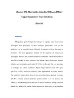
Fig. 1 Proposed flowchart for the management of UUT-UCC
UUT-UCC = urethelial cell carcinoma of the upper urinary tract; MDCTU = multidetector computed tomographic
urography.
3.8 Follow-up
Strict follow-up of UUT-UCC patients after surgical treatment is mandatory to detect metachronous bladder
tumours (in all cases), local recurrence, and distant metastases (in the case of invasive tumours).
When RNU is performed, local recurrence is rare, and the risk of distant metastases is directly
related to the risk factors listed previously. The reported recurrence rate within the bladder after treatment of a
primary UUT-UCC varies considerably from 15% to 50% (10,115,116). Thus the bladder should be observed
in all cases. A prior history of bladder cancer and upper tract tumour multifocality are the risk factors most
often reported for bladder tumours following UUT-UCCs. The surveillance regimen is based on cystoscopy
and urinary cytology for at least 5 yr (10,115,116). A bladder recurrence should not be considered a distant
recurrence.
When conservative treatment is performed, the ipsilateral upper urinary tract requires careful follow-up
due to the high risk of recurrence. Despite notable improvements in endourologic technology, the follow-up of
patients treated with conservative therapy is difficult, and minimally invasive procedures are often necessary
(96,98,117,118). Table 5 lists the recommended follow-up schedules.
Table 5 Guidelines for follow-up of UUT-UCC patients after initial treatment
After RNU, over at least 5 yr GR
Noninvasive tumour
Cystoscopy/urinary cytology at 3 mo and then yearly C
MDCTU every year C
Invasive tumour
Cystoscopy/urinary cytology at 3 mo and then yearly C
MDCTU every 6 mo over 2 yr and then yearly C
Diagnostics evaluation: CT-urography,
urinary cytology, cystoscopy
(± ureteroscopy with biopsies)
- Unifocal tumour
- Size < 1 cm
- Low-grade tumour
- Superficial aspect on MDCTU
Gold standard treatment:
Radical nephroureterectomy
Conservative management:
ureteroscopy, segmental resection,
percutaneous approach
Open
Recurrence
Close and stringent follow-up
Laparoscopic
UUT-UCC
MARCH 2011 11
After conservative management, over at least 5 yr
Urinary cytology and MDCTU at 3 mo, 6 mo, and then yearly C
Cystoscopy, ureteroscopy and cytology in situ at 3 mo, 6 mo, and then every 6 mo over 2 yr, and
then yearly
C
RNU = radical nephroureterectomy; MDCTU = multidetector computed tomographic urography.
4. CONCLUSIONS
These guidelines contain information for the diagnosis and treatment of individual patients according to a
current, standardised approach. When determining the optimal treatment regimen for their patients, physicians
must take into account each individual patient’s specific clinical characteristics with regard to renal function
including medical comorbidities; tumour location, grade, and stage; and molecular marker status.
5. REFERENCES
1. Oosterlinck W, Solsona E, van der Meijden AP, et al. EAU guidelines on diagnosis and treatment of
upper urinary tract transitional cell carcinoma. Eur Urol 2004 Aug;46(2):147-54.
/>2. Phillips B, Ball C, Sackett D, et al. Oxford Centre for Evidence-based Medicine - levels of evidence
(March 2009). Centre for Evidence Based Medicine Web site.
[Access date January 2011]
3. Munoz JJ, Ellison LM. Upper tract urothelial neoplasms: incidence and survival during the last 2
decades. J Urol 2000 Nov;164(5):1523-5.
/>4. Ploeg M, Aben KK, Kiemeney LA. The present and future burden of urinary bladder cancer in the
world. World J Urol 2009 Jun;27(3):289-93.
/>5. Babjuk M, Oosterlinck W, Sylvester R, et al. EAU guidelines on non-muscle-invasive urothelial
carcinoma of the bladder. Eur Urol 2008 Aug;54(2):303-14.
/>6. Jemal A, Siegel R, Ward E, et al. Cancer statistics, 2009. CA Cancer J Clin 2009 Jul-Aug;59(4):225-49.
/>7. Hall MC, Womack S, Sagalowsky AI, et al. Prognostic factors, recurrence, and survival in transitional
cell carcinoma of the upper urinary tract: a 30-year experience in 252 patients. Urology 1998
Oct;52(4):594-601.
/>8. Margulis V, Shariat SF, Matin SF, et al. Outcomes of radical nephroureterectomy: a series from the
Upper Tract Urothelial Carcinoma Collaboration. Cancer 2009 Mar;115(6):1224-33.
/>9. Olgac S, Mazumdar M, Dalbagni G, et al. Urothelial carcinoma of the renal pelvis: a clinicopathologic
study of 130 cases. Am J Surg Pathol 2004 Dec;28(12):1545-52.
/>10. Azemar MD, Comperat E, Richard F, et al. Bladder recurrence after surgery for upper urinary tract
urothelial cell carcinoma: Frequency, risk factors, and surveillance. Urol Oncol 2009 Sep. [Epub ahead
of print]
/>11. Raman JD, Ng CK, Scherr DS, et al. Impact of Tumor Location on Prognosis for Patients with Upper
Tract Urothelial Carcinoma Managed by Radical Nephroureterectomy. Eur Urol 2010 Jun;57(6):1072-
93.
/>12 MARCH 2011
12. Li WM, Shen JT, Li CC, et al. Oncologic Outcomes Following Three Different Approaches to the Distal
Ureter and Bladder Cuff in Nephroureterectomy for Primary Upper Urinary Tract Urothelial Carcinoma.
Eur Urol 2010 Jun;57(6):963-9.
/>13. Novara G, De Marco V, Dalpiaz O, et al. Independent predictors of contralateral metachronous upper
urinary tract transitional cell carcinoma after nephroureterectomy: multi-institutional dataset from three
European centers. Int J Urol 2009 Feb;16(2):187-91.
/>14. Rouprêt M, Catto J, Coulet F, et al. Microsatellite instability as indicator of MSH2 gene mutation in
patients with upper urinary tract transitional cell carcinoma. J Med Genet 2004 Jul;41(7):e91.
/>15. Acher P, Kiela G, Thomas K, et al. Towards a rational strategy for the surveillance of patients with
Lynch syndrome (hereditary non-polyposis colon cancer) for upper tract transitional cell carcinoma.
BJU Int 2010 Aug;106(3):300-2.
/>16. Rouprêt M, Yates DR, Comperat E, et al. Upper urinary tract urothelial cell carcinomas and other
urological malignancies involved in the hereditary nonpolyposis colorectal cancer (lynch syndrome)
tumor spectrum. Eur Urol 2008 Dec;54(6):1226-36.
/>17. Colin P, Koenig P, Ouzzane A, et al. Environmental factors involved in carcinogenesis of urothelial cell
carcinomas of the upper urinary tract. BJU Int 2009 Nov;104(10):1436-40.
/>18. Pommer W, Bronder E, Klimpel A, et al. Urothelial cancer at different tumour sites: role of smoking and
habitual intake of analgesics and laxatives. Results of the Berlin Urothelial Cancer Study. Nephrol Dial
Transplant 1999 Dec;14(12):2892-7.
/>19. Shinka T, Miyai M, Sawada Y, et al. Factors affecting the occurrence of urothelial tumors in dye
workers exposed to aromatic amines. Int J Urol 1995 Sep;2(4):243-8.
/>20. Markovic N, Ignjatovic I, Cukuranovic R, et al. Decreasing incidence of urothelial cancer in a Balkan
endemic nephropathy region in Serbia. A surgery based study from 1969 to 1998. Pathol Biol (Paris)
2005 Feb;53(1):26-9.
/>21. Grollman AP, Shibutani S, Moriya M, et al. Aristolochic acid and the etiology of endemic (Balkan)
nephropathy. Proc Natl Acad Sci U S A 2007 Jul;104(29):12129-34.
/>22. Arlt VM, Stiborova M, vom Brocke J, et al. Aristolochic acid mutagenesis: molecular clues to the
aetiology of Balkan endemic nephropathy-associated urothelial cancer. Carcinogenesis 2007
Nov;28(11):2253-61.
/>23. Laing C, Hamour S, Sheaff M, et al. Chinese herbal uropathy and nephropathy. Lancet 2006
Jul;368(9532):338.
/>24. Lord GM, Cook T, Arlt VM, et al. Urothelial malignant disease and Chinese herbal nephropathy. Lancet
2001 Nov;358(9292):1515-6.
/>25. Tan LB, Chen KT, Guo HR. Clinical and epidemiological features of patients with genitourinary tract
tumour in a blackfoot disease endemic area of Taiwan. BJU Int 2008 Jul;102(1):48-54.
/>26. Rouprêt M, Cancel-Tassin G, Comperat E, et al. Phenol sulfotransferase SULT1A1*2 allele and
enhanced risk of upper urinary tract urothelial cell carcinoma. Cancer Epidemiol Biomarkers Prev 2007
Nov;16(11):2500-3.
/>27. Perez-Montiel D, Wakely PE, Hes O, et al. High-grade urothelial carcinoma of the renal pelvis:
clinicopathologic study of 108 cases with emphasis on unusual morphologic variants. Mod Pathol
2006 Apr;19(4):494-503.
/>28. Stewart GD, Bariol SV, Grigor KM, et al. A comparison of the pathology of transitional cell carcinoma
of the bladder and upper urinary tract. BJU Int 2005 Apr;95(6):791-3.
/>MARCH 2011 13
29. Orsola A, Trias I, Raventos CX, et al. Renal collecting (Bellini) duct carcinoma displays similar
characteristics to upper tract urothelial cell carcinoma. Urology 2005 Jan;65(1):49-54.
/>30. Busby JE, Brown GA, Tamboli P, et al. Upper urinary tract tumors with nontransitional histology: a
single-center experience. Urology 2006 Mar;67(3):518-23.
/>31. Sobin L, Gospodarowicz M, Wittekind C. TNM Classification of Malignant Tumours. Urological
Tumours. Renal Pelvis and Ureter. 7th revised edition, Wiley-Blackwell, uicc 2009:258-261.
/>32. Lopez-Beltran A, Bassi P, Pavone-Macaluso M, et al. Handling and pathology reporting of specimens
with carcinoma of the urinary bladder, ureter, and renal pelvis. Eur Urol 2004 Mar;45(3):257-66.
/>33. Sauter G, Algaba F, Amin M, et al. Tumors of the urinary system in: World Health Organisation
classification of tumors. Pathology and genetics of tumors of the urinary system and male genital
organs. Lyon, France: IARC Press; 2004. p. 110-23.
34. Raman JD, Shariat SF, Karakiewicz PI, et al. Does preoperative symptom classification impact
prognosis in patients with clinically localized upper-tract urothelial carcinoma managed by radical
nephroureterectomy? Urol Oncol 2010 Jan. [Epub ahead of print]
/>35. Dillman JR, Caoili EM, Cohan RH, et al. Detection of upper tract urothelial neoplasms: sensitivity of
axial, coronal reformatted, and curved-planar reformatted image-types utilizing 16-row multi-detector
CT urography. Abdom Imaging 2008 Nov-Dec;33(6):707-16.
/>36. Van Der Molen AJ, Cowan NC, Mueller-Lisse UG, et al. CT urography: definition, indications and
techniques. A guideline for clinical practice. Eur Radiol 2008 Jan;18(1):4-17.
/>37. Wang LJ, Wong YC, Chuang CK, et al. Diagnostic accuracy of transitional cell carcinoma on
multidetector computerized tomography urography in patients with gross hematuria. J Urol 2009
Feb;181(2):524-31; discussion 531.
/>38. Wang LJ, Wong YC, Huang CC, et al. Multidetector computerized tomography urography is more
accurate than excretory urography for diagnosing transitional cell carcinoma of the upper urinary tract
in adults with hematuria. J Urol 2010 Jan;183(1):48-55.
/>39. Ng CK, Shariat SF, Lucas SM, et al. Does the presence of hydronephrosis on preoperative axial
CT imaging predict worse outcomes for patients undergoing nephroureterectomy for upper-tract
urothelial carcinoma? Urol Oncol. 2011 Jan-Feb;29(1):27-32.
/>40. Takahashi N, Glockner JF, Hartman RP, et al. Gadolinium enhanced magnetic resonance urography
for upper urinary tract malignancy. J Urol. 2010 Apr;183(4):1330-65.
/>41. Takahashi N, Kawashima A, Glockner JF, et al. Small (<2-cm) upper-tract urothelial carcinoma:
evaluation with gadolinium-enhanced three-dimensional spoiled gradient-recalled echo MR urography.
Radiology 2008 May;247(2):451-7.
/>42. Brien JC, Shariat SF, Herman MP, et al. Preoperative hydronephrosis, ureteroscopic biopsy grade and
urinary cytology can improve prediction of advanced upper tract urothelial carcinoma. J Urol 2010
Jul;184(1):69-73.
/>43. Luo B, Li W, Deng CH, et al. Utility of fluorescence in situ hybridization in the diagnosis of upper
urinary tract urothelial carcinoma. Cancer Genet Cytogenet 2009 Mar;189(2):93-7.
/>44. Mian C, Mazzoleni G, Vikoler S, et al. Fluorescence In Situ Hybridisation in the Diagnosis of Upper
Urinary Tract Tumours. Eur Urol 2010 Aug;58(2):288-92.
/>45. Nieder AM, Soloway MS, Herr HW. Should we abandon the FISH test? Eur Urol 2007 Jun;51(6):1469-
71.
/>14 MARCH 2011
46. Johannes JR, Nelson E, Bibbo M, et al. Voided urine fluorescence in situ hybridization testing for
upper tract urothelial carcinoma surveillance. J Urol 2010 Sep;184(3):879-82.
/>47. Chen AA, Grasso M. Is there a role for FISH in the management and surveillance of patients with
upper tract transitional-cell carcinoma? J Endourol 2008 Jun;22(6):1371-4.
/>48. Lee KS, Zeikus E, DeWolf WC, et al. MR urography versus retrograde pyelography/ureteroscopy for
the exclusion of upper urinary tract malignancy. Clin Radiol 2010 Mar;65(3):185-92.
/>49. Ishikawa S, Abe T, Shinohara N, et al. Impact of diagnostic ureteroscopy on intravesical
recurrence and survival in patients with urothelial carcinoma of the upper urinary tract. J Urol 2010
sep;184(3):883-7.
/>50. Tavora F, Fajardo DA, Lee TK, et al. Small endoscopic biopsies of the ureter and renal pelvis:
pathologic pitfalls. Am J Surg Pathol 2009 Oct;33(10):1540-6.
/>51. Abouassaly R, Alibhai SM, Shah N, et al. Troubling Outcomes From Population-level Analysis of
Surgery for Upper Tract Urothelial Carcinoma. Urology 2010 Oct;76(4):895-901.
/>52. Jeldres C, Sun M, Isbarn H, et al. A population-based assessment of perioperative mortality after
nephroureterectomy for upper-tract urothelial carcinoma. Urology 2010 Feb;75(2):315-20.
/>53. Langner C, Hutterer G, Chromecki T, et al. pT classification, grade, and vascular invasion
as prognostic indicators in urothelial carcinoma of the upper urinary tract. Mod Pathol 2006
Feb;19(2):272-9.
/>54. Lehmann J, Suttmann H, Kovac I, et al. Transitional cell carcinoma of the ureter: prognostic factors
influencing progression and survival. Eur Urol 2007 May;51(5):1281-8.
/>55. Li CC, Chang TH, Wu WJ, et al. Significant predictive factors for prognosis of primary upper urinary
tract cancer after radical nephroureterectomy in Taiwanese patients. Eur Urol 2008 Nov;54(5):1127-34.
/>56. Fernandez MI, Shariat SF, Margulis V, et al. Evidence-based sex-related outcomes after radical
nephroureterectomy for upper tract urothelial carcinoma: results of large multicenter study. Urology
2009 Jan;73(1):142-6.
/>57. Lughezzani G, Sun M, Perrotte P, et al. Gender-related differences in patients with stage I to III upper
tract urothelial carcinoma: results from the Surveillance, Epidemiology, and End Results database.
Urology 2010 Feb;75(2):321-7.
/>58. Shariat SF, Favaretto RL, Gupta A, et al. Gender differences in radical nephroureterectomy for upper
tract urothelial carcinoma. World J Urol 2010 Oct. [Epub ahead of print]
/>59. Shariat SF, Godoy G, Lotan Y, et al. Advanced patient age is associated with inferior cancer-specific
survival after radical nephroureterectomy. BJU Int 2010 Jun;105(12):1672-7.
/>60. Favaretto RL, Shariat SF, Chade DC, et al. The Effect of Tumor Location on Prognosis in Patients
Treated with Radical Nephroureterectomy at Memorial Sloan-Kettering Cancer Center. Eur Urol 2010
Oct;58(4):574-80.
/>61. Isbarn H, Jeldres C, Shariat SF, et al. Location of the primary tumor is not an independent predictor
of cancer specific mortality in patients with upper urinary tract urothelial carcinoma. J Urol 2009
Nov;182(5):2177-81.
/>62. van der Poel HG, Antonini N, van Tinteren H, et al. Upper urinary tract cancer: location is correlated
with prognosis. Eur Urol 2005 Sep;48(3):438-44.
/>MARCH 2011 15
63. Margulis V, Youssef RF, Karakiewicz PI, et al. Preoperative multivariable prognostic model for
prediction of nonorgan confined urothelial carcinoma of the upper urinary tract. J Urol 2010
Aug;184(2):453-8.
/>64. Kikuchi E, Margulis V, Karakiewicz PI, et al. Lymphovascular invasion predicts clinical outcomes in
patients with node-negative upper tract urothelial carcinoma. J Clin Oncol 2009 Feb;27(4):612-8.
/>65. Kim DS, Lee YH, Cho KS, et al. Lymphovascular invasion and pT stage are prognostic factors in
patients treated with radical nephroureterectomy for localized upper urinary tract transitional cell
carcinoma. Urology 2010 Feb;75(2):328-32.
/>66. Novara G, Matsumoto K, Kassouf W, et al. Prognostic Role of Lymphovascular Invasion in Patients
with Urothelial Carcinoma of the Upper Urinary Tract: An International Validation Study. Eur Urol 2010
Jun;57(6):1064-71.
/>67. Zigeuner R, Shariat SF, Margulis V, et al. Tumour Necrosis Is an Indicator of Aggressive Biology in
Patients with Urothelial Carcinoma of the Upper Urinary Tract. Eur Urol 2010 Apr;57(4) :575-81.
/>68. Seitz C, Gupta A, Shariat SF, et al. Association of tumor necrosis with pathological features and
clinical outcome in 754 patients undergoing radical nephroureterectomy for upper tract urothelial
carcinoma: an international validation study. J Urol. 2010 Nov;184(5):1895-900.
/>69. Remzi M, Haitel A, Margulis V, et al. Tumour architecture is an independent predictor of outcomes
after nephroureterectomy: a multi-institutional analysis of 1363 patients. BJU Int 2009 Feb;103(3):
307-11.
/>70. Wheat JC, Weizer AZ, Wolf JS, Jr., et al. Concomitant carcinoma in situ is a feature of aggressive
disease in patients with organ confined urothelial carcinoma following radical nephroureterectomy.
Urol Oncol 2010 May. [Epub ahead of print]
/>71. Pieras E, Frontera G, Ruiz X, et al. Concomitant carcinoma in situ and tumour size are prognostic
factors for bladder recurrence after nephroureterectomy for upper tract transitional cell carcinoma.
BJU Int. 2010 Nov;106(9):1319-23.
/>72. Rouprêt M, Fromont G, Azzouzi AR, et al. Microsatellite instability as predictor of survival in patients
with invasive upper urinary tract transitional cell carcinoma. Urology 2005 Jun;65(6):1233-7.
/>73. Eltz S, Comperat E, Cussenot O, et al. Molecular and histological markers in urothelial carcinomas of
the upper urinary tract. BJU Int 2008 Aug;102(5):532-5.
/>74. Lughezzani G, Sun M, Perrotte P, et al. Should Bladder Cuff Excision Remain the Standard of Care at
Nephroureterectomy in Patients with Urothelial Carcinoma of the Renal Pelvis? A Population-based
Study. Eur Urol 2010 Jun;57(6):956-62.
/>75. Phé V, Cussenot O, Bitker MO, et al. Does the surgical technique for management of the distal ureter
influence the outcome after nephroureterectomy? BJU Int 2010 Nov.
/>76. Zigeuner R, Pummer K. Urothelial carcinoma of the upper urinary tract: surgical approach and
prognostic factors. Eur Urol 2008 Apr;53(4):720-31.
/>77. Saika T, Nishiguchi J, Tsushima T, et al. Comparative study of ureteral stripping versus open
ureterectomy for nephroureterectomy in patients with transitional carcinoma of the renal pelvis.
Urology 2004 May;63(5):848-52.
/>78. Palou J, Caparros J, Orsola A, et al. Transurethral resection of the intramural ureter as the first step of
nephroureterectomy. J Urol 1995 Jul;154(1):43-4.
/>79. Laguna MP, de la Rosette JJ. The endoscopic approach to the distal ureter in nephroureterectomy for
upper urinary tract tumor. J Urol 2001 Dec;166(6):2017-22.
/>16 MARCH 2011
80. Waldert M, Karakiewicz PI, Raman JD, et al. A delay in radical nephroureterectomy can lead to
upstaging. BJU Int 2010 Mar;105(6):812-7.
/>81. Lughezzani G, Jeldres C, Isbarn H, et al. A critical appraisal of the value of lymph node dissection at
nephroureterectomy for upper tract urothelial carcinoma. Urology 2010 Jan;75(1):118-24.
/>82. Roscigno M, Shariat SF, Margulis V, et al. Impact of lymph node dissection on cancer specific survival
in patients with upper tract urothelial carcinoma treated with radical nephroureterectomy. J Urol 2009
Jun;181(6):2482-9.
/>83. Roscigno M, Shariat SF, Margulis V, et al. The extent of lymphadenectomy seems to be associated
with better survival in patients with nonmetastatic upper-tract urothelial carcinoma: how many lymph
nodes should be removed? Eur Urol 2009 Sep;56(3):512-8.
/>84. Berger A, Haber GP, Kamoi K, et al. Laparoscopic radical nephroureterectomy for upper tract
transitional cell carcinoma: oncological outcomes at 7 years. J Urol 2008 Sep;180(3):849-54;
discussion 854.
/>85. Rassweiler JJ, Schulze M, Marrero R, et al. Laparoscopic nephroureterectomy for upper urinary tract
transitional cell carcinoma: is it better than open surgery? Eur Urol 2004 Dec;46(6):690-7.
/>86. Ong AM, Bhayani SB, Pavlovich CP. Trocar site recurrence after laparoscopic nephroureterectomy.
J Urol 2003 Oct;170(4 Pt 1):1301.
/>87. Rouprêt M, Smyth G, Irani J, et al. Oncological risk of laparoscopic surgery in urothelial carcinomas.
World J Urol 2009 Feb;27(1):81-8.
/>88. Capitanio U, Shariat SF, Isbarn H, et al. Comparison of oncologic outcomes for open and laparoscopic
nephroureterectomy: a multi-institutional analysis of 1249 cases. Eur Urol 2009 Jul;56(1):1-9.
/>89. Favaretto RL, Shariat SF, Chade DC, et al. Comparison Between Laparoscopic and Open Radical
Nephroureterectomy in a Contemporary Group of Patients: Are Recurrence and Disease-Specific
Survival Associated with Surgical Technique? Eur Urol 2010 Aug. [Epub ahead of print]
/>90. Kamihira O, Hattori R, Yamaguchi A, et al. Laparoscopic radical nephroureterectomy: a multicenter
analysis in Japan. Eur Urol 2009 Jun;55(6):1397-407.
/>91. Rouprêt M, Hupertan V, Sanderson KM, et al. Oncologic control after open or laparoscopic
nephroureterectomy for upper urinary tract transitional cell carcinoma: a single center experience.
Urology 2007 Apr;69(4):656-61.
/>92. Simone G, Papalia R, Guaglianone S, et al. Laparoscopic versus open nephroureterectomy:
perioperative and oncologic outcomes from a randomised prospective study. Eur Urol 2009
Sep;56(3):520-6.
/>93. Chen GL, Bagley DH. Ureteroscopic management of upper tract transitional cell carcinoma in patients
with normal contralateral kidneys. J Urol 2000 Oct;164(4):1173-6.
/>94. Gadzinski AJ, Roberts WW, Faerber GJ, et al. Long-term outcomes of nephroureterectomy versus
endoscopic management for upper tract urothelial carcinoma. J Urol 2010 Jun;183(6):2148-53.
/>95. Brown GA, Busby JE, Wood CG, et al. Nephroureterectomy for treating upper urinary tract transitional
cell carcinoma: Time to change the treatment paradigm? BJU Int 2006 Dec;98(6):1176-80.
/>96. Bagley DH, Grasso M, 3rd. Ureteroscopic laser treatment of upper urinary tract neoplasms. World J
Urol 2010 Apr;28(2):143-9.
/>MARCH 2011 17
97. Rouprêt M, Hupertan V, Traxer O, et al. Comparison of open nephroureterectomy and ureteroscopic
and percutaneous management of upper urinary tract transitional cell carcinoma. Urology 2006
Jun;67(6):1181-7.
/>98. Cornu JN, Rouprêt M, Carpentier X, et al. Oncologic control obtained after exclusive flexible
ureteroscopic management of upper urinary tract urothelial cell carcinoma. World J Urol 2010 Apr;
28(2):151-6.
/>99. Rouprêt M, Wallerand H, Traxer O, et al. Checkup and management of upper urinary tract tumours in
2010: An update from the committee of cancer from the French National Association of Urology. Prog
Urol 2010 Apr;20(4):260-71. [Article in French]
100. Jeldres C, Lughezzani G, Sun M, et al. Segmental ureterectomy can safely be performed in patients
with transitional cell carcinoma of the ureter. J Urol 2010 Apr;183(4):1324-9.
/>101. Thompson RH, Krambeck AE, Lohse CM, et al. Elective endoscopic management of transitional cell
carcinoma first diagnosed in the upper urinary tract. BJU Int 2008 Nov;102(9):1107-10.
/>102. Zungri E, Chechile G, Algaba F, et al. Treatment of transitional cell carcinoma of the ureter: is the
controversy justified? Eur Urol 1990;17(4):276-80.
/>103. Palou J, Piovesan LF, Huguet J, et al. Percutaneous nephroscopic management of upper urinary tract
transitional cell carcinoma: recurrence and long-term followup. J Urol 2004 Jul;172(1):66-9.
/>104. Rouprêt M, Traxer O, Tligui M, et al. Upper urinary tract transitional cell carcinoma: recurrence rate
after percutaneous endoscopic resection. Eur Urol 2007;51:709-13; discussion 714.
/>105. Irie A, Iwamura M, Kadowaki K, et al. Intravesical instillation of bacille Calmette-Guerin for carcinoma
in situ of the urothelium involving the upper urinary tract using vesicoureteral reflux created by a
double-pigtail catheter. Urology 2002 Jan;59(1):53-7.
/>106. Nonomura N, Ono Y, Nozawa M, et al. Bacillus Calmette-Guerin perfusion therapy for the treatment of
transitional cell carcinoma in situ of the upper urinary tract. Eur Urol 2000 Dec;38(6):701-4;discussion
705.
/>107. Thalmann GN, Markwalder R, Walter B, et al. Long-term experience with bacillus Calmette-Guerin
therapy of upper urinary tract transitional cell carcinoma in patients not eligible for surgery. J Urol
2002 Oct;168(4 Pt 1):1381-5.
/>108. Audenet F, Yates D, Cussenot O, et al. The role of chemotherapy in the treatment of urothelial cell
carcinoma of the upper urinary tract (UUT-UCC). Urol Oncol 2010 Sep. [Epub ahead of print]
/>109. Hellenthal NJ, Shariat SF, Margulis V, et al. Adjuvant chemotherapy for high risk upper tract
urothelial carcinoma: results from the Upper Tract Urothelial Carcinoma Collaboration. J Urol 2009
sep;182(3):900-6.
/>110. Kaag MG, O’Malley RL, O’Malley P, et al. Changes in Renal Function Following Nephroureterectomy
May Affect the Use of Perioperative Chemotherapy. Eur Urol 2010 Oct;58(4):581-7.
/>111. Lane BR, Smith AK, Larson BT, et al. Chronic kidney disease after nephroureterectomy for upper tract
urothelial carcinoma and implications for the administration of perioperative chemotherapy. Cancer
2010 Jun;116(12):2967-73.
/>112. Matin SF, Margulis V, Kamat A, et al. Incidence of downstaging and complete remission after
neoadjuvant chemotherapy for high-risk upper tract transitional cell carcinoma. Cancer. 2010 Jul 1;
116(13):3127-34.
/>18 MARCH 2011
113. Hall MC, Womack JS, Roehrborn CG, et al. Advanced transitional cell carcinoma of the upper urinary
tract: patterns of failure, survival and impact of postoperative adjuvant radiotherapy. J Urol 1998
Sep;160(3 Pt 1):703-6.
/>114. Czito B, Zietman A, Kaufman D, et al. Adjuvant radiotherapy with and without concurrent
chemotherapy for locally advanced transitional cell carcinoma of the renal pelvis and ureter. J Urol
2004 Oct;172(4 Pt 1):1271-5.
/>115. Raman JD, Ng CK, Boorjian SA, et al. Bladder cancer after managing upper urinary tract transitional
cell carcinoma: predictive factors and pathology. BJU Int 2005 Nov;96(7):1031-5.
/>116. Terakawa T, Miyake H, Muramaki M, et al. Risk factors for intravesical recurrence after surgical
management of transitional cell carcinoma of the upper urinary tract. Urology 2008 Jan;71(1):123-7.
/>117. Chen GL, El-Gabry EA, Bagley DH. Surveillance of upper urinary tract transitional cell carcinoma: the
role of ureteroscopy, retrograde pyelography, cytology and urinalysis. J Urol 2000 Dec;164(6):1901-4.
/>118. Daneshmand S, Quek ML, Huffman JL. Endoscopic management of upper urinary tract transitional
cell carcinoma: long-term experience. Cancer 2003 Jul;98(1):55-60.
/>MARCH 2011 19
6. ABBREVIATIONS USED IN THE TEXT
This list is not comprehensive for the most common abbreviations
EBM evidence based medicine
CIS carcinoma in situ
CT computed tomography
EAU European Association of Urology
EBM evidence-based medicine
FISH fluorescence in situ hybridisation
GR grade of recommendation
HIF hypoxia-inducible factor
HNPCC hereditary nonpolyposis colorectal carcinoma
LE level of evidence
MDCTU multidetector computed tomographic urography
MRI magnetic resonance imaging
MSIs microsatellite instabilities
RNU radical nephroureterectomy
TNM Tumour Node Metastasis
UUT-UCCs upper urinary tract urothelial cell carcinomas
WHO World Health Organization
Conflict of interest
All members of the Upper Urinary Tract Urothelial Cell Carcinomas guidelines working group have provided
disclosure statements of all relationships which they have and which may be perceived as a potential source
of conflict of interest. This information is kept on file in the European Association of Urology Central Office
database. This guidelines document was developed with the financial support of the European Association of
Urology. No external sources of funding and support have been involved. The EAU is a non-profit organisation
and funding is limited to administrative assistance and travel and meeting expenses. No honoraria or other
reimbursements have been provided.
20 MARCH 2011