Báo cáo sinh học: "RNAi and the shape of things to come" ppsx
Bạn đang xem bản rút gọn của tài liệu. Xem và tải ngay bản đầy đủ của tài liệu tại đây (404.11 KB, 5 trang )
When thinking of genetic screens in
Drosophila, the Nobel-prize-winning
screens for embryonic developmental
phenotypes spring to mind. Loss-of-
function mutational analysis has
proved to be a powerful approach for
dissecting complex processes such as
morphogenesis and wing develop-
ment. But historically the fruitfly com-
munity has focused much less on the
study of cells in culture. All that may
now be about to change. In this issue
of Journal of Biology [1], Amy Kiger and
colleagues describe the results of a
powerful strategy for conducting high-
throughput systematic loss-of-function
screens in Drosophila tissue-culture cells
(see ‘The bottom line’ box for a
summary of the work).
Picking apart signaling
pathways
The study was led by Norbert Perri-
mon, a Howard Hughes Medical Insti-
tute investigator who runs a Drosophila
laboratory at Harvard Medical School
in Boston, USA. Perrimon has focused
his lab on developing large-scale
genetic screens to dissect signal trans-
duction pathways by analysis of
mutant phenotypes. The Perrimon lab
has a collection of hundreds of
mutants with interesting developmen-
tal defects, each one waiting for a cre-
ative postdoc to track down the gene
and work out what it does. “One can
assume that genes with similar pheno-
types are related and act in the
same pathways,” says Perrimon. This
approach had allowed the lab to define
groups of mutants, which have then
been used to dissect conserved signal
transduction pathways, such as those
involving the Wnt family of intercellu-
lar signaling molecules and the Jak-Stat
intracellular signal transducers.
Completion of the Drosophila me-
lanogaster genome-sequencing project
three years ago [2] encouraged the
fly community to explore new
approaches. “Now we had 16,000
pieces of the puzzle, and working out
what they each did was going to be an
enormous challenge,” says Perrimon.
When RNA-interference (RNAi) tech-
nology appeared on the scene as a new
way of inactivating genes whose
sequence is known (see the ‘Back-
ground’ box), it seemed an appealing
way to speed up the task of dissecting
complex genetic pathways: genes could
Research news
RNAi and the shape of things to come
Jonathan B Weitzman
BioMed Central
Journal
of Biology
A large-scale screen in Drosophila cells has shown how RNA interference can provide insights into
the pathways controlling cell morphology.
Published: 4 November 2003
Journal of Biology 2003, 2:23
The electronic version of this article is the
complete one and can be found online at
/>© 2003 BioMed Central Ltd
Journal of Biology 2003, 2:23
The bottom line
• Kiger and colleagues have demonstrated that RNA-interference
(RNAi) technology can be used in automated high-throughput screens
in Drosophila cells in culture.
• Systematic loss-of-function analysis of nearly 1,000 predicted cell-
shape regulators has identified sets of genes involved in cellular
morphogenesis.
• Standardized phenotypic annotation defines an RNAi signature for
each gene that can be used to predict the function of unknown genes.
• This ‘proof of principle’ study provides the first steps in a genome-
wide analysis of cell morphology.
be systematically inactivated and loss-
of-function phenotypes studied on a
large scale. But applying the RNAi
approach - which had worked so spec-
tacularly in Caenorhabditis elegans [3,4]
- to Drosophila proved not to be so
straightforward, and early attempts at
injecting interfering RNAs into
embryos were plagued by technical
problems and inconsistent results. It
was not until Jack Dixon’s group at the
University of California in San Diego
showed that gene silencing by RNAi
works well in fly cells in culture [5]
that the stage was set for a screen on
the scale that fly geneticists are used to.
“The cell biology community has a
real ‘nuts and bolts’ view of the way
that nature works,” says Buzz Baum,
who was a postdoctoral fellow in the
Perrimon group. “Lots of cell biolo-
gists were beginning to use RNAi
[when we began this work], but they
still hadn’t learnt to think as geneti-
cists. What I wanted to do when I
joined Norbert’s lab was to take genet-
ics to cell biology.” So, he and Amy
Kiger began to set up the methodolo-
gies for a large-scale screen. “We began
exploring ways in which RNAi in cell
culture could be used in a much more
far-reaching way,” recalls Amy Kiger, a
postdoc in the Perrimon lab and lead
author on the study.
Screening for cell morphology
“We needed to set up a pilot screen to
work out the methodologies,” says Per-
rimon. “We were interested in genes
involved in morphogenesis, so we
decided to focus on changes in cell
morphology and genes that regulate
the cytoskeleton.” They hoped that
some of the genes identified would be
on the same pathways as those that
they had already studied in flies. The
Perrimon group was fortunate to be
surrounded by a number of laborato-
ries with experience of cell-based
screening and robotic technologies,
including automated microscopes.
“The thing that can be difficult
about taking an assay to high through-
put is that the way that the cells and
microscopes behave can be quite dif-
ferent when you scale up,” says Baum,
who now runs his own laboratory at
the Ludwig Institute for Cancer
Research in London, UK. “The most
important thing was finding cell lines
where we could study the sort of things
that we were interested in. We had to
learn quite a bit about fly cell culture.
We got cell lines from all over the
world and tested them with thirty-odd
RNAs. We got all these specific cellular
phenotypes - it was really quite
magical!” They selected two hemocyte
cell lines for the screen; these were
chosen because of their different
shapes - Kc
167
cells are small and
round, whereas the S2R
+
cells are large,
flat and strongly adherent (Figure 1). It
seemed likely that defining the sets of
genes that generated the cell shape in
these two cell lines would give clues
about basic cell morphology and fly
morphogenesis.
Perrimon and colleagues then
scaled up operations by constructing a
library of double-stranded RNAs
(dsRNAs) representing around a thou-
sand genes that could be used as a
‘tool kit’ for exploring cell morphology
phenotypes; the kit included genes
thought to be involved in regulating
the cytoskeleton, such as putative
GTPases and kinases. The library of
dsRNAs was introduced into cells in
384-well optical-bottom plates, to
allow image-based screening. The team
recorded all visible changes three days
after introduction of the RNA, using
automated microscopy. Around 16%
of the genes gave visible phenotypes in
at least one of the two cell lines.
The group then had to work out a
formalized process for dealing with all
the information that came out of the
screen. After familiarizing themselves
with the range of phenotypes that
appeared, they developed a system
of ‘phenotypic annotation’ to record
the effects of silencing each one of
hundreds of genes, starting out by
defining seven major phenotypic cate-
gories on the basis of defects in actin
filaments, microtubules, DNA, cell
shape, cell size, cell number and cell
viability. Each class was then further
subdivided into a number of categories
that describe specific morphological
attributes; for example, changes in cell
shape were categorized using descrip-
tions such as round or flat, retracted,
bipolar, spiky or stretchy. Obviously
these phenotypes overlap in many
cases, so that the effects of each RNAi
can be defined in terms of its pheno-
typic profile. Christophe Antoniewski,
at the Institut Jacques Monod in Paris,
notes that “the use of a ‘phenotype
23.2 Journal of Biology 2003, Volume 2, Issue 4, Article 23 Weitzman />Background
• RNA interference (RNAi) is a technique in which short double-
stranded RNA (dsRNA) species are used specifically to silence the
expression of a targeted (complementary) gene.
• Phenotypic annotation is a way of standardizing the description of
visual phenotypes, as induced by treatment with a specific dsRNA. The
observation of similar RNAi phenotypes with two dsRNAs suggests
that the targeted genes act in the same pathway or molecular machine.
• Cell morphology refers to the shape of a cell that is determined by a
large number of interactions between the cell and the extracellular
matrix, as well as the internal cytoskeleton that is composed of
networks of actin filaments and microtubules.
Journal of Biology 2003, 2:23
matrix’ provides an opportunity for
statistical analysis of the results using
clustering approaches on qualitative
traits instead of quantitative traits.” As
with all genetic screens, genes with
similar profiles might be expected to
participate in common morphogenetic
functions. For example, RNA specific
for the Rho1 GTPase and the Pebble
Rho-GTP exchange factor, as well as for
an uncharacterized predicted kinase
(CG10522), all generated enlarged,
multinucleated cells, reflecting defects
in cytokinesis; Kiger et al. suggest that
CG10522 may be a novel effector,
downstream of Rho1 and with a role
in cytokinesis.
The most commonly seen pheno-
types were changes in actin organiza-
tion and cell shape. Several of the
genes identified are thought to limit
the rate of formation of filamentous
(F) actin and encode proteins with
F-actin capping functions. Some of
these regulators seemed to play similar
roles in both the adherent or non-
adherent fly cell lines. But by compar-
ing the adherent S2R
+
cells with the
round Kc
167
cells, Kiger et al. were able
to study genes involved in maintaining
a particular (round or flat) cell shape.
They expected to identify a set of genes
that were differently expressed in the
two cell lines and were responsible for
their distinct morphologies. Indeed,
they found that 78% of the morpho-
logical RNAi phenotypes were seen in
only one of the cell types. For example,
dsRNAs targeting genes involved in
actin filament formation caused Kc
167
cells to flatten, whereas dsRNAs for
genes involved in cell-matrix adhesion
functions caused S2R
+
cells to round
up and detach. Analysis of the levels of
expression of the integrin adhesion
receptors, which are known to mediate
cell-matrix adhesion, showed that they
are not expressed at lower levels in
Kc
167
cells, although the expression of
the cytoskeletal component talin was
significantly lower. The authors con-
clude that both integrin-mediated
adhesion and reorganization of the
cortical F-actin network are necessary
to determine cell spreading.
Screening frenzy
The Perrimon group was very satisfied
to see how successfully this pilot screen
worked. Having established the ‘proof
of principle’ they have now scaled up
to perform whole-genome screens in
Drosophila cells in culture (see the
‘Behind the scenes’ box for more on the
development of the work). “It’s really
changing completely the way we do
science,” says Perrimon enthusiasti-
cally. “Everything now comes down to
the assays that we design. You can be as
imaginative as you want. We need to
do more and more screens and to
compile databases of annotated infor-
mation so that we can build correla-
tions between components of different
pathways. The long-term experiment is
to see how much of the complexity in
the cell can actually be reduced by
finding these kinds of correlations.”
Others in the field agree. “It’s exciting
to see RNAi technology in cultured cells
being adapted to a high-throughput
format and being used to screen for
genes involved in cell morphogenesis
and cytoskeletal function,” says
Matthew Welch, a cell biologist working
at the University of California, Berkeley.
“Although the classical genetic approach
has been used very successfully for many
years to study cell morphogenesis in
yeast and other organisms, the ability
to systematically silence a large
number of genes in cultured animal
cells using RNAi has brought this
approach to new systems and will help
answer outstanding questions in new
ways.” And Antoniewski concurs. “This
type of approach is fascinating because
it actually combines both ‘forward’
and ‘reverse’ genetics in a single screen.
Kiger et al. have coupled systematic
knock-down technology to a pheno-
typic screen, so systematic ‘reverse
Journal of Biology 2003, Volume 2, Issue 4, Article 23 Weitzman 23.3
Journal of Biology 2003, 2:23
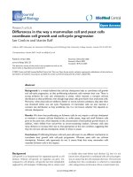
Figure 1
Images of Kc
167
(top) and S2R
+
(bottom) cells, from the study by Kiger et al. [1] (reproduced with permission). Wild-type cells are on the far left.
genetics’ is guided by the functional
assay. This is new and important.”
Baum predicts that deciphering the
meaning of such large datasets will
probably require new ways of thinking
and the help of mathematicians and
physicists. These approaches are being
incorporated into the broad field of
‘systems biology’.
To help the application of this tech-
nology throughout the fly and cell
biology communities, Perrimon and
collaborators have set up the
Drosophila RNAi Screening Center
(DRSC) [6], to make the relatively
expensive and sophisticated technol-
ogy accessible to all interested
researchers; it also has the advantage of
permitting valuable functional com-
parisons across many studies and the
creation of an information database
with a standardized format. The DRSC
is currently performing screens for cell
growth and viability and is accepting
applications for potential collaborative
screens. “We may need many different
assays to dissect a single pathway,”
23.4 Journal of Biology 2003, Volume 2, Issue 4, Article 23 Weitzman />Journal of Biology 2003, 2:23
Behind the scenes
Journal of Biology asked Norbert Perrimon about how and why his group developed the cell-culture-based RNAi
screen.
What motivated you to develop the RNAi screen?
For us it was a pretty logical step to take. We were characterizing mutations of genes involved in signal transduction
that were identified in large classical screens looking for effects on embryonic development in flies. We had come up
with over 600 genes with interesting phenotypes and we were slowly characterizing them and trying to figure out
what they do. The process was pretty slow because one person can only characterize three or so genes during a
four-year postdoc. So when the RNAi technology appeared we started to use it in embryo injections to phenocopy
these mutations. This turned out not to be very easy technically and we were getting a bit depressed. Then Dixon’s
group showed that RNAi worked well in Drosophila cell lines. We decided to shift to a cell culture system and to
scale up to do high-throughput screens to study the pathways that interested us in the flies. It was basically like
setting up a genetic screen in cells and seeing what phenotypes we could find.
How long did it take you to develop the system and what were the steps that ensured your success?
We had to spend quite a bit of time developing a new set of tools and methodologies and we had to devise new
assays that reflected the pathways that we had been studying in vivo. It was really a learning curve and it took almost
three years. To start off we had to do a survey screen of cellular phenotypes to figure out what we could actually
study in these cells. We also needed to develop ways to annotate phenotypes properly so that everyone uses the
same way to describe what they see.
What were your initial reactions to the results and how has this approach been received by others in
the field?
I was worried at the beginning about the technical limitations of such an approach. But I am very happy with the way
that the data are coming out; it’s working extremely well. The community has been very supportive. Many people
have been proposing collaborations and are keen to use the technology. That’s why we set up the Drosophila RNAi
Screening Center (DRSC) [6], to make this technology available to the community. If someone has a good cell-based
assay then they can apply to the center to conduct a genome-wide screen. Our pilot study allowed us to convince
others that this was worth doing on a large scale.
What are the next steps and what does the future hold for such approaches?
One important aspect is that we now have an annotation of all the genes in the fly genome based on their RNAi
signature. We can group genes based on their phenotypes in the different cell-based assays. Matching together
different genes with the same functional profile in RNAi screens allows us to make connections between
components in the same pathway or in the same molecular machine. There is also the issue of merging databases.
We are working on ways to navigate efficiently between RNAi information and databases of protein-protein or
microarray data. This is just the beginning - we have done a full-genome RNAi screen for cell number and by the end
of the year we’ll have finished ten screens on different signal transduction pathways.
says Perrimon. He is interested in any
proposal with a good cell-based assay.
The Center is able to perform a whole-
genome screen in just one week. But at
a cost of US$10,000 per screen the
DRSC will be carefully selecting
screens with a high chance of success.
The cell biology community is keen
to use the RNAi approach to probe basic
questions about cell functions. “This
type of approach, like classical genetics,
is very flexible and can be applied to a
wide range of cellular processes,” says
Welch. “What’s also exciting is that open
access to information from large-scale
efforts like this one will be extremely
useful for other researchers interested in
a range of issues, from the function of
their favorite gene or protein to more
global issues of how gene function is
integrated and coordinated during
complex processes like cell morphogen-
esis.” And as Antoniewski points out,
“in a third of cases, an RNAi phenotype
identified a previously uncharacterized
gene that lacked a corresponding
mutant allele in Drosophila. This is
extremely important, because it shows
that RNAi screens can identify new func-
tions that could not have been identified
by classical genetics using mutagenesis.”
An additional aspect of the potential for
this type of approach is highlighted by
Kiger. “Now that large-scale reverse-
genetic or ‘functional genomic’
approaches are possible in yeast, worms
and fly cells, it is interesting to consider
how we might eventually be able to
make functional comparisons across
species that could shed light on
common (or contrasting) cellular mech-
anisms throughout evolution.”
Other groups are using RNAi tech-
nology in a more focused way to tackle
biological questions. Ronald Vale’s
group at the University of California in
San Francisco has used RNAi targeting
specific gene families to investigate
cytoskeletal function in Drosophila S2
cells. They targeted dynein and all 25
Drosophila kinesins to investigate the
role of molecular motors during
mitosis [7], and also identified sets of
genes important for regulating actin
dynamics during lamella formation
[8]. Rogers et al. [8] suggest, “If proper
cues are provided to these cells, cell
migration and cell polarity may be
amenable to investigation as well.” A
combination of genome-wide screens
and studies focused on specific gene
families will be needed to identify and
then characterize components of the
specific pathways involved in complex
cellular processes. It will also be inter-
esting to apply the tricks of the classic
geneticists’ trade, such as screens for
modifiers and suppressors.
“I think the power of the RNAi
screens will come when we combine
RNAi screening data with data from
other sources, like genomic sequence,
microarray data, proteomic data, and
so on,” adds Baum. “This opens up the
possibility of real systems biology: gen-
erating a more global understanding of
the logical circuitry that underlies cell
behavior. This approach is going to
have a big impact - because I think that
a lot of the apparent complexity in cell
biology is a product of over-expression
studies, and loss-of-function data can
clarify some of these situations.”
“And then we will have to go back
to the fly,” says Perrimon. “The next
step will be to take what we have
learned from the cell-based assays and
validate them in vivo, either using
defined existing mutations or by trying
to generate them using classical
methods or a promising gene-knockout
methodology. Or we can also express
[RNA] hairpins, which basically give an
in vivo RNAi effect.” Many of the lessons
learnt by classical fly genetics have sub-
sequently been confirmed and explored
further in cellular systems. Now it looks
as though researchers can begin by
doing genetics in cells and then return
to the whole fly to investigate further.
References
1. Kiger AA, Baum B, Jones S, Jones MR,
Coulson A, Echeverri C, Perrimon N: A
functional genomic analysis of cell
morphology using RNA-interfer-
ence. J Biol 2003, 2:27.
2. Adams MD, Celniker SE, Holt RA, Evans
CA, Gocayne JD, Amanatides PG,
Scherer SE, Li PW, Hoskins RA, Galle RF,
et al.: The genome sequence of
Drosophila melanogaster. Science 2000,
287:2185-2195.
3. Fraser AG, Kamath RS, Zipperlen P, Mar-
tinez-Campos M, Sohrmann M, Ahringer
J: Functional genomic analysis of
C. elegans chromosome I by system-
atic RNA interference. Nature 2000,
408:325-330.
4. Gonczy P, Echeverri G, Oegema K,
Coulson A, Jones SJ, Copley RR,
Duperon J, Oegema J, Brehm M, Cassin
E, et al.: Functional genomic analysis
of cell division in C. elegans using
RNAi of genes on chromosome III.
Nature 2000, 408:331-336.
5. Clemens JC, Worby CA, Simonson-Leff
N, Muda L, Maehama T, Hemmings BA,
Dixon JE: Use of double-stranded
RNA interference in Drosophila cell
lines to dissect signal transduction
pathways. Proc Natl Acad Sci USA 2000,
97:6499-6503.
6. Drosophila RNAi Screening Center
[]
7. Goshima G, Vale RD: The roles of
microtubule-based motor proteins
in mitosis: comprehensive RNAi
analysis in the Drosophila S2 cell
line. J Cell Biol 2003, 162:1003-1016.
8. Rogers SL, Wiedemann U, Stuurman N,
Vale RD: Molecular requirements for
actin-based lamella formation in
Drosophila S2 cells. J Cell Biol 2003,
162:1079-1088.
Jonathan B Weitzman is a scientist and science
writer based in Paris, France.
E-mail:
Journal of Biology 2003, Volume 2, Issue 4, Article 23 Weitzman 23.5
Journal of Biology 2003, 2:23