Báo cáo sinh học: "The phosphatidylserine receptor has essential functions during embryogenesis but not in apoptotic cell removal" pot
Bạn đang xem bản rút gọn của tài liệu. Xem và tải ngay bản đầy đủ của tài liệu tại đây (2.24 MB, 18 trang )
Research article
The phosphatidylserine receptor has essential functions during
embryogenesis but not in apoptotic cell removal
Jens Böse*, Achim D Gruber
†
, Laura Helming*, Stefanie Schiebe*, Ivonne
Wegener*, Martin Hafner
‡
, Marianne Beales
§
, Frank Köntgen
§
and Andreas
Lengeling*
Addresses: *Junior Research Group Infection Genetics, German Research Center for Biotechnology (GBF), Mascheroder Weg 1, 38124
Braunschweig, Germany.
†
Department of Pathology, School of Veterinary Medicine Hannover, Bünteweg 17, 30559 Hannover, Germany.
‡
Department of Experimental Immunology, German Research Center for Biotechnology (GBF), Mascheroder Weg 1, 38124 Braunschweig,
Germany.
§
Ozgene Pty. Ltd., Canning Vale, WA 6970, Australia.
Correspondence: Andreas Lengeling. E-mail:
Abstract
Background: Phagocytosis of apoptotic cells is fundamental to animal development, immune
function and cellular homeostasis. The phosphatidylserine receptor (Ptdsr) on phagocytes has
been implicated in the recognition and engulfment of apoptotic cells and in anti-inflammatory
signaling. To determine the biological function of the phosphatidylserine receptor in vivo, we
inactivated the Ptdsr gene in the mouse.
Results: Ablation of Ptdsr function in mice causes perinatal lethality, growth retardation and a
delay in terminal differentiation of the kidney, intestine, liver and lungs during embryogenesis.
Moreover, eye development can be severely disturbed, ranging from defects in retinal
differentiation to complete unilateral or bilateral absence of eyes. Ptdsr
-/-
mice with
anophthalmia develop novel lesions, with induction of ectopic retinal-pigmented epithelium in
nasal cavities. A comprehensive investigation of apoptotic cell clearance in vivo and in vitro
demonstrated that engulfment of apoptotic cells was normal in Ptdsr knockout mice, but Ptdsr-
deficient macrophages were impaired in pro- and anti-inflammatory cytokine signaling after
stimulation with apoptotic cells or with lipopolysaccharide.
Conclusion: Ptdsr is essential for the development and differentiation of multiple organs during
embryogenesis but not for apoptotic cell removal. Ptdsr may thus have a novel, unexpected
developmental function as an important differentiation-promoting gene. Moreover, Ptdsr is not
required for apoptotic cell clearance by macrophages but seems to be necessary for the
regulation of macrophage cytokine responses. These results clearly contradict the current view
that the phosphatidylserine receptor primarily functions in apoptotic cell clearance.
BioMed Central
Journal
of Biology
Journal of Biology 2004, 3:15
Open Access
Published: 23 August 2004
Journal of Biology 2004, 3:15
The electronic version of this article is the complete one and can be
found online at />Received: 14 May 2004
Revised: 16 July 2004
Accepted: 21 July 2004
© 2004 Böse et al., licensee BioMed Central Ltd. This is an open-access article distributed under the terms of the Creative Commons Attribution
License ( which permits unrestricted use, distribution, and reproduction in any medium, provided the
original work is properly cited.
Background
Programmed cell death, or apoptosis, is required for the
normal development of almost all multicellular organisms
and is a physiological mechanism for controlling cell
number; as a result, structures that are no longer needed are
deleted during development and abnormal cells are elimi-
nated [1,2]. Most of the cells produced during mammalian
embryonic development undergo physiological cell death
before the end of the perinatal period [3]. Apoptotic cells
are removed rapidly and efficiently as intact cells or apop-
totic bodies by professional phagocytes or by neighboring
cells. This highly regulated process prevents the release of
potentially noxious or immunogenic intracellular materials
and constitutes the fate of most dying cells throughout the
lifespan of an organism [4,5]. Phagocytosis of apoptotic
cells is very distinct from other engulfment processes that
result, for example, in the clearance of microorganisms,
because engulfment of apoptotic cells triggers the secretion
of potent anti-inflammatory and immunosuppressive
mediators, whereas pathogen recognition causes the release
of pro-inflammatory signals [6].
Almost all cell types can recognize, respond to, and ingest
apoptotic cells by using specific sets of phagocytic receptors
that bind to specific ligands on apoptotic cells. Detailed
genetic studies in Drosophila and Caenorhabditis elegans have
recently yielded evidence that basic phagocytic mecha-
nisms and pathways for the recognition and engulfment of
apoptotic cells are highly conserved throughout phylogeny
[7,8]. In vertebrates, a number of receptors have been iden-
tified that can mediate phagocytosis of apoptotic cells.
These include, for example, scavenger receptors and pattern
recognition receptors such as CD36, SR-A and CD14, inte-
grins such as the vitronectin receptor ␣
v

3,
and members of
the collectin family and their receptors CD91 and calretic-
ulin [9-13]. The individual roles of these molecules in
binding, phagocytosis or transduction of anti-inflammatory
signals upon apoptotic cell recognition have not been well
defined, however [5,6,14]. The importance of efficient
mechanisms for apoptotic cell clearance in vivo is sup-
ported by the observation that autoimmune responses can
be provoked in mice when key molecules for apoptotic cell
recognition and uptake are missing. This has been reported
for knockout mice lacking the complement protein C1q
[15], for mice with a mutation in the tyrosine kinase recep-
tor gene Mer [16] and, more recently, in mice lacking trans-
glutaminase 2 or milk fat globule epidermal growth factor 8
(MFG-E8) [17,18].
The exposure of the phospholipid phosphatidylserine (PS)
in the outer leaflet of the plasma membrane of apoptotic
cells has been described as one of the hallmarks of the
induction of apoptosis and is considered to be one of the
most important signals required for apoptotic cell recogni-
tion and removal [19]. A number of cell-surface and bridging
molecules can interact with exposed PS on apoptotic cells.
These include the serum proteins 2-glycoprotein 1 and
protein S [20,21], the growth-arrest-specific gene product
GAS-6 [22], complement activation products [23], the milk
fat globule protein MFG-E8 [24], and annexin I [25]. In most
cases the receptors on phagocytes that recognize these PS-
bridging molecules have not been defined, but it has been
reported that GAS-6 is a ligand for the tyrosine kinase recep-
tor Mer and that MFG-E8 can bind to the vitronectin recep-
tor ␣
v

3
[16,24]. Other molecules that bind PS with varying
specificity are the lectin-like oxidized low-density lipo-
protein receptor-1 (LOX-1) and the scavenger receptors
CD36 and CD68 (for review see [5] and references therein).
The best-characterized molecule so far that binds PS in a
stereo-specific manner is the phosphatidylserine receptor
(Ptdsr) [26]. In vitro, it has been shown that the Ptdsr can
mediate the uptake of apoptotic cells and that such Ptdsr-
mediated phagocytosis can be inhibited through addition of
PS liposomes, the PS-binding molecule annexin V or an anti-
Ptdsr antibody [26]. Moreover, the binding of Ptdsr to PS on
apoptotic cells has been reported to be important for the
release of anti-inflammatory mediators, including transform-
ing growth factor-1 (TGF-1), platelet-activating factor
(PAF), and prostaglandin E2 [26,27]. These data supported
the hypothesis that Ptdsr fulfils a role as a crucial signaling
switch after the engagement of macrophages with apoptotic
cells and is thereby fundamental for preventing local immune
responses to apoptotic cells before their clearance [28].
Very recently, Ptdsr has been found in the cell nucleus. Its
nuclear localization is mediated by five independent
nuclear localization signals, each of which alone is capable
of targeting Ptdsr to the cell nucleus [29]. Moreover, an
additional study performed recently in Hydra showed an
exclusively nuclear localization for the Ptdsr protein [30].
Most interestingly, the nuclear localization of Ptdsr in Hydra
epithelial cells did not change upon phagocytosis of apop-
totic cells. These reports challenge the original hypothesis,
according to which Ptdsr is an exclusively transmembrane
receptor for apoptotic cell recognition and anti-inflamma-
tory signaling.
To examine further the role of Ptdsr in vivo, we performed
gene-expression and gene-targeting studies in mice. A peri-
natally lethal phenotype was observed in Ptdsr-knockout
mice, and Ptdsr-deficient embryos displayed multiple
defects in tissue and organ differentiation. While this work
was in progress, both Li et al. [31] and Kunisaki et al. [32]
also reported the generation and phenotypic characteriza-
tion of Ptdsr-knockout mice. Of note, although some of
15.2 Journal of Biology 2004, Volume 3, Article 15 Böse et al. />Journal of Biology 2004, 3:15
their results were confirmed in our study, we found a funda-
mentally different phenotype with regard to clearance of
apoptotic cells. Moreover, our study revealed marked and
unexpected findings in Ptdsr-deficient mice that are not
related to apoptosis.
Results
Generation of Ptdsr-deficient mice
To investigate in vivo the functions of the phosphatidyl-
serine receptor Ptdsr, we generated a null allele in the mouse
by gene targeting (Figure 1a-c). In contrast to previously
described Ptdsr-knockout mice [31,32], we used Bruce4
embryonic stem (ES) cells for gene targeting [33], thus gen-
erating a Ptdsr-null allele in a pure, isogenic C57BL/6J
genetic background. The newly established knockout mouse
line was named Ptdsr
tm1Gbf
(hereafter referred to as Ptdsr
-/-
).
Heterozygous Ptdsr
+/-
mice were viable and fertile and
showed no obvious abnormalities. Ptdsr
+/-
mice were inter-
crossed to generate homozygous Ptdsr-deficient mice. The
absence of Ptdsr expression in Ptdsr
-/-
embryos was con-
firmed by RT-PCR (data not shown), and by northern and
western blotting analyses (Figure 1d,e). Interbreeding of
heterozygous mice showed that the mutation was lethal,
since homozygous mutants were not detected in over 100
analyzed litters at weaning. To determine the stages of
embryonic development affected by the Ptdsr
tm1Gbf
muta-
tion, timed breedings were followed by PCR genotyping
(Figure 1c) of embryos. We recovered fewer than the
expected number of homozygous embryos from inter-
crosses of Ptdsr
+/-
mice. From a total of 1,031 embryos ana-
lyzed between gestational day (E) 9.5 and E18.5, 198
(19.2%) Ptdsr-deficient homozygous embryos were har-
vested, indicating that the introduced mutation is associated
with a low rate of embryonic lethality in utero.
From E9.5 to E12.5, Ptdsr
-/-
embryos were viable and of
normal size. At E13.5 and thereafter, however, most Ptdsr
-/-
embryos showed morphological abnormalities (Table 1).
All homozygous embryos harvested were growth-retarded
from E13.5 onwards, had a pale appearance, and displayed
multiple developmental dysmorphologies. These included
various head and craniofacial malformations, such as exen-
cephaly, cleft palate and abnormal head shape (Figure 1f,g).
Gross inspection revealed that eye development was
severely affected in 14.1% of homozygous embryos. The
affected animals displayed a complete unilateral or bilateral
absence of the eyes (Table 1) that was never detected in
Ptdsr
+/+
or Ptdsr
+/-
littermates. Furthermore, homozygous
embryos harvested between E12.5 and E15.5 had subcuta-
neous edema (Figure 1f,g). Because we were able to recover
Ptdsr
-/-
embryos until E18.5, we investigated whether Ptdsr-
knockout mice could be born alive. Careful observation of
timed matings allowed us to recover Ptdsr
-/-
neonates, but
homozygous pups died during delivery or within minutes
after birth. Ptdsr-deficient neonates were also growth-
retarded, had a pale appearance and displayed various mal-
formations. These included cleft palate, abnormal head
shape, absence of eyes and edematous skin (Figure 1h).
Thus, deletion of the Ptdsr gene resulted in perinatal lethal-
ity with variable severity and penetrance of phenotypes.
Expression of Ptdsr during embryogenesis and in
adult tissues
The observed perinatal lethality indicates that Ptdsr plays an
important role during development. Analysis by RT-PCR
(data not shown) showed that Ptdsr is expressed early in
development, because we were able to detect Ptdsr tran-
scripts in ES cells and embryos at all developmental stages.
To analyze in more detail the temporal and spatial expres-
sion patterns of Ptdsr, and to correlate expression patterns
with observed pathological malformations, we made use of
a Ptdsr-

-geo gene-trap reporter mouse line generated from a
Ptdsr gene-trap ES cell clone. This line has an insertion of

-galactosidase in the 3´ region of the gene (Figure 2a).
We first examined Ptdsr expression by X-Gal staining in het-
erozygous embryos staged from E9.5 to E12.5. These devel-
opmental stages were chosen so as to investigate Ptdsr
expression in affected organs prior to the onset of patho-
logical malformations in Ptdsr
-/-
embryos. At E9.5 we found
Ptdsr expression in the developing neural tube, somites,
heart, gut and branchial arches (Figure 2b). At E10.5, Ptdsr
expression remained high in the developing nervous
system, with most intense staining in the forebrain, hind-
brain and neural tube. At this stage of embryogenesis, high
levels of Ptdsr expression could also be detected in the
developing limb buds and eyes (Figure 2b). Ptdsr expression
was altered at E12.5, with most intensive -galactosidase
staining in the eyes, developing condensations of the limb
buds, neural tube and brain (Figure 2b). Transverse sections
of X-Gal-stained embryos at E12.5 showed an asymmetric
expression pattern in the neural tube with intense staining
of the central mantle layer but no expression in the dorsal
part of the neural tube (for example, the roof plate; Figure
2c). Expression in dorsal root ganglia lateral to the neural
tube and in the somites was observed; Ptdsr was expressed
throughout the somite structure (myotome, dermatome and
sclerotome; Figure 2d). Expression boundaries between
somites were evident, with no expression in the segmental
interzones, which correspond to the prospective interverte-
bral discs (Figure 2d). Transverse sections of the developing
eye at E12.5 revealed strong Ptdsr expression in the inner
layer of the neural cup, which will later develop into the
neural retina. Furthermore, Ptdsr expression was detected in
the primary lens fiber cells of the developing lens
Journal of Biology 2004, Volume 3, Article 15 Böse et al. 15.3
Journal of Biology 2004, 3:15
15.4 Journal of Biology 2004, Volume 3, Article 15 Böse et al. />Journal of Biology 2004, 3:15
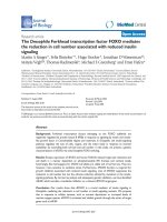
Figure 1
Targeted inactivation of the phosphatidylserine receptor gene. (a) Ptdsr gene-targeting strategy. Homologous recombination in ES cells results in the
deletion of exons I and II of the murine Ptdsr gene through replacement of a loxP-flanked neomycin phosphotransferase gene (neo), thereby ablating
the reading frame of the encoded protein. Coding exons I-VI are shown as filled boxes, and deleted exons are colored green. Restriction sites are:
A, AatII; B, BamHI; EI, EcoRI; EV, EcoRV; K, KpnI; R, RsrII; S, SacII; Sc, ScaI, X, XhoI. The probe sites are red boxes labeled: C, 5´ outside probe;
D, 3´ outside probe. (b) Southern blot analysis of genomic DNA extracted from wild-type (+/+) and Ptdsr
+/-
(+/-) animals, digested with BamHI and
hybridized with the 5´ outside probe to confirm germ-line transmission of the mutant Ptdsr allele. ‘Wild-type’ indicates the BamHI fragment of 17.2
kb from the wild-type Ptdsr allele; ‘mutant’ indicates the BamHI fragment of 11.6 kb from the targeted Ptdsr allele. (c) PCR genotyping of embryos
and animals from intercrosses of heterozygous Ptdsr
+/-
using a wild-type and a mutant allele-specific primer combination, respectively. (d) Northern
blot analysis of total RNA isolated from E13.5 wild-type, Ptdsr
+/-
and Ptdsr
-/-
embryos. (e) Western blot analysis of protein from homogenates of
E13.5 wild-type, Ptdsr
+/-
and Ptdsr
-/-
embryos using a Ptdsr-specific antibody. Developmental abnormalities at (f,g) E15.5 and (h) birth; in this and all
subsequent figures wild-type littermates are located on the left and homozygous mutant mice on the right. The Ptdsr
-/-
embryos show exencephaly (f)
or prosencephalic hernia in the forebrain region (arrowhead, neonate 2; h), uni- or bilateral absence of the eyes (f,g and neonate 2 in h, and arrow,
neonate 3 in h), an abnormal head shape with proboscis (g), edema (arrowheads in f and g), and general anemia (asterisk, neonate 3 in h).
B, EI, X, AEI, X
EI
B S S
X
K EV EI
EI
RS A EI EI EI B
EV
EI X B K
I
ATG
II III IV V VI
TGA
Ptdsr
K B
B, EI, X, A
EI EI EIEV EI
EI, X
neo
B S S
X
K EV EI EI EIEI EI B
EV
EI X B K
EI
neo
Wild-type allele
Targeting vector
Targeted allele
1 kb
X
X
C D
Probes
Southern blot analysis :
BamHI (B)
17.2 kb (wt)
11.6 kb (−/−)
Sc
Sc ScSc
12.4 kb (wt)
ScaI (Sc)
ScSc
ScSc
ScSc
Sc
Sc
Sc Sc
ScSc
ScSc
17.2 kb (−/−)
EI
+/−+/−+/+
Wild-type
Wild-type
Mutant
Mutant
Ptdsr
Ptdsr
Actin
Actin
−/−+/−+/+ −/−+/−+/+ −/−+/−+/+
5 mm 5 mm
1 cm
123
(a)
(b)
(f) (g) (h)
(c) (d) (e)
*
(Figure 2e). We carefully investigated whether Ptdsr is
expressed from E10.5 to E12.5 in the developing kidney and
lungs, but no expression could be detected indicating that
Ptdsr expression is required only at later stages in the devel-
opment of these organs (see below).
Hybridization of a multiple-tissue northern blot revealed a
single transcript of about 1.8 kb in almost every tissue ana-
lyzed in adult mice (Figure 2f). The most prominent expres-
sion was observed in testis, thymus, kidney, liver and skin,
with moderate to low expression in lung, small intestine,
spleen, stomach and skeletal muscle. Thus, Ptdsr is ubiqui-
tously expressed throughout embryogenesis and in adult
tissues, although at different levels.
Ptdsr is required for normal tissue and organ
differentiation
We next examined the role of Ptdsr in organ development.
Serial histological sections of Ptdsr
-/-
and control embryos
were taken to perform a detailed morphological analysis of
all organ systems during development. A significant delay in
organ and tissue differentiation was observed at E16.5 in
lungs, kidneys and intestine. Lungs of control littermates
were properly developed with expanding alveoli (Figure 3a).
Terminal bronchi and bronchioles were already well devel-
oped, and terminally differentiated epithelial cells with cilia
on the luminal cell surface were present. In contrast, almost
no alveoli or bronchioles were present in Ptdsr
-/-
lungs, indi-
cating a delay or arrest in lung sacculation and expansion.
Instead, we observed an abundance of mesenchyme that
appeared highly immature (Figure 3g). A similar delay in
tissue differentiation of Ptdsr
-/-
embryos was found in the
kidneys (Figure 3h). Kidneys from Ptdsr
+/+
embryos were
well developed at E16.5, showing terminally differentiated
glomeruli with Bowman’s capsule and collecting tubules
lined with cuboidal epithelial cells (Figure 3b). In contrast,
Ptdsr-deficient kidneys had only primitive glomeruli at
E16.5, and collecting tubules were less well-developed.
Instead, a large amount of undifferentiated mesenchyme
was present in Ptdsr
-/-
kidneys (Figure 3h). A delay in tissue
differentiation was also found in the intestine at this stage
of development. Ptdsr
-/-
embryos displayed improperly
developed villi and an underdeveloped or absent submu-
cosa (Figure 3i). In wild-type embryos (Figure 3c), intestinal
cellular differentiation was already highly organized, with
intramural ganglion cells between the external and internal
muscular layers. Such neuronal cells were absent from the
intestine of Ptdsr
-/-
embryos (Figure 3i), however.
Some Ptdsr
-/-
mice (4.5 %) also displayed extensive brain
malformations that resulted in externally visible head
abnormalities, with occasional ectopic tissue outside the
skull or exencephaly (Figure 1f,h). Histological analysis
revealed an extensive hyperplasia of brain tissue with herni-
ation of brain tissue either through the skull-cap or through
the ventral skull (Figure 3d,j). In the most severe cases,
expansion of brain tissue in mutant mice resulted in further
perturbations of cortical structures (Figure 3d,j). Of note, a
similar brain phenotype was observed in the Ptdsr-deficient
mouse line generated by Li and colleagues [31].
In contrast to the study of Li et al. [31], however, we
found almost normally developed lungs at birth. Ptdsr
-/-
lungs showed, in comparison to wild-type, only a slight
delay in maturation and were fully ventilated in neonates
in most cases (Figure 3e,k). This demonstrates that Ptdsr-
deficient mice can overcome the delay in embryonic lung
differentiation and display normal lung morphology at
birth. Thus, it would appear highly unlikely that Ptdsr
-/-
mice die from respiratory failure. Consistent with the
observations of Kunisaki and colleagues [32], we found
severely blocked erythropoietic differentiation at an early
erythroblast stage in the liver (Figure 3f,l), suggesting an
explanation for the grossly anemic appearance that we
observed in our Ptdsr
-/-
mice.
Loss of Ptdsr activity is associated with defects in
ocular development and can lead to formation of
ectopic eye structures
By gross morphology we could differentiate two classes of
Ptdsr mutants: those that appeared normal with both eyes
present (Figure 4) and those that were severely affected
and displayed uni- or bilateral anophthalmia (Figure 5).
Journal of Biology 2004, Volume 3, Article 15 Böse et al. 15.5
Journal of Biology 2004, 3:15
Table 1
Penetrance of phenotypes in Ptdsr
-/-
mice from E9.5 to E18.5,
as detected by gross morphology
Dysmorphic phenotypes Ratio in analyzed Penetrance (%)
mice (affected/total)
Head malformations 9/198 4.5
cleft 4/198 2.0
others 5/198 2.5
Edema (E12.5-E15.5) 15/155 9.7
Pale appearance (= E14.5) 72/72 100
Ocular lesions 28/198 14.1
unilaterally absent eyes 21/198 10.6
right 16/198 8.1
left 5/198 2.5
bilaterally absent eyes 7/198 3.5
Subsets of the major categories of malformation are indicated by
indentation.
Analysis of normal or mildly affected embryos revealed no
differences between mutant and wild-type embryos in the
differentiation of the developing eye until E16.5. In both
genotypes, inner and outer layers of the retina displayed a
comparable differentiation status, as shown, for example, at
E12.5 (Figure 4a,e). At day E16.5, however, retinal layers in
Ptdsr
-/-
embryos were much thinner than in wild-type
embryos, contained fewer cells and were greatly reduced in
size (Figure 4b,f). Comparison of the retinal structures of
Ptdsr
+/+
and Ptdsr
-/-
embryos revealed that all four retinal
layers were present in Ptdsr-knockout mice at E16.5 (Figure
4b,f). At E18.5 (Figure 4c,g) and in neonatal animals (post-
natal day P0; Figure 4d,h), the differences in retinal
differentiation between Ptdsr
+/+
and Ptdsr
-/-
mice were still
evident, but the size reduction of the retinal layers was less
pronounced in the knockout mice. Ptdsr-deficient animals
seem to have compensated for the marked delay in cellular
differentiation and expansion of retinal layers. Close exami-
nation of retinal structures revealed that the inner granular
layer was still less expanded in Ptdsr-deficient animals,
however, and that it contained fewer cells and was still
severely underdeveloped in comparison with the corre-
sponding retinal layer in control animals (Figure 4c,g and
4d,h). Thus, even mildly affected Ptdsr
-/-
mutants had ocular
malformations with defects in differentiation of retinal
structures.
We next examined Ptdsr
-/-
embryos that displayed unilateral
or bilateral absence of eyes (Figure 5a) by serial sectioning
of whole embryos. These embryos showed complex malfor-
mations of the optical cup, including absence of the lens
(Figure 5b). Most surprisingly, we found pigmented epithe-
lial cells in the nasal cavity of all Ptdsr-knockout mice with
anophthalmia that were analyzed histopathologically. We
could identify black-colored pigmented cells embedded in
the epithelium of the maxillary sinus that resembled pre-
sumptive retinal-pigmented epithelium (Figure 5b,c). Exam-
ination of consecutive serial sections revealed the formation
of a primitive eye structure, with induction and subsequent
proliferation of ectopic mesenchymal tissue immediately
adjacent to the displaced pigmented epithelium (Figure 5d).
This structure was clearly induced ectopically, and we failed
15.6 Journal of Biology 2004, Volume 3, Article 15 Böse et al. />Journal of Biology 2004, 3:15
Figure 2
Expression analysis of Ptdsr during embryonic development. (a) Schematic representation of the construction of the Ptdsr gene-trap mouse line used for
expression analysis at different embryonic stages. Gray and bright blue boxes represent regulatory elements of the gene-trap, and -geo, the
-galactosidase/neomycin phosphotransferase fusion protein-expression cassette [48,51]. Restriction enzyme nomenclature is as in Figure 1 (b) Whole-
mount -galactosidase staining of heterozygous Ptdsr gene-trap embryos at mid-gestation. Expression of Ptdsr is highest in neural tissues and somites, in
the branchial arches, the developing limbs, the heart, the primitive gut and the developing eye. (c-e) Sectioning of E12.5 -galactosidase-stained embryos
confirms expression of Ptdsr in (c) the neural tube; (inset in c) neural epithelium; (d) somites; and (e) eyes. Expression in the eye is restricted to
developing neural retinal and lens cells. (f) Expression analysis of adult tissues by northern blot. Expression of Ptdsr in the muscle (asterisk) was detected
only on long-term exposures of the filter (> 48 h). A

-actin hybridization was used to confirm equal loading of RNA samples. Scale bar, 100 m.
EV EI
EI
RS A EI EI B
EV
EI
I
ATG
II III IV V VI
TGA
Ptdsr
1 kb
ScSc
ScSc
β-geo
E12.5E10.5E9.5
Brain
Heart
Kidney
Liver
Lung
Muscle
Skin
Small intestine
Spleen
Stomach
Testis
Thymus
2 kb
Ptdsr
-Actin
1.5 kb
2 kb
1.5 kb
*
(a)
(c) (d) (e) (f)
(b)
β
to identify similar changes in any of the wild-type embryos.
In summary, we observed a wide range of ocular malform-
ations in Ptdsr-deficient mice that ranged from differentia-
tion defects in retinal cell layers (for example, the inner
granular layer) in mildly affected homozygotes to anoph-
thalmia in severely affected Ptdsr
-/-
mice that was associated
with induction of ectopic eye structures in nasal cavities.
Phagocytosis and clearance of apoptotic cells is
normal in Ptdsr-deficient mice
We next tested whether Ptdsr is functionally required for the
clearance of apoptotic cells. We started with an investigation
of cell death in vivo in the interdigital areas of the develop-
ing limbs. Apoptosis of interdigital cells in the distal mesen-
chyme of limb buds occurs most prominently from
developmental stages E12.0 to E13.5 and can be easily
examined in situ by whole-mount terminal deoxynucleotide
transferase-mediated UTP end-labeling (TUNEL). We com-
pared the pattern of interdigital cell death in fore and hind
limb buds from Ptdsr
-/-
(n = 3) and Ptdsr
+/+
(n = 3) mice at
E12.5 and E13.5. No differences in accumulation of
TUNEL-positive cell corpses were observed between the two
genotypes (Figure 6a). The kinetics of cell death occurrence
and regression of the interdigital web was similar in wild-
type and mutant littermates, providing no evidence that
Ptdsr-deficiency is associated with impaired clearance of
apoptotic interdigital cells during limb development.
To investigate further whether removal of apoptotic cells is
impaired in Ptdsr
-/-
mice, we stained immunohistochemi-
cally for activated caspase 3 (aCasp3) and analyzed addi-
tional organs and tissues where apoptosis plays a crucial
role in tissue remodeling during development. Starting at
E12.5, we analyzed and compared the number and distribu-
tion of aCasp3-positive cells in over 140 serial sections of
three wild-type and six Ptdsr
-/-
embryos in consecutive and
corresponding sections. The sagittal sections were separated
by 5 m, allowing a detailed analysis of apoptosis in several
Journal of Biology 2004, Volume 3, Article 15 Böse et al. 15.7
Journal of Biology 2004, 3:15
(a) (g)
(b) (h)
(c) (i)
(d) (j)
(e) (k)
(f) (l)
Figure 3
Histological analysis of wild-type and Ptdsr
-/-
organs during
embryogenesis. (a-f) Wild-type embryos and (g-l) Ptdsr
-/-
littermates
were isolated at various embryonic stages, serially sectioned sagittally
and analyzed for developmental abnormalities in detail after H&E
staining. At E16.5, the lungs of (g) Ptdsr
-/-
embryos had sacculation just
starting, and well-formed alveoli (asterisks) or epithelium-lined
bronchioles (arrows) were scarce compared to (a) wild-type lungs. At
E16.5, the glomeruli (arrows) in the kidney of (h) Ptdsr
-/-
embryos were
underdeveloped compared to (b) wild-type, collecting tubules
(arrowheads) were missing and undifferentiated blastemas (asterisks)
were more abundant. The jejunum had no intramural ganglia in Ptdsr
-/-
embryos (i; and arrows in c); and a well-developed submucosa (asterisk
in c) was missing. Brain sections at E18.5 show that (j) Ptdsr
-/-
embryos
may have herniation (arrow) of the hypothalamus through the ventral
skull (secondary palate), most likely through Rathke’s pouch, and a
severe malformation of the cortex (asterisks) compared to (d) wild-type
embryos. At E18.5, (e) wild-type and (k) Ptdsr
-/-
lungs showed normal
sacculation and formation of alveoli (asterisks) and bronchioles (arrow).
(f) Wild-type neonatal liver had significant numbers of megakaryocytes
(arrows), compared to (l) homozygous mutant littermates, and higher
numbers of erythropoietic islands and of mature erythrocytes.
Hepatocellular vacuoles are due to glycogen stores (asterisks) that
were not metabolized in perinatally dying Ptdsr
-/-
animals, in contrast to
wild-type newborns. Scale bar, 100 m, except for (d) and (j), 1 mm.
organs and tissues. Tissue restructuring by programmed cell
death occurred most notably within the ventral part of the
neural tube (Figure 6b,f) and in the developing paravertebral
ganglia (Figure 6d,h) with many apoptotic cells being
present. In these tissues Ptdsr is highly expressed at E12.5
(Figure 2c) but we observed no difference in the number or
distribution of apoptotic cells in Ptdsr
+/+
and Ptdsr
-/-
embryos.
The same was true for the developing kidney: apoptotic cells
were present in Ptdsr
+/+
and Ptdsr
-/-
embryos, in limited
numbers, but we failed to detect any differences in the
number of apoptotic cells between the genotypes (Figure
6c,g). Furthermore, when we continued our analysis of apop-
totic cell clearance in vivo at E16.5, E17.5 and E18.5 of embry-
onic development as well as in neonatal mice, the number
and distribution of apoptotic cells was similar in both geno-
types. As already observed at E12.5, analysis of aCasp3-
stained sections of the developing thymus, heart, diaphragm,
genital ridge, eyes and retina convincingly showed that there
was no impairment in apoptotic cell removal in Ptdsr
-/-
mice.
Moreover, because Li and colleagues [31] reported impaired
clearance of dead cells during lung development in Ptdsr-defi-
cient mice, we examined the rate of apoptosis induction and
cell clearance in our Ptdsr-knockout mice in the lung. Analysis
of aCasp3-stained lung tissue from Ptdsr
+/+
and Ptdsr
-/-
mice at
E17.5 and P0 demonstrated that apoptosis was an extremely
rare event during lung morphogenesis at this stage. In addi-
tion, there were no differences in the number or distribution
of apoptotic cells in Ptdsr
-/-
and Ptdsr
+/+
mice. Furthermore,
we were unable to detect any evidence of tissue necrosis in
lungs from Ptdsr-deficient mice. In contrast to the report of Li
et al. [31], we never observed recruitment of neutrophils or
other signs of pulmonary inflammation at any stage of devel-
opment in our Ptdsr-deficient mice.
To analyze whether macrophages are recruited into areas
where apoptosis is prominent during embryogenesis, we
15.8 Journal of Biology 2004, Volume 3, Article 15 Böse et al. />Journal of Biology 2004, 3:15
Figure 4
Morphology of wild-type and Ptdsr
-/-
retinas. Serial sagittal sections of
(a-d) wild-type and (e-h) Ptdsr
-/-
retina were analyzed for
developmental abnormalities at (a,e) E12.5, (b,f) E16.5, (c,g) E18.5, and
(d,h) P0. Normal patterning of the retina was observed in Ptdsr
-/-
embryos, with an outer granular layer (OGL), outer plexiform layer
(OPL), inner granular layer (IGL) and inner plexiform layer (IPL). Note
that the IGL in Ptdsr
-/-
retinas is less thick than that in wild-type
littermates in comparing (c,g) and (d,h). Morphometric analysis
(numbered lines) of wild-type and Ptdsr
-/-
retinas confirmed the initial
finding of a thinner retina in Ptdsr
-/-
animals than in wild-type (all values
in m). Scale bar, 50 m.
OGL
OPL
IPL
IGL
263.0
285.3
84.2
84.7
187.2
227.3
227.4
(a)
(b)
(c)
(d)
(e)
(f)
(g)
(h)
98.0
Figure 5
Histological analysis of eye development in severely affected eyeless
Ptdsr
-/-
embryos. (a) In anophthalmic Ptdsr
-/-
embryos, unilateral or
bilateral absence of the eyes could be detected. (b-d) Serial H&E-
stained sagittal sections of homozygous mutant embryos at (b) E17.5
and (c,d) E18.5 show complex malformation of the optic cup and lack of
any lens structure. Careful examination of adjacent sections (b-d)
reveals an ectopic misplacement of retinal-pigmented epithelium in the
maxillary sinus. Not only is the deposition of pigment clearly visible
(higher magnification insets) but also the induction of proliferation of
underlying tissues and the change in morphology of the maxillary sinus
(d). Scale bar, 100 m in (b-d).
(a) (b)
(c) (d)
5 mm
stained consecutive serial sections either with the
macrophage surface marker F4/80 or with aCasp3. Surpris-
ingly, there was no co-localization of macrophages with
apoptotic cells. In virtually all embryonic tissues, apoptotic
cells and macrophages were localized in different compart-
ments (Figure 6e,i; and see also Additional data file 1, Figure
S1, with the online version of this article). This suggests that
at this stage of development it is mainly neighboring cells
that are involved in removal of apoptotic cells, rather than
professional macrophages. In summary, our analysis in vivo
did not reveal any impairment in apoptotic cell clearance in
Ptdsr-deficient embryos during development and further sug-
gests that phagocytosis of apoptotic cells is mainly mediated
by non-professional ‘bystander’ cells.
To determine whether macrophages from Ptdsr-knockout
mice were impaired in the efficacy of apoptotic cell uptake in
vitro, we performed phagocytosis assays with fetal-liver-
derived macrophages (FLDMs) and quantified their phago-
cytosis rates. Phagocytosis of apoptotic thymocytes was
investigated at 60, 90 and 120 minutes after addition of
target cells in the absence of serum. Analysis of phagocytosis
rates by flow cytometric analysis (FACS) revealed no differ-
ences in the efficacy of apoptotic cell uptake between Ptdsr
-/-
and Ptdsr
+/+
macrophages and demonstrated no differences
in apoptotic cell engulfment between selected time points
(data not shown). To re-examine and further independently
validate the result of normal apoptotic cell uptake by Ptdsr
-/-
macrophages, we performed phagocytosis assays for 60 min
and determined the percentage of macrophages that had
engulfed apoptotic cells, in a total of at least 300
macrophages counted by fluorescence microscopy. Phago-
cytosed, 5-carboxytetramethylrhodamine- (TAMRA-) labeled
apoptotic cells were identified as being engulfed by inclusion
in F4/80-labeled macrophages. Analysis was done indepen-
dently by three investigators who were not aware of
macrophage genotypes (Ptdsr
-/-
or Ptdsr
+/+
). Again, no differ-
ences were found in the percentage of macrophages that
had engulfed apoptotic cells (Figure 7a,c,e) or in the relative
number of phagocytosed apoptotic cells per macrophage
(phagocytotic index; Figure 7f). Moreover, single Ptdsr
-/-
macrophages could be identified that had engulfed even
more apoptotic target cells than had wild-type macrophages
(Figure 7b,d). Thus, Ptdsr-deficient macrophages had a
normal ability to ingest apoptotic cells and were not
impaired in recognition or phagocytosis of cells that had
undergone programmed cell death.
Ptdsr-deficiency results in reduced production of pro-
and anti-inflammatory cytokines after macrophage
stimulation
In addition to its suggested importance for phagocytosis of
apoptotic cells, it has been proposed that Ptdsr fulfils a
Journal of Biology 2004, Volume 3, Article 15 Böse et al. 15.9
Journal of Biology 2004, 3:15
Figure 6
Analysis of programmed cell death and involvement of macrophages in
the removal of apoptotic cells in wild-type and Ptdsr
-/-
embryos.
(a) Whole-mount TUNEL staining (blue) of limb buds from wild-type
and Ptdsr
-/-
embryos at E13.5 show no differences in the amount or
localization of apoptotic cells during the beginning regression of the
interdigital web. Serial sagittal sections stained for activated caspase 3
(aCasp3; red) in (b-d) wild-type and (f-h) Ptdsr
-/-
embryos at E12.5
show apoptotic cells in the neural tube (b,f), the mesonephros (c,g) and
the developing paravertebral ganglia (d,h). Tissue distribution and total
number of apoptotic cells was indistinguishable between genotypes and
was confirmed by the comparison of consecutive sections of wild-type
and Ptdsr
-/-
embryos from different developmental stages. Analysis of
macrophage numbers and location by F4/80 staining (brown) of
consecutive sections in paravertebral ganglia of (e) wild-type and
(i) homozygous mutant embryos revealed that macrophages (arrows)
are not located close to apoptotic cells during embryonic development.
(For comparison, see also Additional data file 1, Figure S1, with the
online version of this article). Scale bar, 100 m.
(a)
(b) (f)
(c) (g)
(d) (h)
(e) (i)
+/+ −/−
second crucial role in regulating and maintaining a non-
inflammatory environment upon the recognition of apop-
totic cells by macrophages [26]. We therefore tested whether
Ptdsr
-/-
macrophages were able to release anti-inflammatory
cytokines after ingestion of apoptotic cells. We examined
levels of TGF-1 and interleukin-10 (IL-10) after stimula-
tion of FLDMs with lipopolysaccharide (LPS), with and
without co-culture of apoptotic cells. Quantification of
TGF-1 and IL-10 levels after 22 hours of culture demon-
strated that Ptdsr
-/-
macrophages were able to secrete these
anti-inflammatory cytokines upon ingestion of apoptotic
cells, although at a slightly lower level than wild-type
(Figure 8a,b). This indicates that ablation of Ptdsr function
does not compromise in general the ability of macrophages
to release immune-suppressive cytokines after recognition
and engulfment of apoptotic cells.
To analyze whether pro-inflammatory signaling is affected
in Ptdsr
-/-
macrophages, we stimulated FLDMs from Ptdsr
+/+
and Ptdsr
-/-
mice with LPS and measured levels of tumor
necrosis factor-␣ (TNF-␣) at different time points after
stimulation (Figure 8c). Ptdsr
-/-
macrophages produced sig-
nificantly less TNF-␣ than did wild-type macrophages. The
difference in TNF-␣ secretion was first visible after 3 h of
LPS stimulation and became more prominent during the
course of the experiment (for example, after 9 h and 12 h
of LPS stimulation; Figure 8c). To analyze whether TNF-␣
release by Ptdsr
-/-
macrophages can be affected by engulf-
ment of apoptotic cells, we stimulated FLDMs with LPS,
apoptotic cells or both. Quantification of TNF-␣ levels by
ELISA after 22 h showed that Ptdsr-deficient macrophages
release less TNF-␣ after stimulation with LPS alone, and
also after double stimulation of macrophages with LPS and
apoptotic cells (Figure 8d). Moreover, the double stimu-
lation demonstrated that the LPS-induced TNF-␣ release by
Ptdsr
-/-
macrophages could be inhibited by co-administration
of apoptotic cells to an extent comparable to that seen in
wild-type macrophages. Similar results were obtained
when other pro-inflammatory cytokines, such as inter-
leukin-6 and monocyte chemoattractant protein-1, were
analyzed (data not shown). These results indicate that
Ptdsr is not required in macrophages for the inhibition of
pro-inflammatory signaling after recognition and engulf-
ment of apoptotic cells. Ptdsr-deficiency does, however,
affect the overall release of pro- and anti-inflammatory
cytokines after stimulation with LPS and after double
treatment with LPS and apoptotic cells, indicating that
Ptdsr-deficient macrophages have a reduced capacity to
produce or secrete pro- and anti-inflammatory cytokines.
Discussion
Ptdsr is required for the differentiation of multiple
organ systems during development
In this study, we have generated a null mutation in the phos-
phatidylserine receptor (Ptdsr) gene in C57BL/6J mice. We
show that ablation of Ptdsr results in profound differentia-
tion defects in multiple organs and tissues during embryo-
genesis, although with variable penetrance. While this work
was in progress, two other groups reported the generation of
Ptdsr-deficient mice [31,32]. In all three knockout mouse
lines, the first two exons ([31] and this study) or exons one
to three [32] were deleted by replacement with a neomycin-
selection cassette. The Ptdsr-knockout mouse lines differ in
the genetic background in which the mutation was generated
15.10 Journal of Biology 2004, Volume 3, Article 15 Böse et al. />Journal of Biology 2004, 3:15
Figure 7
Phagocytosis of apoptotic cells by fetal liver-derived macrophages
(FLDMs). FLDMs from (a,b) wild-type and (c,d) Ptdsr
-/-
embryos were
cultured for 60 min with TAMRA-stained (red) apoptotic thymocytes
(treated with staurosporine) from C57BL/6J mice and then stained with
F4/80 (green). Macrophages of both genotypes have phagocytosed
apoptotic cells (arrowheads). (e) Quantification of phagocytosis of
apoptotic cells by wild-type or Ptdsr
-/-
macrophages revealed no
differences in the percentage of macrophages that had engulfed
apoptotic cells, whether or not apoptosis had been induced by
staurosporine. Microscopic analysis (b,d) and quantification of the
number of apoptotic cells phagocytosed by single macrophages and
(f) calculation of the average number of cells phagocytosed per
macrophage failed to reveal differences in the efficacy of removal of
apoptotic cells between wild-type and Ptdsr
-/-
FLDMs.
Control Staurosporine
0
5
10
15
20
25
30
35
40
45
+/+
−/−
Percent engulfment
+/+ −/−
0
10
20
30
40
50
60
70
80
90
Phagocytotic index
(a) (b)
(c) (d)
(e) (f)
TAMRA
F4/80
TAMRA
F4/80
and maintained, however. In our case, the Ptdsr-null allele
was generated in an isogenic C57BL/6J background, whereas
Li et al. [31] and Kunisaki et al. [32] investigated the pheno-
type of their Ptdsr-knockout mice in a mixed 129 x C57BL/6
background. The ablation of Ptdsr function results in peri-
natal lethality in all cases, but there are interesting differ-
ences in severity or expressivities of phenotypes among the
different Ptdsr-deficient mouse lines. This might be due
either to differences in genetic background or because the
phenotypes that have been investigated in this study have
not been analyzed in such detail before.
In the Ptdsr-knockout mouse line reported here, growth
retardation started from E12.5 onwards and was associated
with delayed differentiation in several organs in which Ptdsr
is expressed either during embryogenesis or later in adult-
hood. At E16.5 almost no branching morphogenesis of the
lung epithelium was observed in Ptdsr
-/-
lungs. Similarly,
epithelial structures were only partially developed in
mutant kidneys, without terminal differentiation of Bow-
man’s capsule and with a severe reduction in the number of
differentiated collecting tubules. Likewise, the differentia-
tion of the intestine was also severely delayed at this devel-
opmental stage. When compared with wild-type controls,
intestinal tissues of Ptdsr knockout mice appeared unstruc-
tured, with an absence of enteric ganglia and of differenti-
ated smooth muscle tissue. Interestingly, defects in kidney
and intestine differentiation were not described in the Ptdsr-
knockouts generated by Li et al. [31] and Kunisaki et al.
[32]. Surprisingly, when we examined Ptdsr
-/-
embryos
shortly before birth (E18.5) or neonatally, we found only
mild differentiation delays in organs that appeared severely
affected at mid-gestation. This ‘recovery’ was most visible in
Ptdsr
-/-
lungs: at P0 we found expanded lungs in the knock-
out mice that showed normal branching patterns, with dif-
ferentiated alveoli and bronchioles.
We investigated the occurrence of programmed cell death
during lung development in wild-type and Ptdsr-knockout
mice throughout embryogenesis (E16.5 to P0). Compara-
tive immunohistochemistry for aCasp3 revealed that apop-
tosis is a rare event during lung morphogenesis.
Furthermore, we failed to detect any differences in the
number of apoptotic cells in Ptdsr-knockout and wild-type
animals in the rare cases where we could detect apoptotic
cells within lung tissues. These findings are contrary to the
results reported by Li et al. [31], who suggested that
impaired clearance of apoptotic mesenchymal and epithe-
lial cells causes a failure in lung morphogenesis in Ptdsr-
deficient mice. In contrast, our findings are in line with the
current view on lung development during embryogenesis.
Accordingly, formation of the epithelial lung via branching
morphogenesis can be subdivided into a series of sequential
steps that involve: first, formation of the organ anlage in the
form of a placode; second, primary bud formation by
placode invagination; third, branch initiation and branch
outgrowth; fourth, further reiteration of the branching
process; and fifth, terminal differentiation of organ-specific
proximal and distal structures [34,35]. In contrast to other
invagination processes during embryogenesis, such as
mammary gland formation, the lumen of the lungs is
expanded by successive branching events, branch out-
growth and elongation, rather than by apoptosis [34,36].
Finally, because the lungs of Ptdsr
-/-
neonates were almost
fully expanded and appeared normal in structure in com-
parison to wild-type littermates, it is highly unlikely that
Ptdsr mutants die of respiratory lung failure. In addition, Li
and colleagues [31] demonstrated that surfactant expres-
sion is normal in Ptdsr-deficient animals, supporting the
idea of normal maturation of surfactant-producing type II
alveolar epithelial cells and lung function. Other defects
must therefore be responsible for the death of Ptdsr-mutant
mice. The frequently observed subcutaneous edema of
various extents in Ptdsr-deficient homozygotes gave us a
Journal of Biology 2004, Volume 3, Article 15 Böse et al. 15.11
Journal of Biology 2004, 3:15
Figure 8
Cytokine production by FLDMs upon stimulation with
lipopolysaccharide (LPS) and apoptotic cells. FLDMs from wild-type and
Ptdsr
-/-
embryos were incubated (a,b,d) with medium (0), LPS (10 ng/ml),
apoptotic cells (ratio 1:10) or in combination with LPS and apoptotic
cells or (c) with LPS (100 ng/ml) alone. Culture supernatants were
harvested after 22 h (a,b,d) or at the indicated time points (c). TNF-␣
and TGF-1 were quantified by ELISA and IL-10 by cytometric bead
array (CBA) assay. Data are presented as mean ± SEM from at least
three independent experiments, each carried out in triplicate.
*, significant difference between genotypes, p < 0.05; **, significant
difference between genotypes, p < 0.01; Wilcoxon-signed rank test.
0
LPS
Apoptotic
cells
LPS +
apoptotic cells
0
LPS
Apoptotic
cells
LPS +
apoptotic cells
0
LPS
Apoptot
ic
cells
LPS +
apoptotic cells
+/+
−/−
TGF-β1 (pg/ml)
*
*
+/+
−/−
IL-10 (pg/ml)
0
1 h
3 h
6 h
9 h
12 h
+/+
−/−
TNF-α (pg/ml)
**
**
+/+
−/−
TNF-α (pg/ml)
*
0
65
100
32,5
0
350
800
1250
1500
(a) (b)
(c) (d)
0
10
20
30
40
50
60
0
85
175
265
350
440
530
hint that Ptdsr-deficiency and lethality might be associated
with cardiovascular problems. Indeed, very recently we
have obtained strong evidence that Ptdsr-knockout mice
die as a result of defects in heart development that are
associated with specific cardiopulmonary malformations;
(J.E. Schneider, J.B., S.D. Bamfort, A.D.G., C. Broadbent,
K. Clarke, S. Neubauer, A.L. and S. Battacharya, unpub-
lished observations).
In addition, we demonstrate that eye development requires
a functional Ptdsr gene. Ptdsr-deficient embryos can be
roughly divided into two categories. The first, severely
affected group develops anophthalmia that correlates with
formation of ectopic retinal-pigmented epithelium and
induction of proliferation of underlying mesenchyme in the
nasal cavity. This phenotype represents a completely novel
lesion that to our knowledge has not been described before
in any other mouse mutant. The second group shows
normal external eye structures, although in this case retinal
development is temporally delayed during mid-gestation,
with persistent, abnormal morphogenesis of the inner gran-
ular retinal layer at later stages of embryogenesis. A possible
explanation for these two phenotypes can be found in the
expression pattern of the Ptdsr gene. Initially, Ptdsr is
expressed throughout the whole developing nervous
system, with exceptionally high levels in the anterior part of
the forebrain. Later expression becomes more restricted to
the developing retina and lens. Thus, Ptdsr might play an
important role in early events of ocular morphogenesis,
such as establishment and bisection of eye fields and form-
ation of optic cups. These early eye-formation steps are
closely interconnected with development of the forebrain
[37,38] and the nose [39-41].
Interestingly, we occasionally observed serious malform-
ations of forebrain and nasal structures in Ptdsr-knockout
embryos that were associated with bilateral anophthalmia
(see for example the mutant embryo in Figure 1g). This sug-
gests that Ptdsr is involved in the regulation of differentia-
tion processes within forebrain regions, and that ablation of
Ptdsr function might secondarily affect early eye formation.
Li et al. [31] found smaller lenses in Ptdsr-knockout mice
and described the formation of retinal protrusions,
although anophthalmia and specific differentiation defects
of retinal cell layers were not reported in their study. Li et al.
proposed [31] that the eye phenotype they observed could
be explained by failed removal of apoptotic cells during eye
development, but we think that the observed defects are
unrelated to a failure of apoptotic cell clearance. A recent
comprehensive kinetic analysis of apoptosis induction
during mouse retinal development described four major
peaks of apoptotic cell death [42]. This study demonstrated
that there is an initial phase of cell death during the
invagination of the optic cup (E10.5), followed by subse-
quent waves of apoptosis induction immediately before and
after birth (E18.5 to postnatal day P2), and from postnatal
days P9 to P10 and P14 to P16 [42]. Thus, besides the for-
mation of the inner and outer layers of the optic cup in
early eye development, other major phases of retinal cell
apoptosis take place only postnatally and correspond to
important periods in the establishment of neuronal connec-
tions. Furthermore, cell death during normal retinal devel-
opment occurs in retinal layers distinct from the inner
granular layer where we observed the most pronounced dif-
ferentiation defects in the Ptdsr
-/-
mutants described here.
Other studies that connect the postnatal elimination of
apoptotic photoreceptor cells to Ptdsr-mediated macrophage
engulfment [43] should be interpreted with extreme caution
as these studies were based on the monoclonal anti-Ptdsr
antibody mAb 217G8E9 [26,43] (see below).
Consistent with the results of Li et al. [31], we found partic-
ular brain malformations in our Ptdsr
-/-
mice. Exencephaly
and hyperplastic brain phenotypes were observed at a low
penetrance in Ptdsr-mutant mice (less then 4.5% of
homozygotes), but these do not resemble to any extent the
brain-overgrowth phenotypes of caspase- or Apaf1-knockout
mice ([44], and references therein) in that we failed to iden-
tify any differences in the number or distribution of apop-
totic cells or pyknotic cell clusters in the neuroepithelium of
Ptdsr
-/-
and Ptdsr
+/+
mice. Thus, reduced cell death or dimin-
ished clearance of apoptotic neural progenitor cells is
unlikely to be the cause of the brain hyperplasia.
In summary, our studies demonstrate that Ptdsr is required
for normal tissue differentiation, especially during the mid-
gestation period when we observed the most severe differ-
entiation delays in several organs of Ptdsr-knockout mice.
The multiple defects in tissue differentiation cannot be
explained by failure of apoptotic cell clearance, as this
process is normal in our Ptdsr-knockout line. This result
therefore indicates that Ptdsr has a novel, hitherto unex-
pected, role in promoting tissue maturation and terminal
differentiation. Additional studies with conditionally tar-
geted Ptdsr-deficient mice are required to investigate the role
of spatial and temporal Ptdsr expression and function
during tissue differentiation.
Ptdsr is not essential for the clearance of apoptotic
cells
Our studies demonstrate that Ptdsr is not a primary receptor
for the uptake of apoptotic cells. Investigation of apoptotic
cell clearance in vivo in Ptdsr
-/-
embryos conclusively showed
that removal of apoptotic cells is not compromised by
ablation of Ptdsr function. Comparative analysis of ten dif-
ferent tissues and organs in Ptdsr
+/+
and Ptdsr
-/-
animals at
15.12 Journal of Biology 2004, Volume 3, Article 15 Böse et al. />Journal of Biology 2004, 3:15
several stages of embryonic development and in neonates
failed to identify impaired uptake of apoptotic cells at any
time during development. Furthermore, phagocytosis
assays in vitro demonstrated a completely normal uptake of
apoptotic cells by Ptdsr
-/-
macrophages, with some knock-
out macrophages showing loads even higher than wild-type
of engulfed dead cells. These results are contrary to the
expected role of Ptdsr in apoptotic cell clearance and to the
reported findings of Li et al. [31] and Kunisaki et al. [32], as
well as to a study done with a phosphatidylserine receptor
null allele in C. elegans [45]. In previous studies in the
mouse, the distribution and amount of apoptotic cells in
Ptdsr-knockout and control animals were investigated in
only a few tissues and at one [31] or two [32] developmen-
tal stages. Li et al. [31] examined lung, midbrain and retina
at day E17.5 of gestation and identified apoptotic cells by
TUNEL staining. Their findings must be interpreted with
caution because remodeling of cellular structures by apop-
tosis in specific retina layers is known to occur mainly
postnatally [42], and apoptosis plays an important physio-
logical role in the maintenance and homeostasis of lung
epithelium after birth or in pathological conditions involv-
ing pulmonary inflammation and not during lung develop-
ment [46]. This postnatal role for apoptosis is in
accordance with our data, as we rarely observed apoptotic
cells in retina or lung tissue throughout embryogenesis in
Ptdsr
+/+
and Ptdsr
-/-
mice. Kunisaki et al. [32] analyzed
TUNEL-stained sections of liver and thymus at days E13.5
and E16.5 of development in Ptdsr
+/-
and Ptdsr
-/-
embryos
and found reduced rather than increased numbers of
TUNEL-positive cells in Ptdsr-deficient embryos. Using co-
localization of TUNEL-positive cells with F4/80-positive
macrophages they suggested that Ptdsr
-/-
embryos exhibited
a three-fold increase in the frequency of unphagocytosed
TUNEL-positive cells together with a severely reduced
number of F4/80-positive cells. These results must be inter-
preted very carefully, however, as it is technically difficult
to unambiguously identify engulfed target cells in individ-
ual macrophages in solid tissues by fluorescence
microscopy.
In addition, our data suggest that during embryogenesis,
macrophage-mediated clearance of apoptotic cells is not
the only - or even the primary - mechanism for the
removal of apoptotic cells. In many tissues where pro-
grammed cell death occurs as a prominent event during
embryogenesis, such as remodeling of the genital ridge
during gonad morphogenesis and differentiation of the
neural tube, we found almost no co-localization of apop-
totic cells and macrophages. This indicates that in these
cases clearance of apoptotic cells is directly mediated by
neighboring ‘bystander’ cells rather than by macrophages
that have been recruited into areas where apoptosis
occurs. Obviously these in vivo clearance mechanisms are
not compromised by Ptdsr-deficiency in our knockout
mutant. This finding is in line with studies in
macrophageless Sfpi1-knockout embryos that are deficient
for the hematopoietic-lineage-specific transcription factor
PU.1. Here, the phagocytosis of apoptotic cells during
embryogenesis is taken over by ‘stand-in’ mesenchymal
neighbors [47]. As recognition of phosphatidylserine is
thought to be a universal engulfment mechanism for all
cells that are able to phagocytose apoptotic cells, it is very
striking that apoptotic cell clearance mediated by non-
professional bystander cells is also not compromised by
Ptdsr-deficiency.
In contrast to Li et al. [31], we did not observe any impair-
ment in the uptake of apoptotic cells by Ptdsr
-/-
macrophages
in vitro. We performed phagocytosis assays in vitro with fetal-
liver-derived macrophages, while in their assays, Li and col-
leagues used thioglycollate-elicited peritoneal macrophages
after adoptive transfer of Ptdsr
-/-
hematopoietic stem cells.
The different results obtained in the two studies are puzzling;
they might be due to the use of different macrophage or cell
populations. We and Kunisaki et al. [32] found that Ptdsr-
deficiency is to some extent associated with defects in
hematopoiesis. Thus, it seems possible that recruitment and
activation/differentiation of macrophages after adoptive
transfer and thioglycollate elicitation are affected by Ptdsr-
deficiency. We do not think that the different results
observed in Ptdsr-knockout mice in a mixed C57BL/6 x 129
background and in a pure C57BL/6J background can be
attributed to genetic background effects: comparison of
apoptotic cell engulfment efficacies of thioglycollate-elicited
macrophages from 129P2/OlaHsd and C57BL/6J mice did
not show any differences in apoptotic cell uptake (J.B. and
A.L., unpublished observations). Moreover, in contrast to
our studies, neither Li et al. [31] nor Kunisaki et al. [32]
determined phagocytotic engulfment indexes for Ptdsr-
deficient macrophages.
Interestingly, we observed differences between Ptdsr
+/+
and
Ptdsr
-/-
macrophages in the secretion of pro- and anti-
inflammatory cytokines after stimulation with LPS and
apoptotic cells. This provides evidence that cellular
activation and effector mechanisms are impaired in Ptdsr-
deleted macrophages. It remains to be determined which
classical pathways of macrophage activation and function
involve Ptdsr. This is especially important in light of recent
findings that demonstrated nuclear localization of the
Ptdsr protein [29].
Most strikingly, the recently published data regarding the
genetic ablation or perturbation of phosphatidylserine
receptor function in C. elegans are also contradictory.
Journal of Biology 2004, Volume 3, Article 15 Böse et al. 15.13
Journal of Biology 2004, 3:15
Wang et al. [45] reported that psr-1, the C. elegans homolog
of Ptdsr, is important for cell-corpse engulfment, whereas
psr-1 RNAi studies performed by Arur et al. [25] yielded, in
this respect, no phenotype. Moreover, Wang and colleagues
hypothesized on the basis of their data that psr-1 might act
to transduce an engulfment signal upstream of Ced-2 (Crk
II), Ced-5 (Dock 180), Ced-10 (Rac 1) and Ced-12 (Elmo)
in one of the two cell-corpse engulfment pathways in the
worm [45]. But the loss-of-function phenotype of psr-1
mutants and the complementation phenotypes in over-
expressing transgenic worms shown by Wang et al. [45] are
rather weak as compared to the classical C. elegans engulf-
ment mutants [8].
Many previous functional studies that reported a require-
ment for Ptdsr for the phagocytosis of apoptotic cells used
the monoclonal anti-Ptdsr antibody mAb 217G8E9 [26].
This antibody was used in Ptdsr binding and blocking
experiments, as well as in subcellular localization studies,
which led to the conclusion that Ptdsr is a transmembrane
receptor critical for signal transduction at the engulfment
interface. More recently it was used in binding assays to
show that the human and worm Ptdsr molecules can recog-
nize phosphatidylserine [45]. In the course of the study pre-
sented here, we stained immunohistochemically for Ptdsr
with mAb 217G8E9 on wild-type and Ptdsr-deficient
macrophages and fibroblasts (see Additional data file 1,
Figure S2, with the online version of this article; and data
not shown). To our surprise, we observed similar staining
patterns with cells of both genotypes. Furthermore, using a
Ptdsr-peptide array we found that mAb 217G8E9 can bind
weakly to a Ptdsr peptide, explaining the original isolation
of Ptdsr cDNA clones by phage display [26]; but the anti-
body mainly recognizes additional, as-yet unknown, mem-
brane-associated protein(s) (see Additional data file 1,
Figure S2, with the online version of this article). Experi-
ments that have used this antibody should therefore be
interpreted with great caution as they might come to be
viewed in a different light.
Conclusion
Our results demonstrate that Ptdsr is essential for the differ-
entiation and maturation of multiple tissues during
embryogenesis. Ablation of Ptdsr function results in neona-
tal lethality and severe defects in the morphogenesis of
several organs. The developmental malformations cannot
be explained by impaired clearance of apoptotic cells, a
process that proved to be normal in Ptdsr-deficient mice.
This opens up the possibility either that there is an as-yet
unknown Ptdsr receptor, which might act as a primary
phosphatidylserine recognition receptor, or that recognition
of phosphatidylserine and subsequent apoptotic cell engulf-
ment and anti-inflammatory signaling are mainly mediated
through phosphatidylserine bridging proteins and their
cognate receptors. Although Ptdsr
-/-
macrophages were not
impaired in their ability to phagocytose apoptotic cells, they
showed reduced cytokine responses after stimulation.
Further work will be required to determine the molecular
mechanisms of these newly recognized Ptdsr functions
during development.
Materials and methods
Construction of the targeting vector and generation
of Ptdsr-knockout and gene-trap mice
Targeting vector
A Ptdsr-containing bacterial artificial chromosome (BAC)
clone (GenBank accession number AC091694; RP-23-
316F3) was isolated by sequence homology from a
C57BL/6J genomic BAC library (RP-23; BACPAC Resources,
Oakland, USA). A 14.5 kb KpnI/BamHI fragment containing
the entire Ptdsr locus and 5´ and 3´ flanking regions was
subcloned from this BAC clone and a 1.9 kb RsrII/AatII frag-
ment containing exons I and II of the Ptdsr gene was
replaced by a 1.2 kb loxP-flanked neomycin-resistance gene
cassette (neo).
Homologous recombination in ES cells and generation of germ-
line chimeras
Bruce4 ES cells were transfected with KpnI-linearized target-
ing vector and selected with G418. ES-cell clones resistant to
G418 were isolated and analyzed by Southern blot analysis
for homologous recombination events within the Ptdsr
locus. Chimeric mice were produced by microinjection of
two independent homologous recombinant (Ptdsr
+/-
) ES
cells into BALB/c blastocysts and transfer to pseudopregnant
foster mothers followed by development to term. Chimeric
males were mated with C57BL/6J females. From the two
selected ES-cell clones, one successfully contributed to the
germ-line. Germ-line transmission of the mutant allele was
verified by PCR and Southern blot of genomic DNA from
black coat-color F1 offspring.
Ptdsr gene-trap and generation of germ-line chimeras
An ES-cell line carrying a

-geo gene-trap vector in the Ptdsr
locus was identified by searching the BayGenomics data-
base (BayGenomics, San Francisco, USA; [48]) with the
full-length Ptdsr cDNA. A single ES-cell line was identified
carrying the gene-trap in intron V, between exons V and VI
of the Ptdsr gene. Chimeric mice were generated by micro-
injection into CB20 blastocysts and transfer to pseudo-
pregnant foster mothers. Chimeric males were mated with
129P2/OlaHsd females. Germ-line transmission of the
mutant gene-trap allele was verified by expression analysis
using -galactosidase staining and RT-PCR.
15.14 Journal of Biology 2004, Volume 3, Article 15 Böse et al. />Journal of Biology 2004, 3:15
Genotype analysis
The genotypes of embryos or animals were determined by
PCR analysis and confirmed by Southern blot. Genomic
DNA for PCR was prepared from extraembryonic mem-
branes or tail clips using a non-organic tail-DNA extrac-
tion protocol [49]. High molecular weight genomic DNA
for Southern blotting was prepared according to standard
protocols. For PCR analysis the wild-type Ptdsr allele was
detected using forward primer 1 (5’-GACACTGTCCATG-
GCAAACAC-3’) and reverse primer 2 (5’-TAAAGTCGC-
CTTCCAGAAGATT-3’). The primer 1 site is located 5’ to
the deletion and the primer 2 site within the deletion.
This primer pair amplified a fragment of approximately
300 bp from wild-type and Ptdsr
+/-
mice but not from
Ptdsr
-/-
mutants. To detect the mutant Ptdsr allele, genomic
DNA was also amplified using primer 1 and reverse
primer 3 (5’-CCACACGCGTCACCTTAATA-3’), which cor-
responds to a sequence in the neo cassette. In this case, a
500 bp fragment was detected in mice heterozygous or
homozygous for the mutant allele, while no signal was
detected in wild-type mice. For Southern blot analysis,
genomic DNA (30 g) was digested overnight with
BamHI (30 U; Roche Diagnostics GmbH, Mannheim,
Germany) and ScaI (30 U; Roche), fractionated on a 0.8
% agarose gel, transferred to a nylon membrane (Hybond
N; Amersham Biosciences Europe GmbH, Freiburg,
Germany) and hybridized with 5’ and 3’ flanking probes.
The BamHI digest was hybridized with a Ptdsr-specific 5’
flanking probe, and Southern blot gave a single 17.2 kb
band for wild-type (
+/+
), an 11.6 kb band for homozygous
(
-/-
) and both bands for heterozygous (
+/-
) mice. The ScaI
digest was hybridized using a 3’ flanking probe, and
Southern blot gave a single 12.4 kb band for wild-type, a
17.2 kb band for homozygous and both bands for het-
erozygous mice.
Northern blot analysis
Total RNA was isolated from homogenized embryos using
TRIZOL reagent (Invitrogen GmbH, Karlsruhe, Germany).
For northern blots, either total RNA (30 g) was extracted
from embryos, electrophoresed and transferred to a nylon
membrane (Hybond N; Amersham) or a polyA
+
RNA
northern blot (OriGene Technologies Inc., Rockville,
USA) was hybridized using as the probe a Ptdsr fragment
amplified from wild-type cDNA using the forward primer
5’-GTTCCAGCTCGTCAGACTCG-3’ and the reverse primer
5’-TGCCCCTAAGACATGACCAC-3’. In all experiments the
same membrane was re-hybridized with a

-actin probe
(OriGene) to confirm that equivalent RNA levels were
present in each lane. Northern blotting indicated that
homozygous mutant embryos did not express Ptdsr
mRNA and heterozygous mutant embryos expressed only
reduced amounts of Ptdsr mRNA.
Western blot analysis
Embryos (E13.5) for protein isolation were homogenized in
lysis buffer containing 1 ϫ PBS, 1% Nonidet P-40, 0.5%
sodium deoxycholate, 0.1% SDS and protease inhibitor
cocktail (CompleteMini; Roche). Equal amounts (25 g) of
protein lysate were separated by SDS-polyacrylamide gel
electrophoresis and transferred onto a PVDF membrane
(Millipore, Billerica, USA) according to standard protocols.
Western blots were done using a specific antibody to Ptdsr
(PSR N-20, sc-11632; Santa Cruz Biotechnology Inc., Santa
Cruz, USA) and -actin (ab-6276; Abcam, Cambridge, UK)
as described by the supplier. Secondary antibodies conju-
gated to horseradish peroxidase were from Santa Cruz and
Abcam, used as described by the supplier, and detection was
performed with an enhanced chemiluminescence system
(ECLPlus; Amersham).
Animal experiments
Wild-type C57BL/6J and 129P2/OlaHsd mice were
obtained from Jackson Laboratories (Bar Harbor, USA) and
Harlan UK (Bicester, UK), respectively. All mice were
housed in individually ventilated cages in a specific
pathogen-free environment with a 12 h light-dark cycle and
were fed a regular unrestricted diet. The GBF’s routine sur-
veillance program screened for selected pathogens. The
Ptdsr
tm1Gbf
mutant was crossed to C57BL/6J mice to establish
the co-isogenic C57BL/6J-Ptdsr
tm1Gbf
mouse line. All studies
were approved by the appropriate authorities.
Isolation of embryos
Heterozygous male and female mice were intercrossed in
order to obtain Ptdsr-deficient progeny. Females were daily
monitored for vaginal plugs, and noon of the day of plug
detection was defined as E0.5. Embryos at indicated time
points were dissected in sterile PBS, washed in ice-cold PBS
and transferred to cold fixative. Extra-embryonic mem-
branes were kept and used for genotyping. Ptdsr
-/-
embryos
and their wild-type littermates were used for experiments.
Histology, TUNEL staining and
immunohistochemistry
Embryos for histology and immunohistochemistry were
harvested and fixed in 10% neutral-buffered formalin, dehy-
drated through a graded series of alcohol, embedded in
paraffin, sagittally sectioned at 5 m intervals, and every
fifth section was processed for hematoxylin and eosin
(H&E) staining according to standard protocols. Remaining
sections of wild-type and Ptdsr
-/-
specimens were used for
immunohistochemistry. For detection of apoptotic cells and
macrophages, anti-aCasp3 (an antibody specific for acti-
vated caspase 3; R&D Systems, Minneapolis, USA) and anti-
F4/80 (Serotec GmBH, Düsseldorf, Germany; #MCA 1957)
antibodies were used as described by the supplier. Detection
Journal of Biology 2004, Volume 3, Article 15 Böse et al. 15.15
Journal of Biology 2004, 3:15
was performed using indirect streptavidin with biotinylated
secondary antibodies and cobalt-enhanced diaminobenzi-
dine (brown) or fast-red (red) as chromogens. Sections
were counterstained with hematoxylin. For whole-mount
terminal deoxynucleotidyl transferase-mediated UTP end
labeling (TUNEL), limb buds were dissected from E12.5
and E13.5 embryos, fixed in 4% paraformaldehyde and
processed for analysis as previously described [50].
Preparation of fetal liver-derived macrophages
(FLDMs)
Fetal livers were excised from embryos at E12.5 and E13.5,
respectively, washed in PBS and dissociated enzymatically
for 60 min at 37°C. The digestion buffer (150 l per liver)
comprised 0.6 U/ml dispase I (Roche), 0.1% collagenase D
(Roche), 10 U DNase (Roche), and 20% FCS in PBS. X-Vivo
15 medium (Cambrex,
East Rutherford, USA) was added
to the resulting cell suspension, and after centrifugation
(200 ϫ g; 3 min) cells were resuspended in X-Vivo 15
medium supplemented with 50 ng/ml macrophage colony-
stimulating factor (M-CSF; Sigma-Aldrich, St. Louis, USA)
and cultured on non-treated tissue-culture dishes at 37°C
with 5% CO
2
. Every second or third day the medium was
changed by centrifugation. Following withdrawal of M-CSF
on day 6 after excision, adherent cells were cultured for an
additional 24-48 h in X-Vivo 15 medium.
Macrophage phagocytosis assays
For preparation of monolayer cultures of macrophages,
FLDMs were plated on glass coverslips in 24 well plates
(2
x 10
5
cells per well) in X-Vivo 15 medium. For prepara-
tion of apoptotic target cells, primary thymocytes were har-
vested from the thymus of 4- to 8-week-old C57BL/6J mice,
stained with TAMRA for 15 min, and apoptosis was induced
either by treating cells with 5 M staurosporine in medium
for 4 h at 37°C or by culturing cells in medium overnight.
The efficacy of apoptosis induction was compared in thymic
target cells and controls by FACS analysis. On average, 60%
of the cells of the resulting population were apoptotic, with
exposed PS on their surface, and less than 5% of the cells
were necrotic, as confirmed by FITC-annexin V and propid-
ium iodide staining. The apoptotic thymocytes obtained
were washed with PBS and added to the prepared FLDM cul-
tures (ratio 10:1). Phagocytosis was then allowed to proceed
at 37°C and 5% CO
2
. After the indicated time periods, the
uptake of apoptotic cells by FLDMs was stopped by intensive
washing of co-cultures with cold PBS to remove unphagocy-
tosed cells. To measure phagocytosis of apoptotic thymo-
cytes, macrophages were further processed for
immunofluorescence analysis. Cells were fixed in 4%
paraformaldehyde, blocked in 0.5% BSA/PBS and stained
with an anti-F4/80 antibody (Serotec) followed by a sec-
ondary antibody coupled to Alexa 488 (Molecular Probes
Inc., Eugene, USA). Coverslips were mounted on slides and
engulfed thymocytes were enumerated by fluorescence
microscopy. The percentage of phagocytosis was calculated
by counting at least 300 macrophages and determining the
number of macrophages that had engulfed apoptotic thymo-
cytes. The phagocytotic index was calculated according to the
following formula: phagocytotic index = (total number of
engulfed cells/total number of counted macrophages) ϫ
(number of macrophages containing engulfed cells/total
number of counted macrophages) ϫ 100. The experiments
were performed at least three times, each time in triplicate,
and the counting was done by three different investigators.
Measurement of macrophage cytokine production
Monolayer cultures of FLDMs and apoptotic thymocytes
were prepared as described above. FLDMs were incubated
with medium, LPS (10 ng/ml), apoptotic cells (ratio 1:10)
or both for the determination of IL-10, TGF-1 or TNF-␣
levels after co-culture for 22 h. For TNF-␣ quantification at
various time points, FLDMs were cultured with a high con-
centration of LPS (100 ng/ml). Culture supernatants were
harvested and TNF-␣ (Mouse TNF-␣ OptEIA set; BD Bio-
sciences, Heidelberg, Germany) and TGF-1 (Quantikine,
TGF-1 immunoassay; R&D Systems) were measured by
ELISA as described by the supplier. IL-10 in culture super-
natants was determined by a cytometric bead assay (Mouse
inflammation CBA; BD Biosciences) as indicated in the
manual. Data are presented as mean Ϯ SEM from at least
three independent experiments, each carried out in tripli-
cate. Analysis of the results used the Wilcoxon-signed rank
test; p values below 0.05 were considered significant.
Additional data files
The following is provided as an additional data file with the
online version of this article. Additional data file 1 contains:
Figure S1 showing the localization of apoptotic cells and
macrophages in the subcutis of developing embryos; and
Figure S2 showing immunohistochemical staining of the
Ptdsr protein in macrophages derived from wild-type and
Ptdsr-knockout mice.
Acknowledgements
We thank Rudi Balling (GBF Research Center) and Shoumo Bhat-
tacharya (University of Oxford) for many helpful and stimulating discus-
sions. We thank Evi Wollscheid-Lengeling (GBF) for help with harvest
of neonatal mice, Ronald Frank (GBF) for providing Ptdsr peptide
arrays, Maria Ebel (GBF) for ES cell blastocyst injections, Manfred
Rohde (GBF) for electron microscopy, Kurt Dittmar (GBF) for help
with confocal microscopy and Bastian Pasche (GBF) for critical reading
of the manuscript. We thank BayGenomics, a genomic consortium
funded by the US National Heart, Lung, and Blood Institute, for provid-
ing the ES cell gene-trap line RRJ099. This work was supported in part
by the EU project EUMORPHIA, “Understanding human molecular
physiology and pathology through integrated functional genomics in the
mouse model” (QLG2-CT-2002-00930).
15.16 Journal of Biology 2004, Volume 3, Article 15 Böse et al. />Journal of Biology 2004, 3:15
References
1. Jacobson MD, Weil M, Raff MC: Programmed cell death in
animal development. Cell 1997, 88:347-354.
2. Baehrecke EH: How death shapes life during development.
Nat Rev Mol Cell Biol 2002, 3:779-787.
3. Vaux DL, Korsmeyer SJ: Cell death in development. Cell 1999,
96:245-254.
4. Savill J, Fadok V: Corpse clearance defines the meaning of
cell death. Nature 2000, 407:784-788.
5. Lauber K, Blumenthal SG, Waibel M, Wesselborg S: Clearance of
apoptotic cells: getting rid of the corpses. Mol Cell 2004,
14:277-287.
6. Savill J, Dransfield I, Gregory C, Haslett C: A blast from the
past: clearance of apoptotic cells regulates immune
responses. Nat Rev Immunol 2002, 2:965-975.
7. Franc NC, Heitzler P, Ezekowitz RA, White K: Requirement for
croquemort in phagocytosis of apoptotic cells in
Drosophila. Science 1999, 284:1991-1994.
8. Gumienny TL, Brugnera E, Tosello-Trampont AC, Kinchen JM,
Haney LB, Nishiwaki K, Walk SF, Nemergut ME, Macara IG,
Francis R, et al.: CED-12/ELMO, a novel member of the
CrkII/Dock180/Rac pathway, is required for phagocytosis
and cell migration. Cell 2001, 107:27-41.
9. Fadok VA, Warner ML, Bratton DL, Henson PM: CD36 is required
for phagocytosis of apoptotic cells by human macrophages
that use either a phosphatidylserine receptor or the vit-
ronectin receptor
␣␣
v

3
. J Immunol 1998, 161:6250-6257.
10. Platt N, Suzuki H, Kurihara Y, Kodama T, Gordon S: Role for the
class A macrophage scavenger receptor in the phagocyto-
sis of apoptotic thymocytes in vitro. Proc Natl Acad Sci USA
1996, 93:12456-12460.
11. Devitt A, Moffatt OD, Raykundalia C, Capra JD, Simmons DL,
Gregory CD: Human CD14 mediates recognition and
phagocytosis of apoptotic cells. Nature 1998, 392:505-509.
12. Savill J, Hogg N, Ren Y, Haslett C: Thrombospondin cooper-
ates with CD36 and the vitronectin receptor in
macrophage recognition of neutrophils undergoing apop-
tosis. J Clin Invest 1992, 90:1513-1522.
13. Ogden CA, deCathelineau A, Hoffmann PR, Bratton D, Ghebrehi-
wet B, Fadok VA, Henson PM: C1q and mannose binding
lectin engagement of cell surface calreticulin and CD91
initiates macropinocytosis and uptake of apoptotic cells.
J Exp Med 2001, 194:781-795.
14. Grimsley C, Ravichandran KS: Cues for apoptotic cell engulf-
ment: eat-me, don’t-eat-me and come-get-me signals.
Trends Cell Biol 2003, 13:648-656.
15. Botto M, Dell’Agnola C, Bygrave AE, Thompson EM, Cook HT,
Petry F, Loos M, Pandolfi PP, Walport MJ: Homozygous C1q
deficiency causes glomerulonephritis associated with mul-
tiple apoptotic bodies. Nat Genet 1998, 19:56-59.
16. Scott RS, McMahon EJ, Pop SM, Reap EA, Caricchio R, Cohen PL,
Earp HS, Matsushima GK: Phagocytosis and clearance of apop-
totic cells is mediated by MER. Nature 2001, 411:207-211.
17. Szondy Z, Sarang Z, Molnar P, Nemeth T, Piacentini M, Mastrober-
ardino PG, Falasca L, Aeschlimann D, Kovacs J, Kiss I, et al.:
Transglutaminase 2
-/-
mice reveal a phagocytosis-associ-
ated crosstalk between macrophages and apoptotic cells.
Proc Natl Acad Sci USA 2003, 100:7812-7817.
18. Hanayama R, Tanaka M, Miyasaka K, Aozasa K, Koike M, Uchiyama
Y, Nagata S: Autoimmune disease and impaired uptake of
apoptotic cells in MFG-E8-deficient mice. Science 2004,
304:1147-1150.
19. Fadok VA, Voelker DR, Campbell PA, Cohen JJ, Bratton DL,
Henson PM: Exposure of phosphatidylserine on the surface
of apoptotic lymphocytes triggers specific recognition and
removal by macrophages. J Immunol 1992, 148:2207-2216.
20. Balasubramanian K, Schroit AJ: Characterization of phos-
phatidylserine-dependent

2-glycoprotein I macrophage
interactions. Implications for apoptotic cell clearance by
phagocytes. J Biol Chem 1998, 273:29272-29277.
21. Anderson HA, Maylock CA, Williams JA, Paweletz CP, Shu H,
Shacter E: Serum-derived protein S binds to phosphatidyl-
serine and stimulates the phagocytosis of apoptotic cells.
Nat Immunol 2003, 4:87-91.
22. Nakano T, Ishimoto Y, Kishino J, Umeda M, Inoue K, Nagata K,
Ohashi K, Mizuno K, Arita H: Cell adhesion to phosphatidylser-
ine mediated by a product of growth arrest-specific gene 6. J
Biol Chem 1997, 272:29411-29414.
23. Mevorach D, Mascarenhas JO, Gershov D, Elkon KB: Comple-
ment-dependent clearance of apoptotic cells by human
macrophages. J Exp Med 1998, 188:2313-2320.
24. Hanayama R, Tanaka M, Miwa K, Shinohara A, Iwamatsu A, Nagata S:
Identification of a factor that links apoptotic cells to phago-
cytes. Nature 2002, 417:182-187.
25. Arur S, Uche UE, Rezaul K, Fong M, Scranton V, Cowan AE,
Mohler W, Han DK: Annexin I is an endogenous ligand
that mediates apoptotic cell engulfment. Dev Cell 2003,
4:587-598.
26. Fadok VA, Bratton DL, Rose DM, Pearson A, Ezekewitz RA, Henson
PM: A receptor for phosphatidylserine-specific clearance of
apoptotic cells. Nature 2000, 405:85-90.
27. Huynh ML, Fadok VA, Henson PM: Phosphatidylserine-depen-
dent ingestion of apoptotic cells promotes TGF-

1 secre-
tion and the resolution of inflammation. J Clin Invest 2002,
109:41-50.
28. Henson PM, Bratton DL, Fadok VA: The phosphatidylserine
receptor: a crucial molecular switch? Nat Rev Mol Cell Biol 2001,
2:627-633.
29. Cui P, Qin B, Liu N, Pan G, Pei D: Nuclear localization of the
phosphatidylserine receptor protein via multiple nuclear
localization signals. Exp Cell Res 2004, 293:154-163.
30. Cikala M, Alexandrova O, David CN, Proschel M, Stiening B, Cramer
P, Bottger A: The phosphatidylserine receptor from Hydra is a
nuclear protein with potential Fe(II)-dependent oxygenase
activity. BMC Cell Biol 2004, 5:26.
31. Li MO, Sarkisian MR, Mehal WZ, Rakic P, Flavell RA: Phos-
phatidylserine receptor is required for clearance of apop-
totic cells. Science 2003, 302:1560-1563.
32. Kunisaki Y, Masuko S, Noda M, Inayoshi A, Sanui T, Harada M,
Sasazuki T, Fukui Y: Defective fetal liver erythropoiesis and T-
lymphopoiesis in mice lacking the phosphatidylserine recep-
tor. Blood 2004, 103:3362-3364.
33. Köntgen F, Suss G, Stewart C, Steinmetz M, Bluethmann H: Tar-
geted disruption of the MHC class II Aa gene in C57BL/6
mice. Int Immunol 1993, 5:957-964.
34. Affolter M, Bellusci S, Itoh N, Shilo B, Thiery JP, Werb Z: Tube or
not tube: remodeling epithelial tissues by branching mor-
phogenesis. Dev Cell 2003, 4:11-18.
35. Chuang PT, McMahon AP: Branching morphogenesis of the
lung: new molecular insights into an old problem. Trends Cell
Biol 2003, 13:86-91.
36. Debnath J, Mills KR, Collins NL, Reginato MJ, Muthuswamy SK,
Brugge JS: The role of apoptosis in creating and maintaining
luminal space within normal and oncogene-expressing
mammary acini. Cell 2002, 111:29-40.
37. Chow RL, Lang RA: Early eye development in vertebrates.
Annu Rev Cell Dev Biol 2001, 17:255-296.
38. Graw J: The genetic and molecular basis of congenital eye
defects. Nat Rev Genet 2003, 4:876-888.
39. Lagutin OV, Zhu CC, Kobayashi D, Topczewski J, Shimamura K,
Puelles L, Russell HR, McKinnon PJ, Solnica-Krezel L, Oliver G: Six3
repression of Wnt signaling in the anterior neuroectoderm
is essential for vertebrate forebrain development. Genes Dev
2003, 17:368-379.
40. Grindley JC, Davidson DR, Hill RE: The role of Pax-6 in eye and
nasal development. Development 1995, 121:1433-1442.
41. Zhang L, Mathers PH, Jamrich M: Function of Rx, but not Pax6, is
essential for the formation of retinal progenitor cells in
mice. Genesis 2000, 28:135-142.
42. Pequignot MO, Provost AC, Salle S, Taupin P, Sainton KM, Marchant
D, Martinou JC, Ameisen JC, Jais JP, Abitbo M: Major role of BAX
in apoptosis during retinal development and in establish-
ment of a functional postnatal retina. Dev Dyn 2003,
228:231-238.
Journal of Biology 2004, Volume 3, Article 15 Böse et al. 15.17
Journal of Biology 2004, 3:15
43. Hisatomi T, Sakamoto T, Sonoda KH, Tsutsumi C, Qiao H,
Enaida H, Yamanaka I, Kubota T, Ishibashi T, Kura S, et al.:
Clearance of apoptotic photoreceptors: elimination of
apoptotic debris into the subretinal space and
macrophage-mediated phagocytosis via phosphatidyl-
serine receptor and integrin
␣␣
v

3
. Am J Pathol 2003,
162:1869-1879.
44. Zheng TS, Hunot S, Kuida K, Flavell RA: Caspase knockouts:
matters of life and death. Cell Death Differ 1999, 6:1043-1053.
45. Wang X, Wu YC, Fadok VA, Lee MC, Gengyo-Ando K, Cheng LC,
Ledwich D, Hsu PK, Chen JY, Chou BK, et al.: Cell corpse
engulfment mediated by C. elegans phosphatidylserine
receptor through CED-5 and CED-12. Science 2003,
302:1563-1566.
46. Jyonouchi H: Airway epithelium and apoptosis. Apoptosis
1999, 4:407-417.
47. Wood W, Turmaine M, Weber R, Camp V, Maki RA, McKercher
SR, Martin P: Mesenchymal cells engulf and clear apoptotic
footplate cells in macrophageless PU.1 null mouse
embryos. Development 2000, 127:5245-5252.
48. Stryke D, Kawamoto M, Huang CC, Johns SJ, King LA, Harper CA,
Meng EC, Lee RE, Yee A, L’Italien L, et al.: BayGenomics: a
resource of insertional mutations in mouse embryonic
stem cells. Nucleic Acids Res 2003, 31:278-281.
49. The Jackson Laboratory Induced Mutant Resource
[ />50. Conlon RA, Reaume AG, Rossant J: Notch1 is required for the
coordinate segmentation of somites. Development 1995,
121:1533-1545.
51. Mitchell KJ, Pinson KI, Kelly OG, Brennan J, Zupicich J, Scherz P,
Leighton PA, Goodrich LV, Lu X, Avery BJ, et al.: Functional
analysis of secreted and transmembrane proteins critical
to mouse development. Nat Genet 2001, 28:241-249.
52. Frank R: Spot-synthesis: an easy technique for the position-
ally addressable, parallel chemical synthesis on a mem-
brane support. Tetrahedron 1992, 48:9217-9232.
15.18 Journal of Biology 2004, Volume 3, Article 15 Böse et al. />Journal of Biology 2004, 3:15