Báo cáo sinh học: "The curious world of apoptotic cell clearance" doc
Bạn đang xem bản rút gọn của tài liệu. Xem và tải ngay bản đầy đủ của tài liệu tại đây (367.7 KB, 5 trang )
“Curiouser and curiouser!” cried Alice
when she realized the startling effects
of ingesting a small cake on which the
words ‘Eat me’ were beautifully
marked in currants [1]. The world of
apoptosis research is every bit as won-
derful and full of surprises as the
Wonderland that Alice discovered.
Dying cells display enticing ‘eat me’
signals and a collection of colorful
molecular characters to ensure their
digestion. Now, in Journal of Biology
[2], Andreas Lengeling and colleagues
reveal more surprises about the phos-
phatidylserine receptor (Ptdsr) mol-
ecule that was first cloned as a
receptor responsible for the phos-
phatidylserine-specific clearance of
apoptotic cells (see ‘The bottom line’
box for a summary of the work).
Body snatching
Large numbers of cells die by apoptosis
during the development of multi-
cellular organisms (see the ‘Background’
box), and many research groups are
hunting down the molecular culprits
responsible for clearing up the corpses.
Apoptotic cells are removed by a
process involving recognition and
phagocytosis, followed by the induc-
tion of an active anti-inflammatory
response. These events are critical for
efficient corpse elimination and to
prevent the leakage of potentially cyto-
toxic or antigenic cellular contents that
could elicit an autoimmune response;
defects in apoptotic cell clearance are
associated with autoimmune and
inflammatory diseases.
In order to be recognized for
removal, dying cells present signals at
the cell surface that trigger engulfment
either by professional phagocytes
Research news
The curious world of apoptotic cell clearance
Jonathan B Weitzman
BioMed Central
Journal
of Biology
Analysis of knockout mice has brought into question the previously proposed role of the
phosphatidylserine receptor (Ptdsr) in the clearance of apoptotic cell corpses, and has suggested
important functions in regulating differentiation and inflammation.
Published: 29 September 2004
Journal of Biology 2004, 3:13
The electronic version of this article is the
complete one and can be found online at
/>© 2004 BioMed Central Ltd
Journal of Biology 2004, 3:13
The bottom line
• The gene encoding the phosphatidylserine receptor (Ptdsr) was
originally cloned as the antigen recognized by a monoclonal antibody
that prevents macrophages from engulfing dying cells and removing
apoptotic corpses.
• The gene has now been inactivated in mice in three laboratories
independently, to examine its role in apoptotic cell clearance and anti-
inflammatory signaling.
• The newest strain of Ptdsr-deficient mice died around birth and
showed dramatic defects in the development of many tissues including
lungs, kidneys, intestines, and eyes.
• The engulfment and removal of apoptotic cells appears not to be
affected in these Ptdsr-knockout mice, but production of cytokines is
impaired by Ptdsr-deficient macrophages that regulate inflammation.
• It seems that Ptdsr is not required for the clearance of apoptotic cells
but plays unexpected roles, controlling cell differentiation during
development and cytokine production by macrophages.
(macrophages) or by amateurs (neigh-
boring cells). The best known of these
signals is the phospholipid phos-
phatidylserine (PS) [3]. A large
number of proteins have been reported
to bind to exposed PS molecules on
dying cells; some bind to PS directly
and some via bridging molecules.
Working out why there are so many
PS-binding proteins and how they all
work is a major preoccupation of
apoptosis researchers.
A few years ago, Valerie Fadok, Peter
Henson and colleagues, at the National
Jewish Medical and Research Center in
Denver, Colorado, generated mono-
clonal antibodies that prevent phago-
cytosis by human macrophages [4]. The
antibodies also stimulated the produc-
tion of transforming growth factor-
(TGF-) and blocked the production of
the inflammatory cytokine tumor
necrosis factor-␣ (TNF-␣), suggesting
a link between PS recognition and
downregulation of the inflammatory
response after the uptake of apoptotic
cells. Henson’s group used phage
display to clone the antigen recog-
nized by one of these antibodies, mAb
217 [4]. They reported that the mAb
217 recognized a predicted transmem-
brane PS receptor that was similar to
homologs in Caenorhabditis elegans and
Drosophila melanogaster, suggesting con-
servation of function. Henson and col-
leagues proposed this gene as a good
candidate for a PS-specific receptor that
is critical for mediating the uptake of
apoptotic cells, adding a cautionary
note that “we cannot rule out at this
time that it facilitates PS recognition by
some other function which does not
involve direct binding to PS” [4].
At around the same time, Lengeling
was setting up his group at the German
Research Center for Biotechnology
(GBF) in Braunschweig, Germany. The
GBF has a focus on infectious diseases,
and Lengeling was interested in phago-
cytosis by macrophages in different
mouse models (see the ‘Behind the
scenes’ box for more of the rationale
for the work). The Fadok and Henson
paper excited Lengeling, who saw para-
llels between the recognition of
pathogens and the recognition of
apoptotic cells. “In both cases the
phagocytes make use of germline-
encoded receptors,” he notes. “The dif-
ference is that phagocytes recognize
‘self’ antigen molecules on apoptotic
cells instead of the ‘foreign’ molecules
presented by pathogens. But most
importantly, the reaction of a macro-
phage is completely different if it sees
apoptotic cells or a pathogen; pathogens
trigger pro-inflammatory reactions,
whereas apoptotic cells induce strong
anti-inflammatory reactions.”
The PS receptor knockouts
Lengeling’s group was not the only one
keen to figure out what the PS receptor
does: at least two other groups were
also generating and characterizing a
mouse knockout for the PS receptor
(Ptdsr) [5,6]. But comparison of the
reports from the different groups is
puzzling. Li et al. [5] concluded that
the PS receptor is essential for the
removal of apoptotic cells and saw
defective phagocytosis of apoptotic
cells by macrophages derived from
their Ptdsr-knockout mice. They also
found an accumulation of dead cells in
the lung and brain, which they sug-
gested could explain the observed
neonatal lethality. A few months later,
Kunisaki et al. [6] reported that ery-
thropoiesis and T-cell lymphopoiesis
were blocked in their Ptdsr-knockout
mouse strain, and that this mutation
also resulted in impaired clearance of
apoptotic cells in the liver and thymus.
Lengeling’s group was bemused;
they could find no evidence for
impaired clearance using several differ-
ent techniques, both in vitro and in
vivo, looking at different organs and
developmental stages. But their mice
had so many other phenotypes that
they had their hands full [2]. There was
severe perinatal lethality and a large
number of defects in different tissues,
all of which were related to delayed
13.2 Journal of Biology 2004, Volume 3, Article 13 Weitzman />Journal of Biology 2004, 3:13
Background
• Cells that die by the suicide program called apoptosis are
phagocytosed - engulfed and digested by nearby cells - to prevent
harmful leakage of cellular contents. Apoptosis and clearance of dying
cells are essential for normal development.
• Phagocytosis is induced by ‘eat me’ signals expressed on the
apoptotic cell surface that are recognized by receptors on adjacent
cells. The phospholipid phosphatidylserine (PS) is proposed to be a
primary ‘eat me’ signal; it is exposed only on the surface of dying cells.
• Phage display can be used to screen a library of recombinant
peptides expressed on the surface of bacteriophages, and was used to
identify the antigen recognized by a phagocytosis-inhibiting monoclonal
antibody mAb 217 during the cloning of the phosphatidylserine
receptor Ptdsr.
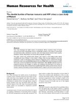
• The genetic background of inbred mouse strains can have a severe
(and unpredictable) effect on the phenotypes of knockout strains.
Commonly used strains, such as C57BL/6J (a black mouse) and 129 (an
agouti brown mouse), have known and unknown differences at
numerous alleles (see Figure 1).
differentiation. Embryos were growth-
retarded, with malformations of the
head, palate, and the developing eye.
The Ptdsr-deficient embryos also had a
delay in tissue differentiation in the
lung, kidney, and intestine. Brain
hyperplasia and a block in erythropoi-
etic differentiation were also observed,
as in the reports from Li et al. [5] and
Kunisaki et al. [6], respectively. One of
the most striking defects was the
absence of eyes in some embryos asso-
ciated with the induction of ectopic
eye structures in nasal cavities. Finally,
they observed reduction in the levels of
macrophage cytokines that had not
been reported by the other groups.
Reconciling the results
The existence of several knockouts of
the same gene with very different
phenotypes is puzzling, intriguing, and
divides researchers in the field about
how to interpret the results. Simon
Brown of the University of Edinburgh,
UK, urges readers to focus on the com-
monality of the three studies. “All three
found the homozygous-null mouse to
be perinatal lethal with clear evidence
of a defect in cell differentiation and
marked effects on tissue and organ
development following the mid-gest-
ation period,” he notes. He admits that
there are some major differences that
draw one’s attention but suspects these
can be explained by differences
in experimental approaches. Most
researchers seem to agree that
Lengeling’s analysis is particularly
careful, but the discrepancies between
the different studies remain perplexing.
Michael Hengartner at the Univer-
sity of Zurich, Switzerland, is unequiv-
ocal. “Lengeling’s results clearly
demonstrate that Ptdsr is most cer-
tainly not a PS receptor, and probably
has nothing to do with apoptotic cell
recognition at all.” He suspects that the
source of the problem is a case of false
identification during the expression-
cloning of the antigen recognized by
mAb 217. This is supported by the
supplementary data provided by Böse
et al. [2], in which mAb 217 is shown
to recognize macrophages derived
from their knockout mouse [2]. “One
wonders why the other two groups did
not perform this control using their
mice,” notes Hengartner. Shigekazu
Nagata from the Osaka University
Medical School, Japan, also feels that
the cloned Ptdsr gene had not been
sufficiently characterized previously.
“Lengeling’s group now shows that
Ptdsr carries an epitope that can be
weakly recognized by the mAb 217.
But the antibody efficiently stains even
the Ptdsr-deficient cells, indicating that
the antibody recognizes a molecule
other than Ptdsr.”
“The PS receptor story is an interest-
ing case of how an excusable error, that
can easily happen in any scientific
pursuit, results in a series of published
data that are guided by prejudice,” says
Angelika Böttger from the Ludwig-
Maximilians-Universität in Munich,
Germany. She is sure that the mAb 217
antibody really does inhibit the phago-
cytosis of apoptotic cells. “The only
problem is that the Ptdsr gene does not
encode the antigen for this antibody.”
She says that the experiment in which
the Ptdsr-deficient cells are stained with
the mAb 217 “should finally convince
everybody that the dogma is wrong.”
Other researchers remain uncon-
vinced. “One should not rush to con-
clude that Ptdsr is not important for
corpse removal based on the analysis
of one mouse Ptdsr-knockout line,”
cautions Ding Xue from the University
of Colorado in Boulder. “The differ-
ences in mutant phenotypes observed
in the three different mouse lines,
including apoptotic corpse removal,
are likely due to the different genetic
backgrounds of the knockout mice or
differences in carrying out various
assays,” he says. “I think that the
Lengeling group should at least
analyze the mouse line from Li et al.
or Kunisaki et al. before making any
definitive conclusions.” Xue cites
numerous precedents in which dif-
ferent genetic backgrounds yield
dramatically different mutant pheno-
types “For example, caspase3-deficient
mice in a C57BL/6J background are
viable, but are nonviable in a 129
background. In such circumstances,
one needs to be cautious about stating
whether the results obtained from one
mouse line are more reliable or credi-
ble than the other lines: most likely,
the results from three groups were all
correct in respect to the mouse lines
that they examined.” Lengeling’s group
used an isogenic C57BL/6J back-
ground, whereas the previous Ptdsr
knockouts were in a mixed 129 x
C57BL/6 background (see Figure 1).
Siamon Gordon, a macrophage
expert at the University of Oxford, UK,
feels that “the main point of this article
is that people were not looking hard
enough before and were jumping to
conclusions.” But Henson himself wel-
comes the different results. “The more
Journal of Biology 2004, Volume 3, Article 13 Weitzman 13.3
Journal of Biology 2004, 3:13
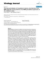
Figure 1
Genetic background variation in mouse
strains, as shown by two adult mice with their
pups. The C57BL/6J mouse (black coat) was
crossed with a chimeric mouse (patchy coat),
consisting of mutant C57BL/6J cells in a
BALB/c white background. The offspring with
a black coat color can then be screened for
germline transmission of the mutant allele.
Image: Ozgene Pty. Ltd.
studies we have on this molecule the
more interesting it gets, and it clearly
has multiple functions.” Henson con-
fides that his group has generated a
fourth knockout strain and prelim-
inary results suggest that the pheno-
types differ from the other three. He
admits that it appears confusing, but is
confident that the data will eventually
fit together.” Gordon suggests that dif-
ferent cells may contribute to clearance
in different scenarios. “Macrophages
are faster and more efficient profes-
sional phagocytes than non-leukocytes,
so they may be the main players during
inflammation or infection.” He notes
that earlier studies had indicated that
macrophages are not essential for
apoptotic clearance during develop-
ment, adding that C. elegans has no
macrophages but can still clear corpses.
More surprises in store
Many experts hope that clues will
come from analysis of the Ptdsr
protein in other species. Xue’s group
has analyzed the role of the Ptdsr gene
in worms and found evidence that it is
involved in removing apoptotic cells
[7]. But Kristin White’s group at MGH-
Harvard in Charlestown, USA, has
studied the PS receptor in flies and
came up with results that fit more with
those in the Lengeling article. “We see
no obvious defect in engulfment of
apoptotic cells in Drosophila embryos
that lack the PS receptor. These
animals are viable, with some subtle
developmental defects,” says White.
Future studies will obviously focus
on functions of the Ptdsr beyond apop-
totic cell clearance. Nagata hopes that
the Lengeling study will trigger investi-
gation of the ‘real’ function of Ptdsr
during mammalian development.
“These have little in common; thus
Ptdsr probably has a specific cellular
role that is required at multiple occa-
sions throughout development,” says
Hengartner. White agrees: “Since we
also see effects on development in the
fly PS receptor mutant, this suggests
that the PS receptor has a biological
13.4 Journal of Biology 2004, Volume 3, Article 13 Weitzman />Journal of Biology 2004, 3:13
Behind the scenes
Journal of Biology asked Andreas Lengeling about the background and
rationale for his study of the phosphatidylserine receptor (Ptdsr) in mice.
What motivated you to generate a Ptdsr knockout mouse?
My group is interested in the function of macrophages in immune
responses and how they defend the body during infection. We were
fascinated by the emerging work on Toll receptors in innate immunity and
the recognition of pathogens. Valerie Fadok’s work introduced the
scientific community to a new receptor, Ptdsr, which could specifically
recognize host apoptotic cells, and exposed PS as a key signal for
phagocyte engulfment. Ptdsr seemed to be crucial for two kinds of
macrophage responses: the engulfment of apoptotic cells and the release
of immunosuppressive mediators. We knocked out the gene encoding
Ptdsr to examine its role in these processes.
How long did the experiments take and what were the steps
that ensured success?
Knocking out the Ptdsr gene turned out not to be too difficult, thanks to
Frank Köntgen at Ozgene. The analysis of the mutant mice turned out to
be more complicated, especially because ablation of Ptdsr was lethal. We
looked hard for differences in the efficacy of apoptotic cell removal in
mutant animals but couldn’t see any. Instead we identified a lot of
interesting phenotypes, which pointed us towards completely novel and
unexpected functions of Ptdsr during development. This success was the
work of a team of gifted specialists, including veterinary pathologist
Achim Gruber.
What was your initial reaction to the results and how were they
received by others?
At first we had a hard time believing that there was actually no
impairment in apoptotic cell removal in our knockout mice. Eventually we
realized that Ptdsr functions as a differentiation-promoting factor in many
different organs and tissues during embryogenesis. Surprisingly, this was
received quite openly in the community. It turned out that scientists
working in other model systems, such as flies, worms, and even Hydra,
also had evidence for alternative Ptdsr functions.
What are the next steps?
There are two major things that need to be followed up. First, as Ptdsr is
not the major receptor for apoptotic cells, the question remains whether
there is a specific PS receptor out there or whether PS is only recognized
by ‘bridging molecules’. This will require elegant interdisciplinary
approaches that compare different animal model systems, such as mice,
flies, and worms. Second, we want to investigate the primary function of
Ptdsr by generating conditional alleles of Ptdsr, by investigating
downstream pathways via gene-expression array profiling, and by doing a
lot of biochemistry.
function that is more general than the
recognition of apoptotic cells. The use
of different biological systems and
approaches to dissect the role of this
protein should help us to understand
this more general role.”
There are likely to be more sur-
prises in store. “The Ptdsr protein
carries a domain called the Jumonji C
(JmjC) domain,” notes Nagata, specu-
lating that, like other proteins with this
domain, Ptdsr may play a role in chro-
matin remodeling and the stability of
heterochromatin. Edinburgh’s Brown
makes similar predictions and indeed,
there is evidence that the Ptdsr protein
may be located in the nucleus [8,9].
Böttger, whose lab studies the PS recep-
tor from Hydra, suggests “it could
modify nuclear proteins, transcription
factors or maybe proteins maintaining
nuclear architecture and thus have
these profound effects on differentia-
tion during early mouse development.”
Alice’s adventures came to an end
when she awoke from her dream. But
our apoptotic adventures are likely to
continue for some time as we learn
more about the surprises that govern
when and how cells die and who is
responsible for clearing up the remains.
References
1. Carroll L: Alice’s Adventures in Wonderland.
London: Macmillan; 1865.
2. Böse J, Gruber AD, Helming L, Schiebe
S, Wegener I, Hafner M, Beales M,
Köntgen F, Lengeling A: The phos-
phatidylserine receptor has essen-
tial functions during embryogenesis
but not in apoptotic cell removal.
J Biol 2004, 3:15.
3. Fadok VA, Voelker DR, Campbell PA,
Cohen JJ, Bratton DL, Henson PM:
Exposure of phosphatidylserine on
the surface of apoptotic lympho-
cytes triggers specific recognition
and removal by macrophages.
J Immunol 1992, 148:2207-2216.
4. Fadok VA, Bratton DL, Rose DM,
Pearson A, Ezekewitz RA, Henson PM: A
receptor for phosphatidylserine-
specific clearance of apoptotic cells.
Nature 2000, 405:85-90.
5. Li MO, Sarkisian MR, Mehal WZ, Rakic P,
Flavell RA: Phosphatidylserine recep-
tor is required for clearance of apop-
totic cells. Science 2003, 302:1560-1563.
6. Kunisaki Y, Masuko S, Noda Z, Inayoshi
A, Sanui T, Harada M, Sasazuki T, Fukui,
Y: Defective fetal liver erythro-
poiesis and T lymphopoiesis in mice
lacking the phosphatidylserine
receptor. Blood 2004, 103:3362-3364.
7. Wang X, Wu YC, Fadok VA, Lee MC,
Gengyo-Ando K, Cheng LC, Ledwich D,
Hsu PK, Chen JY, Chou BK, et al.: Cell
corpse engulfment mediated by
C. elegans phosphatidylserine recep-
tor through CED-5 and CED-12.
Science 2003, 302:1563-1566.
8. Cikala M, Alexandrova O, David CN,
Pröschel MBS, Cramer P, Böttger A: The
phosphatidylserine receptor from
Hydra is a nuclear protein with
potential Fe(II)-dependent oxyge-
nase activity. BMC Cell Biol 2004, 5:26.
9. Cui P, Qin B, Liu N, Pan G, Pei D: Nuclear
localization of the phosphatidylser-
ine receptor protein via multiple
nuclear localization signals. Exp Cell
Res 2004, 293:154-163.
Jonathan B Weitzman is a scientist and science
writer based in Paris, France.
E-mail:
Journal of Biology 2004, Volume 3, Article 13 Weitzman 13.5
Journal of Biology 2004, 3:13