Báo cáo sinh học: "Astrocytes derived from glial-restricted precursors promote spinal cord repair" ppt
Bạn đang xem bản rút gọn của tài liệu. Xem và tải ngay bản đầy đủ của tài liệu tại đây (11.78 MB, 21 trang )
Research article
Astrocytes derived from glial-restricted precursors promote
spinal cord repair
Jeannette E Davies*, Carol Huang*, Christoph Proschel
‡
, Mark Noble
‡
,
Margot Mayer-Proschel
‡
and Stephen JA Davies*
†
Addresses: *Department of Neurosurgery, Baylor College of Medicine, 1709 Dryden Street, Suite 750, Houston, Texas 77030, USA.
†
Department of Neuroscience, Baylor College of Medicine, 1 Baylor Plaza, Houston, Texas 77030, USA.
‡
Department of Biomedical
Genetics, University of Rochester Medical Center, 601 Elmwood Avenue, Rochester, New York 14642, USA.
Correspondence: Stephen JA Davies. Email:
Abstract
Background: Transplantation of embryonic stem or neural progenitor cells is an attractive
strategy for repair of the injured central nervous system. Transplantation of these cells alone
to acute spinal cord injuries has not, however, resulted in robust axon regeneration beyond
the sites of injury. This may be due to progenitors differentiating to cell types that support
axon growth poorly and/or their inability to modify the inhibitory environment of adult
central nervous system (CNS) injuries. We reasoned therefore that pre-differentiation of
embryonic neural precursors to astrocytes, which are thought to support axon growth in the
injured immature CNS, would be more beneficial for CNS repair.
Results: Transplantation of astrocytes derived from embryonic glial-restricted precursors
(GRPs) promoted robust axon growth and restoration of locomotor function after acute
transection injuries of the adult rat spinal cord. Transplantation of GRP-derived astrocytes
(GDAs) into dorsal column injuries promoted growth of over 60% of ascending dorsal
column axons into the centers of the lesions, with 66% of these axons extending beyond the
injury sites. Grid-walk analysis of GDA-transplanted rats with rubrospinal tract injuries
revealed significant improvements in locomotor function. GDA transplantation also induced a
striking realignment of injured tissue, suppressed initial scarring and rescued axotomized CNS
neurons with cut axons from atrophy. In sharp contrast, undifferentiated GRPs failed to
suppress scar formation or support axon growth and locomotor recovery.
Conclusions: Pre-differentiation of glial precursors into GDAs before transplantation into
spinal cord injuries leads to significantly improved outcomes over precursor cell
transplantation, providing both a novel strategy and a highly effective new cell type for
repairing CNS injuries.
BioMed Central
Journal
of Biology
Journal of Biology 2006, 5:7
Open Access
Published: 27 April 2006
Journal of Biology 2006, 5:7
The electronic version of this article is the complete one and can be
found online at />Received: 5 November 2005
Revised: 21 March 2006
Accepted: 22 March 2006
© 2006 Davies et al.; licensee BioMed Central Ltd.
This is an Open Access article distributed under the terms of the Creative Commons Attribution License ( />which permits unrestricted use, distribution, and reproduction in any medium, provided the original work is properly cited.
Background
Traumatic injury to the adult central nervous system (CNS)
is associated with multiple different types of damage, all of
which pose substantial challenges to attempts to carry out
tissue repair. Promoting regenerative growth of severed
motor and sensory axons requires the provision of appro-
priate substrates and/or the overriding of a variety of
inhibitors that prevent axon regeneration. The expression of
molecular inhibitors of axon growth has been extensively
characterized in fibrotic, glial scar tissue [1-4] and in CNS
myelin [5-7]. In particular, adult astrocytes at sites of injury
have been shown to express proteoglycans that inhibit axon
growth [4,8,9] and have a major role in the formation of
misaligned scar tissue [10], which lacks the linear organiza-
tion of adult CNS white matter thought to be required for
rapid, long-distance axon growth [11-14].
A wide range of approaches have now been applied follow-
ing CNS injury to promote regenerative growth of both
sensory and motor axons, with a particular focus on the
transplantation of a variety of cell types, often in combina-
tion with other therapies. Cell-based transplantation strate-
gies for promoting axon growth across spinal cord injuries
[15] have included the use of neural stem cells, neonatal
brain astrocytes, fibroblasts, bone-marrow derived cells and
peripheral nervous system glia such as Schwann cells and
olfactory ensheathing cells. Although transplants of some
cell types have provided more benefit than others, the
general lack of significant axon regeneration beyond sites of
injury has led to the combination of cellular transplant
strategies with delivery of neurotrophic factors, treatments
designed to override or degrade the scar, and/or with the
use of biomaterials to offer both potential substrates and
organized tissue structures [16,17]. Such combinations have
resulted in varying degrees of successful axon regeneration.
We have been interested in the possibility that repair of
adult CNS injuries might be particularly enhanced with the
introduction of cells from the immature CNS, a tissue that
has a far greater regenerative capacity than the adult CNS
(reviewed in [18]). One possible approach is to transplant
embryonic stem cells or neural progenitor cells. Although
these cells have been shown to promote limited behavioral
recovery via remyelination of host axons [19-22], their
transplantation directly into or adjacent to traumatic spinal
cord injuries has not resulted in the regeneration of signifi-
cant numbers of endogenous axons across the site of injury
[21,23-25]. This may be due to the failure of the majority
of these cells to differentiate [26] or because the inflamma-
tory environment of adult CNS injuries directs undifferen-
tiated neural stem cells or glial progenitors to a ‘scar
astrocyte’-like phenotype [27] that is poorly supportive of
axon growth [8,28].
An alternative to allowing the lesion environment to reg-
ulate differentiation of stem or progenitor cells is to trans-
plant a cell type from the immature CNS that is known to
be supportive of axon growth. In this regard, embryonic
astrocytes have long been thought of as an attractive cell
type for repair of the adult CNS [29]. Establishing astro-
cytic cultures directly from the embryonic CNS, however,
generates cell populations containing mixed astrocytic phe-
notypes contaminated with glial progenitors and microglia,
and such populations have yielded relatively modest
success in promoting axon regeneration after transplant-
ation to adult CNS injuries [30,31]. Isolating embryonic
astrocytes directly from the embryonic CNS is also very
challenging, owing to the relatively low abundance of these
cells in vivo. The generation of postnatal astrocytic cultures
is normally associated with prolonged growth in con-
ditions in vitro that allow aging of these cells to a less sup-
portive phenotype [32], which has also resulted in minimal
axon growth after their transplantation to adult spinal cord
injuries [33].
To address the above problems, we have explored the alter-
native approach of pre-differentiating embryonic glial pre-
cursors to specific astrocytes in vitro, a technique that
permits the rapid generation of sufficiently large, homoge-
neous populations of embryonic astrocytes of a desired
phenotype for transplantation to adult CNS injuries. In
applying this approach, we have generated pure popula-
tions of astrocytes directly from glial-restricted precursor
(GRP) cells [34-36], the earliest arising progenitor cell pop-
ulation restricted to the generation of glia. Astrocytes were
generated by exposing GRP cells to bone morphogenetic
protein-4 (BMP-4), which induces astrocyte generation
from embryonic neural precursor cells and GRP cells both
in vitro and in vivo [34,37] and is thought to have important
roles in regulating glial differentiation in vivo [38]. GRP-
derived astrocytes (GDAs) generated by BMP exposure fall
within the population of cells defined by their antigenic
phenotype as type-1 astrocytes. Studies in vitro of type-1
astrocytes purified from the postnatal CNS have shown that
they promote extensive neurite growth from a variety of
neurons [39,40], express high levels of molecules that
support axon growth, such as laminin and fibronectin [41]
and nerve growth factor (NGF) or neurotrophin-3 (NT-3)
[42] and also show minimal immunoreactivity to chon-
droitin sulfate proteoglycans (CSPG) [41]. Moreover, the
directed generation of astrocytes from embryonic GRP cells
may provide cells that show the beneficial axon-growth-
promoting properties that characterize the early CNS.
Our study shows that transplantation of GDAs into acute
spinal cord injuries promotes levels of tissue reorganization,
axon regeneration and locomotor recovery that previously
7.2 Journal of Biology 2006, Volume 5, Article 7 Davies et al. />Journal of Biology 2006, 5:7
have been achieved only by combining cell transplantation
with multiple therapeutic approaches. We also show, in iden-
tical lesions, that transplanted GRP cells are not supportive of
axon growth or functional recovery, thus demonstrating the
critical importance of pre-differentiating progenitor cells
before transplantation to the injured adult CNS.
Results
Regeneration of endogenous dorsal column axons
Transplantation of GDAs into stab-wound lesions of the
dorsal column white matter pathways of adult rat spinal
cord (Figure 1a-c) resulted in the growth of the majority of
the cut ascending dorsal column axons into the lesion center
(Figures 2 and 3a), with 66% of these axons extending
further beyond the lesion site into adjacent white matter
(Figures 2 and 3a,b,e,f). In order to minimize labeling of
spared axons, a discrete population of ascending axons
aligned with the lesion site was traced en passage with a
single biotinylated dextran amine (BDA) injection caudal to
GDA-transplanted or control stab injuries of the right-hand
dorsal column cuneate and gracile white matter pathways
(Figure 1c; see the Glossary box for an explanation of
terms). Previous studies have shown that about 30-40% of
ascending dorsal column axons projecting to the dorsal
column nuclei arise from postsynaptic dorsal column
neurons in spinal laminar 4 and that 25% of ascending
dorsal column axons are also propriospinal in origin
[43,44]. Indeed, it is thought that only 15% of primary
afferents of dorsal root ganglion (DRG) neurons entering
Journal of Biology 2006, Volume 5, Article 7 Davies et al. 7.3
Journal of Biology 2006, 5:7
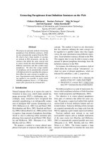
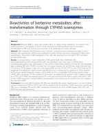
Figure 1
The models of spinal cord injury in adult rats used in this study. Schematic illustrations of (a-c) white matter of the dorsal column and (d) the
dorsolateral funiculus white-matter pathways of the spinal cord. (a,d) Dorsal views of the rat brain and spinal cord. (b) horizontal and (c) sagittal
views of the dorsal column white matter pathways at the C1/C2 cervical vertebrae of the spinal cord. (a) Dorsal column white matter on the right
side was transected (shaded area) at the C1/C2 spinal level, and the ability of either BDA-labeled endogenous axons or axons from
microtransplanted GFP-expressing adult sensory neurons (DRGs) to cross injuries bridged with GDAs or GRPs was assayed. (b) Injections of GDA
or GRP cells (black diamonds) suspended in medium were made directly into the centers of the injury sites as well as their rostral and caudal
margins in the cervical spinal cord. (c) A discreet population of endogenous ascending axons within the cuneate and gracile white matter pathways of
dorsal columns was labeled by BDA injection at the C3/C4 spinal level (5 mm caudal to the lesion site, shaded). Alternatively, microtransplants of
GFP
+
DRGs were injected 500 m caudal to the injury site. (d) The right-side dorsolateral funiculus white matter containing descending axons of the
rubrospinal tract was transected at the C3/C4 spinal level and GDAs or GRPs were transplanted as described for dorsal column injuries. To trace
axotomized rubrospinal tract axons, BDA was injected into the left-side red nucleus (RN) 8 days before the end of each experiment. CC, central
canal; Cf, cuneate fasciculus; CST, corticospinal tract; DF, dorsolateral funiculus; Gf, gracile fasciculus; GM, gray matter; RST, rubrospinal tract; T1,
level of the first thoracic vertebra.
C1/C2
GDAs
or GRPs
BDA
Rostral
Cf/Gf
Cf/Gf
Cf/Gf
CST
CC
GM
GM
C1/C2
GDAs
or GRPs
C1/C2
GDAs
or GRPs
C3/C4
BDA
DRGs
or
Horizontal view
Sagittal view
C3/C4
C8
T1
C3/C4
C8
T1
Rostral
GDAs
or GRPs
Dorsal midline
//
//
//
(a) (b) (d)
(c)
RN
RN
BDA
DF/RST
the spinal cord at lumbar levels reach the cervical spinal
cord and that most leave dorsal column white matter within
two to three segments of entering [45]. Therefore our en
passage labeling of dorsal column axons at the cervical level
included significant proportions of axons from both DRG
neurons and CNS spinal neurons.
Sample counts from every third parasagittal section at 8 days
after injury revealed similar numbers of BDA-labeled
axons 0.5 mm caudal to the injury site in both control and
experimental spinal cords, with averages of 107 ± 47
versus 101 ± 45 axons sampled per animal, respectively
(see also Figure 2). In GDA-transplanted cords, on average
61% (standard deviation (s.d.) ± 11) of caudal labeled
axons extended into the lesion center, 39% (s.d. ± 15) of
caudal axons extended 0.5 mm beyond the lesion center
into adjacent white matter and 28% (s.d. ± 7) extended
7.4 Journal of Biology 2006, Volume 5, Article 7 Davies et al. />Journal of Biology 2006, 5:7
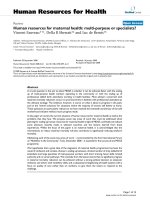
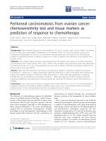
Figure 2
Quantification of numbers of regenerating BDA
+
axons in
GDA-transplanted versus control dorsal column white matter at 8 days
after injury and transplantation. BDA-labeled axons were counted in
every third sagittally oriented section within the lesion center and at
points 0.5 mm, 1.5 mm, and 5 mm rostral to the injury site, up to and
including the dorsal column nuclei (DCN). Note that 61% of BDA
+
axons had reached the centers of GDA-transplanted lesions and 39% to
0.5 mm beyond injury sites, compared with just 4% (lesion center) and
3.8% (0.5 mm rostral) present in controls. The steady decline in
numbers of BDA
+
axons within rostral white matter indicates a
staggered front of maximum axon growth beyond sites of injury in
GDA-transplanted groups at this time point. Note the total absence of
axons at 5.0 mm rostral and in dorsal column nuclei in controls. Counts
of BDA
+
axons labeled in all adjacent sagittally oriented sections in
representative GDA-treated and control lesioned cords revealed totals
of 372 and 330 axons, respectively, at 0.5 mm caudal to the injury site.
Increases in numbers of BDA
+
axons in GDA-treated animals compared
with controls were statistically significant (p < 0.01) in all rostral spinal
cord regions. Error bars indicate ± 1 standard deviation.
Percentage of total BDA
+
axons
0.5 mm
caudal
Lesion
center
0.5 mm
rostral
1.5 mm
rostral
5.0 mm
rostral
DCN
GDA
Control
0
20
40
60
80
100
Glossary
Astrogliosis Injury-induced changes in the
morphology of adult astrocytes characterized by
hypertrophy of their cell bodies and processes.
Axon sparing Axons that are not severed by
trauma to the spinal cord.
Axon sprouting Growth of collateral branches
from injured or spared axons.
Axotomized Describes a severed axon.
Bregma The junction of the coronal and sagittal
suture lines on the surface of the skull.
Dorsal columns Dorsal medial white matter
pathways. Contain ascending cuneate and gracile
sensory pathways and descending corticospinal
motor pathways in rats.
Dorsolateral funiculus Dorsal/lateral white matter
of the spinal cord; contains the rubrospinal tract.
En passage Within a pathway.
Gray matter CNS tissue containing the majority of
neuron cell bodies and a relatively low density of myelin.
Pial surface or pia mater Connective tissue at the
CNS outer surface named for the astrocytic ‘end
feet’ (pia) processes attached to capillaries on its
inner surface.
Propriospinal neurons Widely distributed in
spinal cord gray matter, these neurons form long-
and short-distance connections that are thought to
coordinate limb motion and mediate control of
reflexes.
Reactive astrocyte An astrocyte that has responded
to CNS injury or degeneration; typically displays a
swollen or hypertrophic cell body and processes.
Red nucleus Pigmented midbrain nucleus that
among other functions relays motor-control signals
from cortical and subcortical regions of the brain,
for example, cerebral cortex, cerebellum and
thalamus, to the spinal cord. Neurons within the
magnocellular and parvicellular subdivisions of the
red nucleus give rise to the rubrospinal tract.
Rubrospinal tract White matter pathway
containing axons descending from the red nucleus.
Axons innervate motor control circuits in cervical
and lumbar enlargements of the spinal cord. It
regulates coordinated, fine motor control in rats.
Spinal laminar 4 A region of dorsal spinal cord
gray matter that contains CNS neurons with
ascending axons within the dorsal column white
matter pathways.
White matter Highly myelinated CNS axon
pathways that contain large numbers of glial cells
(astrocytes, oligodendrocytes, microglia and glial
progenitors) and very few neurons.
Journal of Biology 2006, Volume 5, Article 7 Davies et al. 7.5
Journal of Biology 2006, 5:7
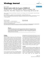
Figure 3 (see legend on the following page)
LC
Rostral
BDA hPAP
BDA GFAP
BDA
(a)
(b) (c) (d)
(e)
(f)
1.5 mm beyond the lesion site. Even at the relatively short
8-day time point, small numbers of axons extended still
further, with averages of seven BDA
+
axons (s.d. ± 5; 7%)
detected per animal at 5 mm rostral to the injury site and
four axons (s.d. ± 3; 4%) in the dorsal column nuclei in
GDA-transplanted animals. In contrast, in four out of five
control animals, no axons were observed within the lesion
centers or within white matter beyond the lesion. In just
one out of five control animals, six BDA
+
axons (4%) were
found in the ventral-most regions of the lesion site (that is,
at the ventral margin), effectively rostral to the caudal
lesion margin and therefore aligned with the lesion center.
These were most likely to be due to a limited axonal
sparing and/or sprouting in this animal, resulting in the
presence of these axons in the ventral white matter of the
cuneate pathway at the interface with gray matter. The fact
that no BDA
+
axons were observed beyond 1.5 mm rostral
to the lesion in this animal (Figure 2), or were observed
crossing the injury site near the pial surface or within
GFAP-negative regions of the lesion center proper in all
control animals, supports the hypothesis that these six
axons had sprouted around the injury at the gray/white
matter interface rather than having been spared. Overall,
approximately 99% of the cut ends of BDA
+
axons in
control cords remained within caudal lesion margins and
had dystrophic endings (Figure 3c). In sharp contrast, very
few dystrophic axons were observed at the caudal interface
of GDA transplants with adjacent white matter compared
with control injury sites (compare Figure 3c and d).
GDA ‘bridge’ supports axon growth
To further demonstrate the capacity of transplanted GDAs
to support axon growth in an adult rat model of spinal cord
injury that eliminates the possibility of axon sparing, we
examined the ability of axons growing from adjacent trans-
plants of adult DRG sensory neurons to cross identical
spinal cord stab injuries bridged with GDAs. In these experi-
ments, immediately after injury rats received microtrans-
plants of adult mouse sensory neurons expressing green
fluorescent protein (GFP) within dorsal column white
matter 400-500 m caudal to GDA-transplanted stab
injuries (Figures 1c and 4a) or control stab injuries injected
with media alone. In these experiments we also examined
the ability of transplantation of GRP cells themselves to
promote regeneration (Figures 1c and 4c).
Newly growing axons from the transplanted neurons failed
to cross GRP-transplanted injuries (Figures 4c and 5c) or
lesions injected with medium (data not shown). In contrast,
53% (s.d. ± 3) of rostrally directed GFP
+
axons grew into the
center of GDA-transplanted injuries, 62% of axons at the
lesion center reached 0.5 mm beyond lesion sites, 42%
reached 1.5 mm into rostral white matter, and small
numbers of axons extended up to 2 mm beyond the injury
site (Figure 4a). Comparison of endogenous BDA
+
and
GFP
+
axons from the two separate experiments (Table 1,
experiments 1 and 2) revealed a remarkably similar effi-
ciency of axon growth (66% and 62%, respectively) exiting
GDA-filled injuries. Thus, transplantation of GDAs was able
to promote axon growth across acute dorsal column
injuries, but transplantation of GRP cells (from which GDAs
are derived) had no such effect.
There was a striking correlation between the extent of
axonal growth and the degree of occupancy (bridging) of
the lesion by GDAs. In two GDA-transplanted animals in
which GDAs did not completely fill the lesion site, very few
GFP
+
axons penetrated the GDA-poor caudal lesion margins
and GFP
+
axons within lesion centers were confined to areas
containing GDAs (Figure 4b). In areas of the lesions devoid
of GDAs, GFP
+
axons formed dystrophic endings within
caudal lesion margins (Figure 4b). In these cases, no axons
were observed to cross the site of injury and enter rostral
white matter. GFP
+
axon growth was not fasciculated and
was often aligned with human placental alkaline phos-
phatase (hPAP)-positive processes of GDAs and parallel
with the host GFAP
+
astrocyte processes in the rostral and
caudal lesion margins (Figure 5a). Similarly, BDA
+
endoge-
nous axons were often aligned with hPAP
+
GDAs within
rostral (Figure 3b) and caudal (Figure 3d) lesion margins.
7.6 Journal of Biology 2006, Volume 5, Article 7 Davies et al. />Journal of Biology 2006, 5:7
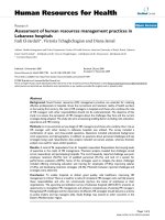
Figure 3 (see figure on the previous page)
Endogenous sensory axon regeneration across GDA-transplanted dorsal column injuries at 8 days after lesion and transplantation. (a) A montaged,
low-magnification confocal image scanned from a single 25-m thick sagittal section, showing BDA-labeled ascending dorsal column axons (green)
that have entered, grown within and exited a hPAP
+
(red) GDA-transplanted dorsal column lesion. LC, lesion center. (b) A high-magnification image
of a rostral graft/host interface showing BDA
+
axons exiting the GDA graft and entering host white matter. A few axons were observed to have
turned away from the interface and grown back towards the lesion center (arrowhead). (c) In control lesions, the vast majority of BDA
+
axons have
formed dystrophic endings and failed to leave the caudal margins of the lesion, marked by hypertrophic GFAP
+
astrocytes (red).
(d) A high-magnification image showing numerous BDA
+
axons that have successfully crossed the host/graft interface at the caudal lesion margin. A
few cut axons (arrowheads) have, however, failed to leave the caudal lesion interface and can be seen to have turned and/or formed dystrophic
endings, particularly in regions containing few hPAP
+
GDAs (red). (e) BDA
+
axons located near the pial surface and ventral regions of cuneate white
matter at 1.5 mm rostral to a GDA-bridged lesion site. (f) BDA
+
axon growth cones in white matter 1.5 mm rostral to the lesion site often display
streamlined growth cones indicative of rapid growth. Scale bars: (a,c) 100 m; (b-e) 50 m; (f) 5 m (top) and 10 m (bottom).
Journal of Biology 2006, Volume 5, Article 7 Davies et al. 7.7
Journal of Biology 2006, 5:7
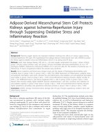
Figure 4
A comparison of the ability of GDA versus GRP transplants to promote axon growth across dorsal column injuries from adjacent microtransplanted
adult sensory neurons at 8 days after injury and transplantation. (a) A montaged, confocal image scanned from a single 75-m thick sagittally
oriented section showing GFP
+
axons (green) entering and exiting a dorsal column lesion bridged with hPAP
+
(red) GDAs. (b) In two cases in which
GDA transplants did not adequately fill the injury site or migrate into lesion margins, GFP
+
sensory axons failed to cross the caudal lesion margin and
instead formed dystrophic endings identical to those in control untreated injuries. LC, lesion center. (c) Confocal montage showing the complete
failure of transplanted GRPs to support the growth of GFP axons across a dorsal column injury. Note that, despite the ability of transplanted GRPs
to span the injury site, the majority of GFP
+
axons have formed dystrophic endings within the caudal lesion margin. Scale bars: (a) 300 m;
(b) 100 m; (c) 200 m.
LC
GFP GDAs
GFP GDAs
GFP GRPs
Rostral
(a)
(b)
(c)
As variation in lesion size has previously been observed
after spinal cord injuries in different strains of adult mice
[46], we conducted a qualitative assessment of lesion size
and shape resulting from stab injuries of the dorsal columns
in both Fischer 344 and Sprague Dawley rats, which
revealed a degree of variability in Fischer 344 rats that was
not observed in Sprague Dawleys (data not shown). This
variation in lesion size and shape between individual
Fischer 344 rats therefore precluded their use in quantitative
tracing studies of endogenous axon regeneration (see Mat-
erials and methods section for further details). Both rat
strains, however, showed equally consistent failure of GFP
+
axons to cross control lesions and both strains also showed
successful axon growth across dorsal column injuries
bridged with GDAs. Thus, treatment of dorsal column
lesions with GDAs in two different strains of rat resulted in
robust axon growth across sites of injury and failure of
axons to traverse control injuries.
7.8 Journal of Biology 2006, Volume 5, Article 7 Davies et al. />Journal of Biology 2006, 5:7
Figure 5
A comparison of GFP
+
axon and host astrocyte alignment in GDA- versus GRP- transplanted lesion margins at 8 days after injury. (a) A high-
magnification image showing aligned axon growth (green) associated with aligned GFAP
+
host astrocytic processes (red) in the caudal margin of a
GDA-transplanted lesion. (b) In contrast, GFAP
+
astrocytic processes (green) are misaligned in the caudal margin of a GRP-transplanted lesion (red).
(c) A high-power confocal image showing GFP
+
axons displaying tortuous, misaligned patterns of growth and dystrophic end bulbs (arrowhead) within
the astrogliotic caudal margin of a GRP-transplanted lesion. Scale bars: (a) 25 m; (b,c) 50 m.
GFAP hPAPGRP GFP GFAP
GFP GFAP
(a)
(b) (c)
Alignment of host tissue
In both dorsal column axon regeneration experiments, the
linearity of axonal growth we observed, particularly within
lesion margins (Figures 3c,d and 5a), prompted us to
examine the underlying tissue organization. Transplanta-
tion of dissociated GDAs was associated not only with a
significant reduction in astrogliosis but also with a striking
reorganization of host astrocyte cell bodies and processes
within lesion margins (Figures 5a and 6b,d and Additional
data file 1). To examine host astrocytes, we took advantage
of an unexpected downregulation of GFAP in the trans-
planted GDAs at 4 and 8 days after transplantation (Figure
6b) to identify host astrocytes with anti-GFAP immunos-
taining. Intra-lesion GDAs did, however, remain positive for
the astrocyte lineage markers S100 and vimentin (Addi-
tional data file 2) and did not express the oligodendrocyte
lineage antigens NG2 (Figure 7e,h) or proteolipid protein
(data not shown). GFAP
+
host astrocytes within the margins
of control medium-injected lesions (Figures 3c, 6a,c and
Additional data file 3), and in animals receiving GRP cell
transplants (Figure 5b,c) exhibited the characteristic hyper-
trophic cell bodies of adult reactive astrocytes and formed a
dense mass of numerous, ramified, misaligned processes
typical of astrogliotic scar tissue. In contrast, in animals
receiving GDA transplants, host GFAP
+
astrocyte processes
within lesion margins were now oriented toward lesion
centers (Figures 5a and 6b,d and Additional data file 1).
Quantitative analysis of the alignment of host GFAP
+
astro-
cytic processes in the lesion margins revealed considerable
differences between GDA-transplanted and control injury
sites. Control lesion margins had an average angle of 59.4°
(s.d. ± 22, median = 61°) between adjacent pairs of astro-
cytic processes. In contrast, GDA-filled lesions had average
angles of only 11.6° (s.d. ± 12.6, median = 7°) between
adjacent host GFAP
+
processes within lesion margins
(Figure 6e). Moreover, GDAs within lesion margins often
interweaved with endogenous GFAP
+
astrocytes (Figure 6b),
creating an aligned environment of glial cell surfaces, thus
providing a directional guidance of axon growth across the
interfaces of GDA-bridged lesions and adjacent white matter
(Figures 5a and 6b,d and Additional data file 1).
Suppression of inhibitory proteoglycans
GDA transplantation was also associated with a delayed
expression of axon-growth-inhibitory proteoglycans in
dorsal column lesions. The margins of control dorsal
column lesions examined 4 days after injury displayed a
high density of neurocan immunoreactivity associated with
numerous, fine, GFAP-negative processes (Figure 7a), which
we have previously shown to be primarily associated with
NG2
+
glia [4]. In addition, NG2 immunoreactivity in
control lesions was predominantly associated with invading
meningeal fibroblasts and blood vessels in the center of
control lesions (Figure 7d,g; see also [4]). In contrast, the
margins of lesions containing GDA grafts at 4 days after
injury showed a marked reduction in overall neurocan
immunoreactivity (Figure 7b versus 7a), resembling instead
the pattern of neurocan expression previously observed 2
days after injury in control lesions [4]. GDA-transplanted
injury sites also showed reduced NG2 immunoreactivity
compared with controls at 4 days after injury (Figure 7e,f).
At the 8-day time point, however, neurocan immunoreactiv-
ity in the margins of GDA-transplanted lesions was similar
in intensity and distribution to neurocan detected in control
lesions at 8 days after injury (Figure 7c), indicating that the
effect of the GDA transplant was to delay the expression of
neurocan in lesion margins. Significantly, however, even at
Journal of Biology 2006, Volume 5, Article 7 Davies et al. 7.9
Journal of Biology 2006, 5:7
Table 1
Numbers of animals per experimental group
Experiment Details Strain Time point Control lesion +GDA +GRP
1 Analysis of endogenous sensory axon regeneration, Sprague Dawley 4 days 4 6
CSPG expression and GDA phenotype 8 days 5 6
2 Analysis of axon growth from GFP
+
transplanted Fischer 344 8 days 6 9
sensory neurons Sprague Dawley 8 days 4 4
8 days 4 6
3 Analysis of RST axon growth, red nucleus and Sprague Dawley 8 days 6 + cyc 6 + cyc
behavioral recovery 5 weeks 9 + cyc 9 + cyc
5 weeks 7
5 weeks 7 (sham)
4 Analysis of acute behavioral recovery Sprague Dawley 14 days 6 + cyc 5 + cyc 6 + cyc
Cyc, cyclosporine
7.10 Journal of Biology 2006, Volume 5, Article 7 Davies et al. />Journal of Biology 2006, 5:7
Figure 6
Reorganization of lesion margins by GDAs. (a,c) Control lesions; (b,d) transplanted lesions. Control lesions at (a) 4 days and particularly at
(c) 8 days after injury have a dense meshwork of hypertrophic cell bodies and processes of endogenous astrocytes within lesion margins that is
typical of forming glial scar tissue. (b) At 4 days after injury and transplantation, ‘flares’ of hPAP
+
GDAs (green) are interwoven with realigned host
GFAP
+
astrocytes within lesion margins (the caudal margin is shown). Processes of both transplanted GDAs and host astrocytes are oriented
towards the lesion center. Note that hPAP
+
GDAs are not GFAP
+
. (d) At 8 days after injury and transplantation, GDAs have effected a reduction
in host astrogliosis and a striking realignment of host GFAP
+
astrocytes compared with the control (c). (e) Quantification of the alignment of host
GFAP
+
processes in lesion margins. The angles measured between each pair of GFAP
+
processes in control (n = 100) and GDA-transplanted lesion
margins (n = 100) are graphically displayed in a histogram. Each bin along the x-axis represents the angle between a pair of processes: 0° is parallel
and 90° is perpendicular. The y-axis indicates the number of pairs of GFAP
+
processes within each bin. Note the striking difference in alignment of
GFAP
+
host astrocytic processes in margins of GDA-transplanted lesions versus controls. GDA-transplanted lesions have an average angle of just
11.6° (median 7°) between paired processes, versus 59.4° (median 61°) for control lesion margins. Statistical analysis: p < 0.0001, t-test.
Scale bars: (a,c,d) 100 m; (b) 50 m.
Pairs of GFAP
+
processes
0-10
11-20
21-30
31-40
41-50
51-60
61-70
71-80
81-90
Angle (degrees) between adjacent processes
Control
GDA
GFAP
GFAP
GFAP
GFAP
GDA
(a) (b)
(c)
(e)
(d)
0
25
50
75
Journal of Biology 2006, Volume 5, Article 7 Davies et al. 7.11
Journal of Biology 2006, 5:7
Figure 7
GDA transplantation suppresses neurocan and NG2 immunoreactivity. (a) At 4 days after injury, control lesion margins display dense neurocan
immunoreactivity (green) mainly associated with fine, GFAP
–
processes and to a lesser extent with GFAP
+
astrocyte cell bodies (red). (b) Neurocan
immunoreactivity at 4 days after injury and transplantation is greatly reduced in margins of hPAP
+
GDA-transplanted lesions. (c) At 8 days after
injury and GDA transplantation, neurocan immunoreactivity within lesion margins has increased compared with the 4-day time point. Note,
however, that intra-lesion hPAP
+
GDAs continue not to be immunoreactive to neurocan. (d-f) GDA-transplanted lesion centers (e,f) at 4 days after
injury show a marked reduction in NG2 immunoreactivity (red) compared with (d) control lesions. hPAP
+
cells are stained green. (g-i) Although
overall NG2 immunoreactivity has increased within the center of GDA-transplanted lesions (h,i) at 8 days after injury compared with the (e,f) 4-day
time point, it is reduced compared with the more uniformly distributed NG2 immunoreactivity within the center of control lesions at 8 days after
injury. Scale bars: (a,b,g) 100 m; (c,h,i) 50 m; (d-f) 200 m.
NG2
hPAP
NG2 NG2
NG2 NG2
(a) (b) (c)
(d) (e) (f)
(g) (h) (i)
NG2
hPAP
Neurocan
hPAP
Neurocan
hPAP
Neurocan
GFAP
the 8-day time point GDAs within lesion margins and
centers displayed little or no neurocan immunoreactivity
(Figure 7c). Unlike the more uniform density of NG2
immunoreactivity within lesion centers at 8 days in control
cords, NG2 immunostaining within GDA-transplanted
injuries had a more patchy distribution (Figure 7h,i). This
almost certainly reflected a chimeric mix of NG2
–
GDAs
with host NG2
+
tissue at lesion centers (compare Figure 7g
and h,i), as we did not find hPAP
+
NG2
+
cells in the lesions
even 8 days after transplantation.
GDA transplantation promotes rubrospinal axon
regeneration and suppression of red nucleus neuron
atrophy
GDA transplantation was also beneficial for CNS neurons,
as demonstrated by analysis of rubrospinal tract (RST)
axons within injuries to the right-side dorsolateral funiculus
of the spinal cord and their corresponding neuronal cell
bodies within the left-side red nucleus of the brain (Figure
1d). Severe injury to this descending, somatic motor control
pathway disrupts the ability of rats to step rhythmically and
coordinate accurate fore- and hind-limb placement. In
animals in which the dorsolateral funiculus was transected,
GDAs again filled the site of injury, integrated into host
tissue and realigned host astrocytes. In animals receiving no
GDAs, there was a complete absence of RST axons within
the lesion centers (Figure 8b). The majority of BDA
+
axons
in control injury sites had dystrophic endings and remained
between 500 and 800 m from lesion centers (Figure 8b).
In sharp contrast, in four out of six animals receiving GDA
transplants, BDA-labeled RST axons were readily observable
within lesion centers (Figure 8a) and also within caudal
white matter up to 1.5 mm beyond the site of injury. In
addition, the majority of axotomized RST axons within
GDA-transplanted animals were observed interacting with
GDAs in rostral lesion margins and had sprouted to within
300 m of lesion centers (Figure 8a). Those axons that had
grown into caudal white matter in GDA-bridged injuries
were invariably observed in the ventral half of the injury
sites, which correlated with regions of GDA transplants that
more often continuously spanned the injury site (Figure
8a). In the two out of six GDA-recipient animals in which
GDA grafts did not span sites of injury (see also Figure 4b),
no BDA axons were observed within white matter beyond
the site of injury (data not shown).
We also examined the effects of GDA transplants at longer
time points associated with ongoing behavioral recovery (as
discussed later) and found that a transient presence of
GDAs was apparently sufficient to have significant pro-
longed effects. At 5 weeks after injury, hPAP
+
GDAs were no
longer detectable but BDA
+
RST axons were still observed
within lesion centers and had extended further within
caudal white matter up to 3 mm beyond sites of injury.
Notably, in six of nine rats, RST axons were also now
observed to have sprouted up within the more dorsal
regions of lesion centers and to have even grown out into
dorsal roots (Figure 8c,d). The presence of growth cones
within caudal white matter (Figure 8e) clearly demonstrates
that successful GDA transplants had stimulated RST axon
regeneration beyond sites of injury. In two out of nine rats,
a few widely dispersed axon arborizations, similar in mor-
phology to those seen for axons innervating terminal fields,
were also now detected in gray matter at distances of
1-2 mm beyond the injury site (Figure 8f). Ventral margins
of GDA-transplanted injuries, which at earlier time points
showed more effective spanning of the lesion, continued to
show a rostro-caudal alignment of host astrocyte processes,
thus demonstrating that the tissue alignment associated
with GDA transplantation did not require the continued
presence of GDAs for maintenance of this effect. The proba-
ble importance of GDAs in this effect was indicated by
observations that rostro-caudal alignment of host astrocytes
was less pronounced within dorsal lesion margins where
GDA colonization was less complete at earlier time points.
The dorsal host astrocytes also appeared more hypertrophic
than the ventral host astrocytes (Figure 8c). Nonetheless,
even dorsally, host astrocytes were more aligned and less
reactive than in control animals. In contrast to the benefi-
cial effects associated with GDA transplantation, in control
rats that received lesions and cyclosporine but no GDA
transplants the RST axons remained in the rostral lesion
margins at 5 weeks after injury and had dystrophic endings
(data not shown).
GDA transplants to dorsal lateral funiculus lesions also
resulted in a suppression of atrophy of neurons within the
injured red nucleus (Figure 9a and Additional data file 4).
Atrophy of significant numbers of red nucleus neurons
begins 1 week after RST transection [47]. We similarly
found that the number of neurons with a cell body diameter
greater than 20 m in the injured left-side red nucleus in
rats not receiving GDA transplants fell to 52% of the values
in the uninjured right-side nucleus at 5 weeks after injury.
Design-based stereological analysis (see Materials and
methods) revealed, however, that the injured left-side
nucleus in GDA-transplanted animals contained 81% as
many large-diameter neurons as found in the uninjured
right-side nucleus, effectively an approximately 65%
increase in numbers of neurons that had maintained a cell
body diameter of greater than 20 m above that observed
for control, injured red nuclei (Figure 9a).
GDA transplantation promotes behavioral recovery
A further indication of the efficacy of GDAs in promoting
CNS recovery was seen behaviorally. Transection of the
7.12 Journal of Biology 2006, Volume 5, Article 7 Davies et al. />Journal of Biology 2006, 5:7
Journal of Biology 2006, Volume 5, Article 7 Davies et al. 7.13
Journal of Biology 2006, 5:7
Figure 8
Transplanted GDAs promote regeneration of rubrospinal axons. (a) Confocal montage scanned through a depth of 60 m, showing a small
population of BDA
+
rubrospinal tract (RST) axons (green) that have traversed a GDA-bridged (red) lesion of the dorsolateral funiculus and entered
caudal white matter at 8 days after injury. The majority of RST axons, however, have sprouted to within 300 m of the lesion center (LC) but failed
to extend beyond the site of injury. Note the absence of BDA-labeled axons within the dorsal-most regions of the injury site. (b) Confocal montage
showing the complete failure of axotomized BDA
+
RST axons to cross control lesions at 8 days after injury and that the majority of axons have
remained within rostral lesion margins at a distance of 500-800 m from the lesion center (LC). (c) At 5 weeks after injury and transplantation, a
small population of BDA
+
RST axons have traversed GDA-bridged injury sites and extended within caudal white matter. Note that BDA
+
axons have
also sprouted into the dorsal regions of the lesion center and even extended beyond the pial surface (arrowhead; see also the high-power image in
(d)). Note the lower levels of GFAP immunoreactivity (red) in more ventral regions of the injury margins and center, coincident with the presence
of BDA
+
axons. (e) Two examples of RST axons displaying growth cones within white matter 2 mm caudal to a GDA-treated lesion, at 5 weeks
after transplantation. Note the collateral branch (asterisk). (f) Confocal image of a BDA
+
terminal field-like axonal plexus within layer 5 spinal cord
gray matter, immediately adjacent to the dorsolateral funiculus white matter at 5 weeks after injury and transplantation. In contrast, in all
GDA-transplanted rats and controls injected with medium alone at 8 days after injury, no BDA labeling was observed within gray matter beyond the
injury site. Scale bars: (a-c) 200 m; (d) 100 m; (e) 5 m; (f) 10 m.
LC
LC
BDA hPAP
BDA GFAP
BDA GFAP
BDA GFAP
(a)
(b)
(c)
(d) (e)
(f)
Caudal
dorsolateral funiculus severs descending, supraspinal axons
and results in chronic deficits in both fore- and hind-limb
motor function [48], which can be detected by the grid-walk
behavioral test [49]. Following transection of the dorso-
lateral funiculus, rats that received GDA transplants per-
formed significantly better than controls at all post-surgery
time points (Figure 9b) and their behavior improved signifi-
cantly between 3 days and 28 days after injury (two-way
repeated measures ANOVA, p < 0.05). Rats that received
GDA transplants made an average of 4.7 mistakes at 3 days
after injury and transplantation and improved to an average
of 2.9 mistakes at 28 days after injury. In contrast, control
lesioned rats made on average 6.0 mistakes at 3 days after
injury, and showed no statistically significant improvement
at any later time point, with an average of 5.1 mistakes at 28
days after injury. Before surgery, rats in control and treated
groups performed equally well, with a baseline average of
2.0 mistakes. Thus, the average number of mistakes made
by GDA-transplanted animals had improved to just 0.9
points above baseline, compared with no recovery of
control lesioned rats at 28 days after injury. Moreover,
analysis of individual rats at 28 days showed that four out
of nine lesioned animals that received GDA transplants had
scores that were now statistically identical to their pre-
surgery baseline scores (data not shown). As discussed later,
it thus appears that GDA transplantation was associated
both with reductions in the extent of neurological deficit at
3 days after injury and with further recovery in the 4 weeks
following injury.
Our data clearly demonstrate the failure of transplanted GRP
cells to suppress scar formation and support axon growth
across acute spinal cord injuries. In the light of a recent
study showing the ability of GRPs to suppress glutamate-
mediated neurotoxicity in vitro [50], there remained,
however, the potential for acutely transplanted GRPs still to
promote functional recovery via this mechanism, an effect
7.14 Journal of Biology 2006, Volume 5, Article 7 Davies et al. />Journal of Biology 2006, 5:7
Control
Percent of uninjured
red nucleus neurons
GDA
Missed steps
Days
−1
3
7
10
14
17
21
23
28
GDA + cs
Lesion only
Lesion + cs
Sham
Missed steps
−1
3
7
10
14
Days
GDA + cs
Lesion + cs
GRP + cs
100
80
60
40
20
0
1
2
3
4
5
6
7
8
1
2
3
4
5
6
7
(a)
(b)
(c)
Figure 9
GDA transplantation suppresses atrophy of red nucleus neurons and
promotes robust behavioral recovery. (a) Injured left-side red nuclei
in control rats contained an average of 52% of the neurons counted in
uninjured right-side red nuclei at 5 weeks after injury. The numbers of
neurons in the injured left-side red nuclei of GDA-transplanted
animals, however, was 81% of total neuron numbers in uninjured
right-side nuclei (*p < 0.01). (b) Grid-walk analysis of locomotor
recovery. Graph showing the average number of mistakes per
experimental group at different time points after injury for
GDA-transplanted rats versus the control-lesion and sham-operated
groups. GDA-transplanted animals (green) performed significantly
better than lesioned controls at all post-injury time points (p < 0.05).
(c) Transplanted GRPs do not promote locomotor recovery. Graph
showing the average number of grid-walk mistakes per experimental
group from 1 day before injury (baseline pre-lesion) to 2 weeks after
injury for a separate series of matched RST-lesioned rats that received
either GRP or GDA transplants versus lesion-only control rats. Note
the complete failure of locomotor recovery in GRP-transplanted
animals compared with lesion-only controls at all time points and
confirmation of significant locomotor recovery in response to GDA
transplantation (p < 0.05). cs, cyclosporine.
that would be detectable during the first week after injury.
To investigate this hypothesis, we conducted an analysis of
grid-walk performance at time points ranging from 3 days
to 2 weeks after injury in a further series of matched RST-
lesioned rats that received transplants of GRP cells, GDAs or
control medium injections. In accordance with previous
results, GDA-transplanted rats once again showed a signifi-
cant recovery of locomotor function compared with con-
trols (Figure 9c) at all time points after injury (two-way
repeated measures ANOVA, p < 0.05). Notably, at 14 days
after injury, GDA-transplanted rats had an average score of
1.5 (± 0.2) mistakes compared with 5.9 (± 0.2) mistakes for
lesion-only controls (Figure 9c). In sharp contrast, GRP-
transplanted animals showed no recovery of locomotor
function compared with controls at all time points after
injury (Figure 9c).
Discussion
We have demonstrated that astrocytes derived from embry-
onic spinal cord GRP cells can promote axon regeneration
and functional recovery after transplantation into acute adult
spinal cord injuries. The ability of GDAs to fill the injury site,
suppress astrogliosis, realign host tissues and delay expres-
sion of axon-growth-inhibitory proteoglycans suggests that
these cells are unusually effective in providing an environ-
ment that supports axon growth within acute CNS injuries.
These attributes, in combination with their ability to reduce
atrophy of axotomized CNS neurons and promote a signifi-
cant behavioral recovery, make GDAs an attractive novel cell
type with which to repair the damaged CNS.
The effects of GDAs on the growth of both ascending dorsal
column axons and descending RST axons beyond sites of
injury compare favorably with previous transplant-based
spinal cord injury therapies. Intra-lesion sciatic nerve grafts
have proven to be relatively poorly supportive of sensory
axon reentry into white matter rostral to dorsal column
transection injuries without additional treatments that
support axon growth [51]. Intra-lesion transplantation of
marrow stromal cells alone has not resulted in any sensory
axons exiting the rostral margins of grafts [52] and,
although the ability of regenerating sensory axons to cross
dorsal column or dorsal root entry zone injuries bridged by
olfactory ensheathing cells is still contentious [53-56], it is
generally accepted that intra-lesion Schwann cell and
neonatal astrocyte grafts are also poorly supportive of axon
reentry into host white matter [33,57]. Although the growth
of RST axons into and beyond GDA-bridged lesions was less
efficient than that observed for ascending axons of the
dorsal columns, the ability of RST axons to exit GDA-
bridged lesions and extend up to 3 mm beyond the injury
nonetheless offers a marked improvement over the complete
failure of RST axons to cross spinal cord injuries bridged
with olfactory ensheathing cells [58].
Our experiments demonstrate the value of pre-differentiation
of precursor cells prior to transplantation and show that the
signals encountered within the lesion site are not able to
convert GRP cells themselves to a population that supports
axon growth. This was confirmed by the complete failure of
transplanted GRP cells to suppress scar formation and
provide a bridge that supports axon growth. The fact that
pre-differentiation of these cells to GDAs is required for
repair of acute spinal cord injuries was also confirmed by
the inability of transplanted GRP cells to support locomotor
recovery in our RST-lesion model. These data are consistent
with previous studies showing a failure of undifferentiated
GRP cells to promote supraspinal axon regeneration [24] or
behavioral recovery [19] after transplantation to spinal cord
injuries. Whether this is because the adult lesion environ-
ment lacks the signaling molecules required to generate
GDA-like cells from transplanted GRP cells in vivo or
whether those signals are overridden by other influences,
such as inflammatory cytokines, remains to be investigated.
Determination of the exact mechanisms by which any cell
type provides benefit after transplantation to the traumati-
cally injured CNS is challenging given the wide range of pos-
sible effects of such procedures, and there are a variety of
means by which GDA transplantation could have contributed
to the significant recovery of locomotion we observed in our
grid-walk experiments. The early onset of recovery at the
3-day time point suggests an initial neuroprotective effect
associated with GDA transplantation, consistent with our
observed rescue of red nucleus neurons from atrophy.
Rescue of red nucleus neurons from atrophy has been
achieved through provision of brain-derived neurotrophic
factor (BDNF [59]). Ongoing analyses of gene expression in
GDAs shows readily detectable levels of BDNF mRNA (C.P.,
unpublished observations). In contrast, previous studies
indicate that postnatal type-1 astrocytes (derived from the
cortex) do not make BDNF [42], revealing another advantage
of GDAs. Thus, it is apparent that the antigenic category of
type-1 astrocytes [60] in which GDAs were originally placed
[35,36] is too broadly defined and needs refinement.
The significant increases in behavioral recovery from 3
days onwards observed in GDA-treated animals in two
separate experiments also suggests that axon regeneration
and/or plasticity of connection may have contributed to
overall functional recovery. As suppression of atrophy of
axotomized red nucleus neurons has also been associated
with regeneration of their axons into sciatic nerve grafts
[59], the significantly greater number of neurons with cell
body diameters over 20 m in the injured red nucleus of
Journal of Biology 2006, Volume 5, Article 7 Davies et al. 7.15
Journal of Biology 2006, 5:7
GDA-treated animals at 5 weeks after injury, combined
with the further elongation of RST axons in caudal white
matter observed between 8 days and 5 weeks, supports a
possible contribution of RST axon growth and plasticity to
overall behavioral recovery. The relatively modest extent of
RST axon growth, however, and the formation of terminal-
like structures within adjacent gray matter at the spinal
level of cervical vertebra C4, makes it likely that effects of
GDAs on other pathways also contributed to functional
recovery. Previous studies have shown that recovery of
locomotor function after dorsal spinal cord hemisection
injuries is associated with plasticity of corticospinal inner-
vation of surviving propriospinal pathways, which in turn
form new connections with denervated motor neurons
[61]. The ability of GDAs to provide benefit to both
ascending dorsal column and RST axons raises the possi-
bility that GDAs will also be found to support the recovery
of other axon populations relevant to locomotion, such as
those in the descending reticulospinal and lateral cortico-
spinal pathways [49].
We have previously shown that decorin-mediated suppres-
sion of the levels of the core proteins and glycosamino-
glycan side chains of CSPGs can render spinal cord injuries
more permissive for axon growth [62]. The delayed expres-
sion of inhibitory CSPGs associated with GDA transplant-
ation therefore seems likely to have had an important role
in enabling regenerative axon growth. The absence of neuro-
can and NG2 immunoreactivity shown by GDAs within
sites of injury indicates that intra-lesion GDAs may be
refractory to signaling molecules known to induce expres-
sion of neurocan in neonatal astrocyte cultures [63]. Thus,
intra-lesion GDAs maintained an axon-growth-supportive
phenotype with respect to CSPG expression. Moreover, the
presence of GDAs within lesions also modified the host
response to injury and resulted in a significant reduction in
NG2 expression within lesion centers and a delay in neuro-
can expression at lesion margins, which together may have
created a window of opportunity for axons not only to
enter but also to exit the injury site. In this context, the
greater extent of initial RST axon retraction from the injury
site compared with ascending dorsal column axons may
mean that all but the fastest-responding RST axons missed
this window of inhibitor suppression. The ability of
decorin infusion to maintain a significant reduction in the
levels of multiple axon-growth-inhibitory CSPGs within
acute spinal cord injuries at 8 days after injury [62] suggests
that a combined treatment with GDA and decorin may
extend the window of opportunity for acute RST axon
regeneration.
Also of potential importance to the ability of GDAs to
promote axonal regeneration, and perhaps one of the most
interesting effects of GDA transplantation, was the extent of
linear tissue organization induced by these cells at sites of
injury. Although previous studies have demonstrated that
alignment of host astrocytic processes alone is not sufficient
to promote axon growth across CNS injuries in the presence
of inhibitory CSPGs [12,64], clearly the efficiency of axon
growth across an injury site with reduced inhibitor expression
will be enhanced if axons are not required to negotiate a
maze of misaligned cellular processes, that is, if they can take
the shortest route. The fact that a dissociated suspension of
GDAs is able to effect a linear alignment within acute adult
spinal cord injuries without the addition of an aligned bio-
matrix suggests that the creation of such tissue organization
is a fundamental aspect of the biology of these cells.
Conclusions
In summary, GDA transplantation into the injured spinal
cord promoted levels of axon regeneration, alignment of
host tissue, suppression of scar formation, neuronal rescue
and locomotor recovery that have not been associated with
transplantation of other cell types. Critically, these benefits
were dependent upon pre-differentiation of glial precursors
to a desired astrocytic phenotype prior to transplantation
and were not observed with transplantation of glial-
restricted precursors. Our study demonstrates that the envi-
ronment of acute, adult spinal cord injuries does not
promote differentiation of glial precursors into the most
advantageous cell type for tissue repair and functional recov-
ery. Achieving such linear tissue organization, robust axonal
growth and functional recovery in the absence of additional
biomaterials, cell modification, or delivery of adjunctive
bioactive therapies leads to great interest in now determining
whether the beneficial effects of transplanted astrocytes
derived from embryonic precursors can be enhanced still
further by the application of rational combination therapies.
Materials and methods
Isolation of GRPs and generation of GDAs
GRPs labeled with A2B5 antibody (a cell-surface glycolipid
unique to this cell type during early stages of rat spinal
development) were isolated by fluorescence-activated cell
sorting of dissociated cell suspensions from spinal cord of
embryonic day 13.5 transgenic Fischer 344 rat embryos
expressing the gene for hPAP under the control of ROSA26
promoter (transgenic rat line:TgN(R26ALPP)14EPS) [65].
GRPs were maintained in culture with DMEM-F12 media
(Gibco/Invitrogen, Carlsbad, USA) with 10 ng/ml basic
fibroblast growth factor (bFGF; Sigma, St. Louis, USA) and
N2 tissue culture supplement (Gibco/Invitrogen) on a
mixed laminin/fibronectin substrate and exposed to
10 ng/ml of human recombinant BMP-4 (R&D Systems) for
7.16 Journal of Biology 2006, Volume 5, Article 7 Davies et al. />Journal of Biology 2006, 5:7
7 days in culture to differentiate them into A2B5-negative
astrocytes. The following culture conditions were controlled
and consistent between batches of GDAs: growth substrate,
cell density, growth media, cell feeding schedule, the con-
centrations and source of growth factors, the total length of
time in culture and the number of passages of GRPs before
initiating the differentiation protocol.
Characterization of the GDA phenotype in vitro
Cells in culture were labeled live with A2B5 monoclonal anti-
body (Chemicon, Temecula, USA; for GRPs or type-2 astrocytes)
or anti-NG2 antibodies (Chemicon; for oligodendrocyte pre-
cursors), then fixed with cold acid and alcohol and labeled
with antibodies for GFAP (Sigma; for astrocytes), FGF
receptor 3 (Sigma; for type-1 astrocytes), or proteolipid
protein (plp)/DM20 (Chemicon; for oligodendrocyte
lineage cells). Secondary antibodies were purchased from
Jackson Immunologicals (West Grove, USA) and Molecular
Probes (Eugene, USA). BMP-4-induced GDAs were uni-
formly immunoreactive for human alkaline phosphatase in
vitro. Although no NG2
+
or plp/DM20
+
oligodendrocyte
precursors were detected in these cultures, undifferentiated
GRPs (A2B5
+
GFAP
–
) or astrocytes of a type-2 phenotype
(A2B5
+
GFAP
+
) were occasionally detected after BMP-4
treatment. These cell types represented less than 1% of the
total cell population. To ensure GDA suspensions for trans-
plantation did not contain undifferentiated GRPs or cells
with the phenotype of type-2 astrocytes, potentially
contaminating cell types were removed from the suspen-
sion by immuno-panning with the anti-A2B5 antibody. A
small volume of the resulting suspension was plated onto
glass coverslips and labeled with antibodies to A2B5 and
GFAP to verify a uniform type-1 astrocyte phenotype. For
transplantation, 100% GFAP
+
A2B5
–
GDAs were suspended
in Hanks Balanced Salt Solution (HBSS) at a density of
30,000 cells/l.
Lesion models and cell transplantation
Adult female Sprague Dawley or Fischer 344 rats (3 months
old, Harlan, Indianapolis, USA) were anesthetized by injec-
tion of a cocktail containing ketamine (42.8 mg/ml),
xylazine (8.2 mg/ml) and acepromazine (0.7 mg/ml). For
dorsal column injuries (Figure 1a-c), the right-side dorsal
column was unilaterally transected between cervical verte-
brae 1 and 2 using a 30-gauge needle as a blade (see also
[4,11,62]). Lesions extended to a depth of 1 mm and
extended laterally 1 mm from the midline. For rubrospinal
tract lesions, unilateral transections of the right-side dorso-
lateral funiculus including the rubrospinal pathway were
conducted at the C3/C4 spinal cord level with micro-scissors
(Fine Science Tools, Foster City, USA). Lesions extended to a
depth of 1 mm and extended medially 1 mm from the
lateral pial surface of the spinal cord (Figure 1d).
A total of 4 l of GDA or GRP suspension (30,000 cells/l;
120,000 cells) per animal was acutely transplanted into six
different sites in dorsal column lesions (two injections each
into medial and lateral regions of the rostral and caudal
lesion margins, and two injections into medial and lateral
regions of the lesion center; Figure 1b). All dorsal column
in vivo experiments were conducted in the absence of
immunosuppressants. GDA or GRP transplants were
injected in an identical pattern into lesions of the dorso-
lateral funiculus and a total of 6 l of GDA or GRP suspen-
sion (30,000 cells/l; 180,000 cells) injected per injury
site. Control lesion rats were injected with 6 l HBSS. One
set of rats in the dorsolateral funiculus lesion-only group
and all rats that received GDA or GRP transplants and
dorsolateral funiculus lesions were administered daily
injections of cyclosporine (1 mg per 100 g body weight)
beginning the day before injury and transplantation
through to experimental endpoints. Sham-operated rats in
which the spinal cord was exposed but not lesioned, and
rats that received a lesion but no cyclosporine, were
included as control groups (Table 1).
Our previous studies have characterized scar formation and
CSPG expression after spinal cord injury in adult Sprague
Dawley rats, a strain commonly used in CNS regeneration
studies. Unilateral dorsal column stab injuries identical to
those in the present study reliably generated lesions of
uniform size and induced consistent, quantifiable changes
in CSPG expression in adult female Sprague Dawley rats
[4,62]. As the GDAs were derived from hPAP Fischer 344
rats, however, we conducted an initial pilot series of intra-
lesion GDA transplants versus untreated controls in dorsal
column injuries of both adult female Fischer 344 and
Sprague Dawley rats and assayed the ability of axons
growing from adjacent DRG neuron transplants to cross
sites of injury versus controls that did not receive GDAs.
Although GFP
+
axons consistently failed to cross control
lesions in both strains of rats, we observed significant vari-
ations in lesion size and margin morphology in control
Fischer 344 rats, a phenomenon that we did not observe in
Sprague Dawley rats. The greater variation in lesion size and
rostro-caudal distances of lesion margins from lesion
centers in Fischer 344 rats precluded accurate quantification
of the numbers of endogenous, BDA-labeled axons at set
distances from lesion centers in this strain of rats. Therefore,
a separate study to investigate and quantify regeneration of
BDA
+
endogenous ascending dorsal column axons and
CSPG expression in GDA-transplanted versus control dorsal
column lesion animals was conducted in Sprague Dawley
rats (see Table 1 for the rat strains and numbers of animals
used in each study). Thus, bridging dorsal column lesions
with GDAs in two different strains of rat, in two separate
axon regeneration experiments, both resulted in robust
Journal of Biology 2006, Volume 5, Article 7 Davies et al. 7.17
Journal of Biology 2006, 5:7
axon growth across sites of injury and failure of axons to tra-
verse control injuries.
Adult DRG neuron transplantation
Single-cell suspensions of adult mouse sensory neurons
were prepared from 10- to 12-week-old transgenic mice
expressing the gene for enhanced GFP [66] as previously
described [11,12,62]. No growth factors were added to the
neuron suspension. 500 nl of the neuron suspension (about
1,500 neurons/l) was acutely microtransplanted into
dorsal column white matter approximately 500 m caudal
to the lesion (Figure 1c).
Histology
At 4 days, 8 days and 5 weeks after surgery, animals were
deeply anesthetized and transcardially perfused with 0.1 M
PBS followed by 4% paraformaldehyde in 0.1 M PBS. For
frozen sectioning, dissected spinal cords were cryoprotected
in a 30% sucrose/PBS solution at 4°C overnight. Tissue was
embedded in OCT (Sakura Finetek, Torrance, USA) and
quickly frozen. Serial 25-m thick frozen sections were cut
in the sagittal plane and air-dried onto gelatin-coated glass
slides. For vibratome sectioning, dissected spinal cords
were post-fixed in 4% paraformaldehyde overnight, then
embedded in 5% gelatin/5% agar. Serial 75-m thick sagit-
tal sections were collected and processed as free-floating
sections. All tissue sections were washed in PBS, blocked
with 4% normal goat serum in solution with 0.1%
triton/PBS for 30 min, then incubated with appropriate
primary antibodies in the blocking solution overnight at
4°C. Secondary antibody incubations were for 1 h at room
temperature.
The following primary antibodies were used: monoclonal
anti-GFAP (Sigma) and polyclonal anti-GFAP (Sigma);
monoclonal anti-vimentin (Chemicon); monoclonal anti-
plp/DM20 (Chemicon); polyclonal anti-NG2 (Chemicon);
polyclonal anti-GFP (Molecular Probes); monoclonal anti-
hPAP (Sigma); polyclonal anti-hPAP (Fitzgerald, Concord,
USA). Secondary antibodies conjugated with Cy5, Cy2
(Jackson), Alexa-488 or Alexa-594 (Molecular Probes) were
used to visualize primary antibody binding. All secondary
antibodies were pre-absorbed against rat serum. To control
for nonspecific binding of secondary antibodies, adjacent
sections were also processed as described above without
primary antibodies. Labeled sections were examined using
an Olympus BX60 fluorescence light microscope and a Leica
TCS SP2 confocal microscope. Molecule co-localization and
cellular associations were determined with Leica three-
dimensional analysis software. All immunohistological
images were acquired with confocal microscopy (Leica TCS
SP2) of sections cut in the sagittal plane. Spinal cord rostral
to the lesion is shown to the left in all figures.
Tracing endogenous dorsal column or rubrospinal
axons
In both lesion models, endogenous axons were traced by
injection of 10% BDA in sterile PBS (Molecular Probes)
8 days before an experimental endpoint. In the dorsal
column lesion model, ascending endogenous axons were
traced by BDA injection to a depth of 0.5 mm into the right-
side cuneate and gracile white matter at the C3/C4 spinal
level (Figure 1c). Descending RST axons were traced in the
dorsolateral-funiculus lesion model by injection of BDA
into the magnocellular region of the left-side red nucleus
(coordinates: 6.04 mm posterior, +0.7 mm lateral and
7.6 mm below bregma). For histological analysis of BDA-
labeled axons, 25 m serial sagittal sections were collected
and processed for immunohistochemistry, as described
above. BDA was visualized by incubating tissue sections
with the VectastainABC solution (Vector Labs, Burlingame,
USA), and further intensified with the Tyramide-Alexa 488
reagent (Molecular Probes).
Quantification of endogenous ascending dorsal
column axons
The number of BDA-labeled axons was counted in every third
tissue section spanning the medial-lateral extent of dorsal-
column injury sites at the following locations: 0.5 mm caudal
to the injury; directly at the injury center; 0.5 mm, 1.5 mm
and 5 mm rostral to the injury site; and within the dorsal
column nuclei. To control for differences in axon tracing
and labeling efficiency between animals, the numbers of
BDA-labeled axons counted within the lesion center and at
all rostral sites were normalized to the number of BDA-
labeled axons detected 0.5 mm caudal to the lesion site for
each tissue section examined. The normalized values from
each tissue section for each separate animal (control and
GDA-transplanted) were averaged to generate values for each
animal. The values for each animal (n = 6 GDA-transplanted,
5 control) were then averaged and displayed graphically.
ANOVA or t-tests were performed as appropriate, with a
value of p < 0.01 taken to be significant. For separate experi-
ments analyzing the growth of GFP
+
axons from microtrans-
planted sensory neurons, identical methods were used to
count GFP
+
axons from alternate sagittally orientated 75 m
vibratome sections.
Quantification of alignment of host GFAP
+
astrocyte
processes
Confocal images were generated from scanning through
30-m thick sagittal oriented sections of caudal and ventral
dorsal column lesion margins immunostained for GFAP to
show host astrocytic processes. Five sections were selected
from the lateral to medial center of lesions in three control
and three GDA-transplanted rats (see Additional data files 1
and 3). Within each confocal image, GFAP
+
processes were
7.18 Journal of Biology 2006, Volume 5, Article 7 Davies et al. />Journal of Biology 2006, 5:7
randomly selected within the lesion margin and ‘best fit’
lines traced over them using Image Pro Plus software
(Media Cybernetics, Silver Spring, USA). Then an immedi-
ately adjacent GFAP
+
process was identically traced and the
angle between the lines calculated with the Image Pro Plus
software. In all, 20 pairs of GFAP
+
host astrocytic processes
from each confocal image (5 images per group) were
analyzed and the mean and median angles were deter-
mined. A t-test was performed to determine the statistical
significance of the difference in measured angles between
astrocytic processes for GDA and control groups, with a
highly significant p value of < 0.0001.
Grid-walk behavioral analysis
Two weeks prior to surgery, rats were trained to walk across
a horizontal ladder (foot misplacement apparatus, Colum-
bus Instruments, Columbus, USA) and only rats that consis-
tently crossed without stopping were selected for
experiments. The grid-walk test is a sensitive measure of the
ability of rats to step rhythmically and to coordinate accu-
rate placement of both fore- and hind-limbs. For analysis of
acute to long-term recovery of locomotor function in GDA-
transplanted versus untreated lesion controls (Table 1,
Experiment 3), trained rats were randomly assigned to one
of four groups: RST lesion + GDA + cyclosporine (n = 9);
RST lesion + suspension media + cyclosporine (n = 9); RST
lesion only (n = 7); and sham operation (n = 7). One day
before surgery (baseline) and at time points of 3, 7, 10, 14,
17, 21, 24, and 28 days after surgery, each rat was tested
three times and the number of missteps from each trial was
averaged to generate a daily score for each animal. To
compare the effects of GRP and GDA transplants on recov-
ery of locomotor function (Table 1, Experiment 4), a further
series of trained rats were randomly assigned to one of three
groups: RST lesion + GRPs + cyclosporine (n = 6); RST
lesion + GDAs + cyclosporine (n = 5); and RST lesion +
medium injection + cyclosporine (n = 6). Grid-walk perfor-
mance was tested in an identical fashion to that used for
rats in Experiment 3 at time points of 3, 7, 10, and 14 days
after injury. Two-way repeated measures ANOVA and the
Tukey post test (with a significance level of p < 0.05) were
used to analyze both datasets.
Quantification of red nucleus neurons
At 5 weeks after injury and transplantation, 25 m serial
frozen sections were cut in the coronal plane from the brains
of rats that had undergone behavioral analysis. Every third
section through the rostro-caudal extent of the red nucleus
was stained with 0.2% cresyl violet. Standard, design-based
stereology methods (CAST software, Olympus, Melville,
USA) were used to quantify numbers of neurons in both red
nuclei of RST-lesioned, GDA-transplanted (n = 6) and
control (medium injection + cyclosporine, n = 6) rats. An
optical fractionator was applied to left- and right-side red
nuclei from every sixth section. Cell bodies greater than
20 m in diameter and characteristic neuronal morphology
were counted. The numbers of neurons counted in the left-
side injured red nucleus were normalized to counts obtained
for the uninjured right-side nucleus for each animal. The
values for each animal within a group were averaged and
displayed graphically. A t-test was performed to determine
the statistical significance of the difference between the
groups (with a value of p < 0.01 taken to be significant).
All procedures were performed under the guidelines of the
National Institutes of Health and approved by the Institu-
tional Animal Care and Utilization Committee of Baylor
College of Medicine, Houston, USA.
Additional data files
The following files are available: a figure showing the align-
ment of host GFAP
+
processes in animals that have received
GDA transplants (Additional data file 1); a figure showing
the expression of astrocytic markers by GDAs in vivo (Addi-
tional data file 2); a figure showing misaligned host astro-
cytic processes in control lesions (Additional data file 3);
and a figure showing suppression of atrophy of axotomized
red nucleus neurons by intra-spinal transplanted GDAs
(Additional data file 4).
Acknowledgements
This work was supported by funding from the Christopher Reeve
Foundation, NIH RO1-NS046442, NIH RO1-NS42820, the New York
State Department of Health Spinal Injury Research Program grants
CO19772 and CO16889 and the New York State Center of Research
Excellence for Spinal Cord Injury. The 1F6 anti-neurocan antibody was
obtained from the Developmental Hybridoma Bank developed under
the auspices of the NICHID and maintained by the University of Iowa,
Department of Biological Sciences.
References
1. Bundesen LQ, Scheel TA, Bregman BS, Kromer LF: Ephrin-B2
and EphB2 regulation of astrocyte-meningeal fibroblast
interactions in response to spinal cord lesions in adult
rats. J Neurosci 2003, 23:7789-7800.
2. De Winter F, Oudega M, Lankhorst AJ, Hamers FP, Blits B,
Ruitenberg MJ, Pasterkamp RJ, Gispen WH, Verhaagen J: Injury-
induced class 3 semaphorin expression in the rat spinal
cord. Exp Neurol 2002, 175:61-75.
3. Moreau-Fauvarque C, Kumanogoh A, Camand E, Jaillard C,
Barbin G, Boquet I, Love C, Jones EY, Kikutani H, Lubetzki C, et
al.: The transmembrane semaphorin Sema4D/CD100, an
inhibitor of axonal growth, is expressed on oligodendro-
cytes and upregulated after CNS lesion. J Neurosci 2003,
23:9229-9239.
4. Tang X, Davies JE, Davies SJ: Changes in distribution, cell
associations, and protein expression levels of NG2, neuro-
can, phosphacan, brevican, versican V2, and tenascin-C
during acute to chronic maturation of spinal cord scar
tissue. J Neurosci Res 2003, 71:427-444.
Journal of Biology 2006, Volume 5, Article 7 Davies et al. 7.19
Journal of Biology 2006, 5:7
5. Chen MS, Huber AB, van der Haar ME, Frank M, Schnell L,
Spillmann AA, Christ F, Schwab ME: Nogo-A is a myelin-
associated neurite outgrowth inhibitor and an antigen for
monoclonal antibody IN-1. Nature 2000, 403:434-439.
6. McKerracher L, David S, Jackson DL, Kottis V, Dunn RJ, Braun PE:
Identification of myelin-associated glycoprotein as a major
myelin-derived inhibitor of neurite growth. Neuron 1994,
13:805-811.
7. Wang KC, Koprivica V, Kim JA, Sivasankaran R, Guo Y, Neve RL,
He Z: Oligodendrocyte-myelin glycoprotein is a Nogo
receptor ligand that inhibits neurite outgrowth. Nature
2002, 417:941-944.
8. McKeon RJ, Schreiber RC, Rudge JS, Silver J: Reduction of neurite
outgrowth in a model of glial scarring following CNS injury
is correlated with the expression of inhibitory molecules on
reactive astrocytes. J Neurosci 1991, 11:3398-3411.
9. McKeon RJ, Jurynec MJ, Buck CR: The chondroitin sulfate
proteoglycans neurocan and phosphacan are expressed by
reactive astrocytes in the chronic CNS glial scar. J Neurosci
1999, 19:10778-10788.
10. Berry M, Maxwell WL, Logan A, Mathewson A, McConnell P,
Ashhurst DE, Thomas GH: Deposition of scar tissue in the
central nervous system. Acta Neurochir Suppl (Wien ) 1983,
32:31-53.
11. Davies SJ, Goucher DR, Doller C, Silver J: Robust regeneration
of adult sensory axons in degenerating white matter of
the adult rat spinal cord. J Neurosci 1999, 19:5810-5822.
12. Davies SJ, Fitch MT, Memberg SP, Hall AK, Raisman G, Silver J:
Regeneration of adult axons in white matter tracts of the
central nervous system. Nature 1997, 390:680-683.
13. Davies SJ, Field PM, Raisman G: Long interfascicular axon
growth from embryonic neurons transplanted into adult
myelinated tracts. J Neurosci 1994, 14:1596-1612.
14. Pettigrew DB, Crutcher KA: White matter of the CNS sup-
ports or inhibits neurite outgrowth in vitro depending on
geometry. J Neurosci 1999, 19:8358-8366.
15. Reier PJ: Cellular transplantation strategies for spinal cord
injury and translational neurobiology. NeuroRx 2004, 1:424-451.
16. Bunge MB: Bridging areas of injury in the spinal cord.
Neuroscientist 2001, 7:325-339.
17. Verma P, Fawcett J: Spinal cord regeneration. Adv Biochem Eng
Biotechnol 2005, 94:43-66.
18. Nicholls J, Saunders N: Regeneration of immature mammalian
spinal cord after injury. Trends Neurosci 1996, 19:229-234.
19. Cao Q, Xu XM, Devries WH, Enzmann GU, Ping P, Tsoulfas P,
Wood PM, Bunge MB, Whittemore SR: Functional recovery in
traumatic spinal cord injury after transplantation of
multineurotrophin-expressing glial-restricted precursor
cells. J Neurosci 2005, 25:6947-6957.
20. Keirstead HS, Nistor G, Bernal G, Totoiu M, Cloutier F, Sharp K,
Steward O: Human embryonic stem cell-derived oligo-
dendrocyte progenitor cell transplants remyelinate and
restore locomotion after spinal cord injury. J Neurosci 2005,
25:4694-4705.
21. McDonald JW, Liu XZ, Qu Y, Liu S, Mickey SK, Turetsky D,
Gottlieb DI, Choi DW: Transplanted embryonic stem cells
survive, differentiate and promote recovery in injured rat
spinal cord. Nat Med 1999, 5:1410-1412.
22. Cummings BJ, Uchida N, Tamaki SJ, Salazar DL, Hooshmand M,
Summers R, Gage FH, Anderson AJ: Human neural stem
cells differentiate and promote locomotor recovery in
spinal cord-injured mice. Proc Natl Acad Sci USA 2005,
102:14069-14074.
23. Han SS, Liu Y, Tyler-Polsz C, Rao MS, Fischer I: Transplantation
of glial-restricted precursor cells into the adult spinal
cord: survival, glial-specific differentiation, and preferen-
tial migration in white matter. Glia 2004, 45:1-16.
24. Hill CE, Proschel C, Noble M, Mayer-Proschel M, Gensel JC,
Beattie MS, Bresnahan JC: Acute transplantation of glial-
restricted precursor cells into spinal cord contusion
injuries: survival, differentiation, and effects on lesion
environment and axonal regeneration. Exp Neurol 2004,
190:289-310.
25. Hofstetter CP, Holmstrom NA, Lilja JA, Schweinhardt P, Hao J,
Spenger C, Wiesenfeld-Hallin Z, Kurpad SN, Frisen J, Olson L:
Allodynia limits the usefulness of intraspinal neural stem
cell grafts; directed differentiation improves outcome.
Nat Neurosci 2005, 8:346-353.
26. Cao QL, Howard RM, Dennison JB, Whittemore SR: Differen-
tiation of engrafted neuronal-restricted precursor cells is
inhibited in the traumatically injured spinal cord. Exp
Neurol 2002, 177:349-359.
27. Alonso G: NG2 proteoglycan-expressing cells of the adult
rat brain: possible involvement in the formation of glial
scar astrocytes following stab wound. Glia 2005, 49:318-338.
28. Groves AK, Entwistle A, Jat PS, Noble M: The characterization
of astrocyte cell lines that display properties of glial scar
tissue. Dev Biol 1993, 159:87-104.
29. Silver J: Transplantation strategies using embryonic
astroglial cells to promote CNS axon regeneration in
neonatal and adult mammals. Clin Res 1988, 36:196-199.
30. Kliot M, Smith GM, Siegal JD, Silver J: Astrocyte-polymer
implants promote regeneration of dorsal root fibers into
the adult mammalian spinal cord. Exp Neurol 1990, 109:57-69.
31. Wunderlich G, Stichel CC, Schroeder WO, Muller HW: Trans-
plants of immature astrocytes promote axonal regener-
ation in the adult rat brain. Glia 1994, 10:49-58.
32. Smith GM, Rutishauser U, Silver J, Miller RH: Maturation of
astrocytes in vitro alters the extent and molecular basis of
neurite outgrowth. Dev Biol 1990, 138:377-390.
33. Wang JJ, Chuah MI, Yew DT, Leung PC, Tsang DS: Effects of
astrocyte implantation into the hemisected adult rat
spinal cord. Neuroscience 1995, 65:973-981.
34. Gregori N, Proschel C, Noble M, Mayer-Proschel M: The tri-
potential glial-restricted precursor (GRP) cell and glial
development in the spinal cord: generation of bipotential
oligodendrocyte-type-2 astrocyte progenitor cells and
dorsal-ventral differences in GRP cell function. J Neurosci
2002, 22:248-256.
35. Rao MS, Mayer-Proschel M: Glial-restricted precursors are
derived from multipotent neuroepithelial stem cells. Dev
Biol 1997, 188:48-63.
36. Rao MS, Noble M, Mayer-Proschel M: A tripotential glial pre-
cursor cell is present in the developing spinal cord. Proc
Natl Acad Sci USA 1998, 95:3996-4001.
37. Gomes WA, Mehler MF, Kessler JA: Transgenic over-
expression of BMP4 increases astroglial and decreases
oligodendroglial lineage commitment. Dev Biol 2003,
255:164-177.
38. Mekki-Dauriac S, Agius E, Kan P, Cochard P: Bone morpho-
genetic proteins negatively control oligodendrocyte pre-
cursor specification in the chick spinal cord. Development
2002, 129:5117-5130.
39. Baehr M, Bunge RP: Growth of adult rat retinal ganglion cell
neurites on astrocytes. Glia 1990, 3:293-300.
40. Noble M, Fok-Seang J, Cohen J: Glia are a unique substrate for
the in vitro growth of central nervous system neurons.
J Neurosci 1984, 4:1892-1903.
41. Gallo V, Bertolotto A: Extracellular matrix of cultured glial
cells: selective expression of chondroitin 4-sulfate by
type-2 astrocytes and their progenitors. Exp Cell Res 1990,
187:211-223.
42. Condorelli DF, Salin T, Dell’ Albani P, Mudo G, Corsaro M,
Timmusk T, Metsis M, Belluardo N: Neurotrophins and their
trk receptors in cultured cells of the glial lineage and in
white matter of the central nervous system. J Mol Neurosci
1995, 6:237-248.
43. Chung K, Langford LA, Coggeshall RE: Primary afferent and
propriospinal fibers in the rat dorsal and dorsolateral
funiculi. J Comp Neurol 1987, 263:68-75.
44. Giesler GJ, Jr., Nahin RL, Madsen AM: Postsynaptic dorsal
column pathway of the rat. I. Anatomical studies. J Neuro-
physiol 1984, 51:260-275.
45. Smith KJ, Bennett BJ: Topographic and quantitative descrip-
tion of rat dorsal column fibres arising from the lumbar
dorsal roots. J Anat 1987, 153:203-215.
7.20 Journal of Biology 2006, Volume 5, Article 7 Davies et al. />Journal of Biology 2006, 5:7
46. Inman D, Guth L, Steward O: Genetic influences on sec-
ondary degeneration and wound healing following spinal
cord injury in various strains of mice. J Comp Neurol 2002,
451:225-235.
47. Egan DA, Flumerfelt BA, Gwyn DG: Axon reaction in the red
nucleus of the rat. Perikaryal volume changes and the
time course of chromatolysis following cervical and tho-
racic lesions. Acta Neuropathol (Berl) 1977, 37:13-19.
48. Muir GD, Whishaw IQ: Red nucleus lesions impair over-
ground locomotion in rats: a kinetic analysis. Eur J Neurosci
2000, 12:1113-1122.
49. Schucht P, Raineteau O, Schwab ME, Fouad K: Anatomical cor-
relates of locomotor recovery following dorsal and ventral
lesions of the rat spinal cord. Exp Neurol 2002, 176:143-153.
50. Maragakis NJ, Rao MS, Llado J, Wong V, Xue H, Pardo A, Herring J,
Kerr D, Coccia C, Rothstein JD: Glial restricted precursors
protect against chronic glutamate neurotoxicity of motor
neurons in vitro. Glia 2005, 50:145-159.
51. Oudega M, Hagg T: Neurotrophins promote regeneration of
sensory axons in the adult rat spinal cord. Brain Res 1999,
818:431-438.
52. Lu P, Yang H, Jones LL, Filbin MT, Tuszynski MH: Combinatorial
therapy with neurotrophins and cAMP promotes axonal
regeneration beyond sites of spinal cord injury. J Neurosci
2004, 24:6402-6409.
53. Ramer LM, Richter MW, Roskams AJ, Tetzlaff W, Ramer MS:
Peripherally-derived olfactory ensheathing cells do not
promote primary afferent regeneration following dorsal
root injury. Glia 2004, 47:189-206.
54. Li Y, Carlstedt T, Berthold CH, Raisman G: Interaction of
transplanted olfactory-ensheathing cells and host astro-
cytic processes provides a bridge for axons to regenerate
across the dorsal root entry zone. Exp Neurol 2004, 188:300-
308.
55. Riddell JS, Enriquez-Denton M, Toft A, Fairless R, Barnett SC:
Olfactory ensheathing cell grafts have minimal influence
on regeneration at the dorsal root entry zone following
rhizotomy. Glia 2004, 47:150-167.
56. Andrews MR, Stelzner DJ: Modification of the regenerative
response of dorsal column axons by olfactory ensheathing
cells or peripheral axotomy in adult rat. Exp Neurol 2004,
190:311-327.
57. Plant GW, Bates ML, Bunge MB: Inhibitory proteoglycan
immunoreactivity is higher at the caudal than the
rostral Schwann cell graft-transected spinal cord inter-
face. Mol Cell Neurosci 2001, 17:471-487.
58. Ramer LM, Au E, Richter MW, Liu J, Tetzlaff W, Roskams AJ:
Peripheral olfactory ensheathing cells reduce scar and
cavity formation and promote regeneration after spinal
cord injury. J Comp Neurol 2004, 473:1-15.
59. Kobayashi NR, Fan DP, Giehl KM, Bedard AM, Wiegand SJ, Tetzlaff W:
BDNF and NT-4/5 prevent atrophy of rat rubrospinal
neurons after cervical axotomy, stimulate GAP-43 and
Talpha1-tubulin mRNA expression, and promote axonal
regeneration. J Neurosci 1997, 17:9583-9595.
60. Raff MC, Abney ER, Cohen J, Lindsay R, Noble M: Two types
of astrocytes in cultures of developing rat white matter:
differences in morphology, surface gangliosides, and
growth characteristics. J Neurosci 1983, 3:1289-1300.
61. Bareyre FM, Kerschensteiner M, Raineteau O, Mettenleiter TC,
Weinmann O, Schwab ME: The injured spinal cord spon-
taneously forms a new intraspinal circuit in adult rats.
Nat Neurosci 2004, 7:269-277.
62. Davies JE, Tang X, Denning JW, Archibald SJ, Davies SJ: Decorin
suppresses neurocan, brevican, phosphacan and NG2
expression and promotes axon growth across adult rat
spinal cord injuries. Eur J Neurosci 2004, 19:1226-1242.
63. Asher RA, Morgenstern DA, Fidler PS, Adcock KH, Oohira A,
Braistead JE, Levine JM, Margolis RU, Rogers JH, Fawcett JW:
Neurocan is upregulated in injured brain and in cytokine-
treated astrocytes. J Neurosci 2000, 20:2427-2438.
64. Davies SJ, Field PM, Raisman G: Regeneration of cut adult
axons fails even in the presence of continuous aligned glial
pathways. Exp Neurol 1996, 142:203-216.
65. Kisseberth WC, Brettingen NT, Lohse JK, Sandgren EP: Ubiqui-
tous expression of marker transgenes in mice and rats.
Dev Biol 1999, 214:128-138.
66. Okabe M, Ikawa M, Kominami K, Nakanishi T, Nishimune Y:
‘Green mice’ as a source of ubiquitous green cells. FEBS Lett
1997, 407:313-319.
Journal of Biology 2006, Volume 5, Article 7 Davies et al. 7.21
Journal of Biology 2006, 5:7