Báo cáo sinh học: "The short coiled-coil domain-containing protein UNC-69 cooperates with UNC-76 to regulate axonal outgrowth and normal presynaptic organization in Caenorhabditis elegans" ppt
Bạn đang xem bản rút gọn của tài liệu. Xem và tải ngay bản đầy đủ của tài liệu tại đây (2.26 MB, 26 trang )
Research article
The short coiled-coil domain-containing protein UNC-69
cooperates with UNC-76 to regulate axonal outgrowth and
normal presynaptic organization in Caenorhabditis elegans
Cheng-Wen Su
1,2
*, Suzanne Tharin
3,4,10
*, Yishi Jin
5
, Bruce Wightman
6
,
Mona Spector
4
, David Meili
1,7,11
, Nancy Tsung
8,12
, Christa Rhiner
1,2
,
Dimitris Bourikas
2,7
, Esther Stoeckli
2,7
, Gian Garriga
9
, H Robert Horvitz
8
and Michael O Hengartner
1,2
Addresses:
1
Institute for Molecular Biology, University of Zurich, Winterthurerstrasse 190, CH-8057 Zurich, Switzerland.
2
Neuroscience
Center Zurich, ETH and University of Zurich, Winterthurerstrasse 190, CH-8057 Zurich, Switzerland.
3
Program in Genetics, SUNY at Stony
Brook, Stony Brook, NY 11794, USA.
4
Cold Spring Harbor Laboratory, Cold Spring Harbor, NY 11724, USA.
5
Howard Hughes Medical
Institute, Department of Molecular, Cellular and Developmental Biology, Sinsheimer Laboratories, University of California, Santa Cruz,
CA 95064, USA.
6
Biology Department, Muhlenberg College, Allentown, PA 18104, USA.
7
Zoological Institute, University of Zurich,
Winterthurerstrasse 190, CH-8057 Zurich, Switzerland.
8
Howard Hughes Medical Institute, Department of Biology, Massachusetts Institute
of Technology, Cambridge, MA 02139, USA.
9
Department of Molecular and Cell Biology, University of California, Berkeley, CA 94720, USA.
Current Addresses:
10
Department of Neurosurgery, Brigham and Women’s Hospital, Children’s Hospital and Harvard Medical School,
300 Longwood Avenue, Boston, MA 02115, USA.
11
Abteilung für Klinische Chemie und Biochemie, Universitäts-Kinderklinik,
Steinwiesstrasse 75, CH-8032 Zürich, Switzerland.
12
Clinigen Inc., 400 W. Cummings Park #5700, Woburn, MA 01801, USA.
*These authors contributed equally to this work.
Correspondence: Michael O Hengartner. Email:
Abstract
Background: The nematode Caenorhabditis elegans has been used extensively to identify
the genetic requirements for proper nervous system development and function. Key to this
process is the direction of vesicles to the growing axons and dendrites, which is required
for growth-cone extension and synapse formation in the developing neurons. The
contribution and mechanism of membrane traffic in neuronal development are not fully
understood, however.
Results: We show that the C. elegans gene unc-69 is required for axon outgrowth, guidance,
fasciculation and normal presynaptic organization. We identify UNC-69 as an evolutionarily
conserved 108-amino-acid protein with a short coiled-coil domain. UNC-69 interacts
physically with UNC-76, mutations in which produce similar defects to loss of unc-69 function.
BioMed Central
Journal
of Biolo
gy
Journal of Biology 2006, 5:9
Open Access
Published: 25 May 2006
Journal of Biology 2006, 5:9
The electronic version of this article is the complete one and can be
found online at />Received: 16 March 2005
Revised: 23 December 2005
Accepted: 5 April 2006
© 2006 Su and Tharin et al.; licensee BioMed Central Ltd.
This is an Open Access article distributed under the terms of the Creative Commons Attribution License ( />which permits unrestricted use, distribution, and reproduction in any medium, provided the original work is properly cited.
Background
At its simplest, a neuron is composed of three major struc-
tures, a central cell body and two networks of extensively
branched membrane structures, the dendrite and the axon.
Growing axons respond to a wide variety of extracellular
attractive and repulsive signals that direct migration to a
fated location. Although many guidance receptors have
been identified on extending growth cones, little is known
about how activation of receptors mediates coordinated
neurite extension. In addition to signaling cues in the extra-
cellular matrix, neurite elongation and growth-cone exten-
sion depend on a concerted effort of vesicular transport and
regulated membrane addition. For growth cones to extend,
vesicles derived from the Golgi apparatus fuse with the
plasma membrane by a process of regulated exocytosis [1].
Likewise, synapse formation also requires transport of pre-
and post-synaptic components supplied in membranous
organelles [2,3]. These vesicles are not only transported but
are also differentially sorted into dendrites or axons [4,5].
To fulfill these tasks, intrinsic cytosolic factors are required
to regulate transport of the vesicles [6] and to differentially
control dendritic versus axonal growth and morphogenesis.
The nematode Caenorhabditis elegans has been extensively
used to study vesicular transport in neuronal development.
For example, monomeric kinesin UNC-104/KIF1A,
UNC-116/kinesin heavy chain (KHC), kinesin light chain
KLC-2, and various cytoplasmic dynein complex compo-
nents regulate various vesicle trafficking events [7-9]. KLC-2
might regulate the transport of various axonal and synaptic
cargos by recruiting adaptor and regulatory proteins such as
UNC-16, UNC-14 and UNC-51 [9,10]. In the absence of
UNC-16 (a JNK-scaffolding protein), a glutamate receptor
and synaptic vesicles containing the synaptobrevin
homolog SNB-1 dislodge from the post- and pre-synaptic
terminals [7]. UNC-16 binds directly to the tetratrico-
peptide repeat (TPR) domain of KLC-2, whereas the RUN-
domain-containing protein UNC-14 associates with
UNC-16 in the presence of KLC-2 [9]. UNC-14 interacts
physically with the serine/threonine kinase UNC-51, and
both proteins are required for axonal outgrowth [10,11].
Noticeably, although membranous structures with variable
size accumulate within axons in unc-51 [12,13] and unc-14
[13] mutants, suggesting that both genes are involved in
axonal transport, synaptic vesicles are normally clustered in
presynaptic terminals in these mutants [13].
C. elegans UNC-76 and its homologs have been impli-
cated in both axonal outgrowth and synaptic transport via
association with the heavy chain of Kinesin-1. In worms
mutant for unc-76, the nervous system is disorganized: the
axons fail to extend and axonal bundles are defasciculated
[13,14]. In Drosophila, Unc-76 interacts with the tail of
KHC and is important for transporting synaptic cargos in
the axons [15]. The mechanism of UNC-76-mediated
transport remains elusive, although there is some evi-
dence that secondary modification by protein kinase C
(PKC) or polyubiquitination of the fasciculation and
elongation protein zygin/zeta 1 (FEZ1), one of the mam-
malian UNC-76 homologs, contributes to its neurite out-
growth activity [16,17].
In this study we report the cloning and characterization of
UNC-69, a small, evolutionarily conserved coiled-coil
domain-containing protein that acts as a novel binding
partner of UNC-76 in C. elegans. Whereas a weak reduction-
of-function allele of unc-69 results in a selective defect in
mislocalization of a synaptic vesicle marker, strong unc-69
mutants show extensive defects in axonal outgrowth, fascic-
ulation and guidance. Mutations in UNC-69 preferentially
disrupt membrane traffic within axons. We show that
UNC-69 and UNC-76 participate in a common genetic
pathway necessary for axon extension and cooperate to reg-
ulate the size and position of synaptic vesicles in axons.
Moreover, both proteins colocalize as puncta in neuronal
processes. We propose that UNC-69 and UNC-76 form a
conserved protein complex in vivo to regulate axonal trans-
port of vesicles.
9.2 Journal of Biology 2006, Volume 5, Article 9 Su and Tharin et al. />Journal of Biology 2006, 5:9
In addition, a weak reduction-of-function allele, unc-69(ju69), preferentially causes
mislocalization of the synaptic vesicle marker synaptobrevin. UNC-69 and UNC-76 colocalize
as puncta in neuronal processes and cooperate to regulate axon extension and synapse
formation. The chicken UNC-69 homolog is highly expressed in the developing central
nervous system, and its inactivation by RNA interference leads to axon guidance defects.
Conclusions: We have identified a novel protein complex, composed of UNC-69 and
UNC-76, which promotes axonal growth and normal presynaptic organization in C. elegans.
As both proteins are conserved through evolution, we suggest that the mammalian homologs
of UNC-69 and UNC-76 (SCOCO and FEZ, respectively) may function similarly.
Results
unc-69 encodes a conserved short coiled-coil
domain-containing protein
unc-69 was identified in a large-scale behavioral screen for
uncoordinated (Unc) mutants [18]. unc-69 loss-of-function
(lf) mutants move poorly, coil ventrally and are phenotypi-
cally similar to other coiler Unc mutants, many of which are
defective in axonal outgrowth and guidance. Additionally,
unc-69 mutant hermaphrodites lay more eggs in the absence
of food than wild-type worms do (see Additional data file 1,
available with the online version of this article), suggesting
a defect in the hermaphrodite-specific neurons (HSNs),
which control egg-laying behavior.
Previous genetic data placed unc-69 between lin-12 and tra-1
on chromosome III, 0.12 map units to the left of ced-9 [19].
Using cosmid rescue, we were able to identify the predicted
gene T07A5.6a (previously named T07C4.10b) as unc-69
(Figure 1a). The unc-69 gene encodes a 108-amino-acid
protein and contains a short coiled-coil domain in its car-
boxyl terminus (Figure 1b). Although UNC-69 could possi-
bly form a homodimer via its coiled-coil domain, we failed
to detect any homophilic interactions of UNC-69 (see Addi-
tional data file 1).
The original alleles of unc-69, unc-69(e587) and
unc-69(e602), are both nonsense mutations in the carboxy-
terminal half of the protein (see Figure 1b). The
unc-69(e602) mutation causes a T-to-A transversion and
replaces a leucine with an amber stop codon at position 77;
unc-69(e587) results in a C-to-T transition, changing a gluta-
mine to an amber stop codon at position 86; both of these
mutations lie within the well conserved coiled-coil domain.
Both unc-69(e602) and unc-69(e587) are candidate genetic
null alleles, as the axon extension and branching defects of
the neurons named ALM and AVM were not enhanced sig-
nificantly when either of these two alleles was placed in
trans to the deficiency nDf40 (Table 1, Figure 2).
We also isolated a hypomorphic allele, ju69, which results in
a G-to-A transition at the start codon and changes the initi-
ator methionine to an isoleucine. Theoretically, the M-to-I
substitution (M1I) should abolish translation initiation and
hence synthesis of the UNC-69 protein. As the phenotype of
unc-69(ju69) mutants is much weaker than that of the other
two alleles, however, we suspect that a small amount of
UNC-69 functional protein is still being produced, either by
leaky translation initiation at the original site, or through
initiation at the internal, in-frame ATG site at residue 49,
which would leave the coiled-coil domain intact. Indeed,
overexpression of a mutant fusion protein of UNC-69 with
green fluorescent protein (UNC-69(M1I)::GFP) or a carboxy-
terminal fragment of UNC-69 (residues 41-108) could
partially suppress the locomotion defect of the unc-69(e587)
mutants (data not shown, and see Additional data file 1).
Finally, we analyzed a small deletion, ok339, which com-
pletely eliminates the unc-69 locus. Unfortunately, this dele-
tion also removes the essential neighboring gene T07A5.5
and was therefore not studied further (see Additional data
file 1). Expressed sequence tag (EST) analysis suggested that
the unc-69 locus encodes two splice variants (see Figure 1a
and see Additional data file 1). Northern blot analysis of
poly(A)
+
RNA from mixed-stage worms as well as from
embryos revealed a 0.65 kb major transcript (Figure 1c),
consistent with the predicted size of the T07A5.6a transcript.
UNC-69 is conserved from single-celled eukaryotes
to complex metazoans
We found that UNC-69 is highly conserved through evolu-
tion and encodes the C. elegans homolog of mammalian
SCOCO (short coiled-coil protein), a protein recently found
to interact with dominant-negative ARF-like 1 (ARL1)
protein in a yeast two-hybrid screen [20]. The Saccharomyces
cerevisiae UNC-69 homolog, Slo1p (SCOCO-like open
reading frame protein), has been shown to interact with
Arl3p, a homolog of mammalian ARFRP1, another ARF-like
protein, which is involved in endoplasmic reticulum-Golgi
and post-Golgi transport [21,22]. Uncharacterized
UNC-69/SCOCO homologs can also be found in many
other animal species (Figure 3a and Additional data file 1).
All of the UNC-69 homologs are predicted to form a coiled-
coil structure near their carboxyl termini (the underlined
region in Figure 3a). In an alignment of the S. cerevisiae,
C. elegans, C. briggsae, mosquito, fly, Fugu, zebrafish,
Xenopus, mouse and human protein sequences, identity over
the coiled-coil regions is 32.6% (Figure 3a). The identity
in the coiled-coil region jumps to 73.9% if the yeast
sequence is excluded. Except for yeast, an acidic region
immediately upstream of the coiled-coil domain as well as a
serine/ threonine-rich region and a basic region downstream
appear also to be highly conserved. In contrast, the amino
terminus of UNC-69 and its homologs is highly divergent,
both in length and in amino-acid sequence. The function of
UNC-69 proteins seems to be conserved, since expression of
human SCOCO as a transgene under the unc-69 promoter
restored locomotion to unc-69 mutants (Figure 3c).
We assessed the tissue distribution of human SCOCO tran-
scripts by probing a human fetal tissue northern blot. This
probe detected a single transcript of approximately 2.1 kb in
all tissues examined (brain, lung, liver and kidney; Figure
3b). Human SCOCO mRNA appeared to be enriched in
fetal brain, possibly hinting at a role for SCOCO in mam-
malian nervous system development.
Journal of Biology 2006, Volume 5, Article 9 Su and Tharin et al. 9.3
Journal of Biology 2006, 5:9
UNC-69 is expressed in the nervous system and
other tissues from early embryogenesis to
adulthood
We generated transgenic animals expressing either
amino- or carboxy-terminally gfp-tagged unc-69 fusion
constructs under the control of the endogenous unc-69
promoter. Both translational fusion constructs rescued
the Unc phenotype of unc-69 mutants, suggesting that the
fusion proteins were correctly expressed and biologically
functional. UNC-69::GFP expression was first detectable
9.4 Journal of Biology 2006, Volume 5, Article 9 Su and Tharin et al. />Journal of Biology 2006, 5:9
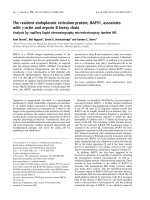
Figure 1
The unc-69 locus encodes a 108-amino-acid protein with a short coiled-coil domain. (a) Genetic and physical maps of chromosome III in the vicinity
of the unc-69 locus. unc-69 is close to and left of ced-9. Cosmids and subclones able to rescue the locomotion defect of unc-69(e587) mutants are
shown in bold. B: BamHI; H: HindIII; M: MluI; P: PstI; R: EcoRI; S: SacI. Introduction of a frameshift mutation at the BamHI site in the second exon
(denoted with an x) abrogated rescue by the minimal PstI-SacI rescuing fragment. Both splice variants, T07A5.6a and T07A5.6b, are contained within
this fragment. (b) The UNC-69 protein sequence. The boxed region is predicted to form a coiled-coil domain. Arrows indicate the positions of the
three known unc-69 mutations. Additional amino acids encoded by T07A5.6b are shown in italics (see Additional data file 1). (c) Northern-blot
analysis of unc-69 revealed a single major transcript of 0.65 kb (arrow).
lin-12 unc-69 ced-9 unc-49
0.5 map unit
unc-69 rescue
R01H10
C30B11
10 kb
2 kb
250 bp
M1 I (ju69)
T07A5.6a
T07A5.6b
AT G ATA
HMR
PstI EcoRI BamHI MIuI SacI
0.65 kb
Mixed stages
Embryos
RRHRBMSP
C15B3
C41B4
F11D2
F46H1
W08C6
tra-1
−
−
−
−
+
+
+
+
−
−
+
+
+
−
(a) (c)
(b)
in embryos (Figure 4a,b). In immature neurons, we
observed expression of UNC-69::GFP in the processes
and growth cones of developing neurites (arrowhead in
Figure 4c). In older larvae and adults, UNC-69::GFP was
expressed in neurons of the anterior, lateral, ventral and
retro-vesicular ganglia in the head, and in neurons of the
preanal, dorso-rectal and lumbar ganglia in the tail. The
fusion protein was also present in the ventral nerve cord
(VNC), in the dorsal nerve cord (DNC), in the dorsal and
ventral sublateral nerve cords, and in commissural axons
(Figure 4d-f). The reporter was expressed in the neurons
named CAN, HSN, ALM, PLM, AVM, PVM, BDU, and
SDQR, as evidenced by its localization to the cell bodies
of these neurons. Expression of unc-69 in these latter cells
Journal of Biology 2006, Volume 5, Article 9 Su and Tharin et al. 9.5
Journal of Biology 2006, 5:9
Figure 2
Schematic diagram of the ALM and AVM neurons in C. elegans. The
different parts of the neurons are given designated letters; see Table 1
for details. Anterior is to the left.
ALM-FL
ALM-E
AVM-E
AVM-B
ALM-B
ALM-NR
AVM-V
AVM-NR
ALML
AVM-FL
AVM
Table 1
Axon outgrowth and guidance defects in unc-69 mutants
ALM defect (%)
Genotype B NR E FL n
Wild type 0 0 0 12 113
unc-69(e602) 12 36 77 84 77
unc-69(e587) 15 45 85 89 80
unc-69(e602) (m+z-) 0 4.3 39 91 70
unc-69(e602)/nDf40 (m+z-) 0 7.2 20 72 69
unc-69(e587) (m+z-) 1.4 7.2 43 87 69
unc-69(e587)/nDf40 (m+z-) 0 8.3 20 82 60
unc-69(e602)* 19 48 62 95 113
unc-69(e602); opEx[P
mec-7
::unc-69]* 112 2 5 81
unc-69(e602); opEx[P
mec-7
::unc-69]* 59 6 10 79
unc-69(e602); opEx[P
mec-7
::unc-69]* 8 29 0 10 85
AVM defect (%)
Genotype B NR E FL V n
Wild type 0 0 1 8 0 106
unc-69(e602) 32 27 72 73 2.7 77
unc-69(e587) 64 70 86 87 0 80
unc-69(e602) (m+z-) 1.4 4.3 57 86 0 70
unc-69(e602)/nDf40 (m+z-) 0 0 56 80 0 69
unc-69(e587) (m+z-) 2.9 8.8 56 85 0 69
unc-69(e587)/nDf40 (m+z-) 0 3.6 76 95 0 60
unc-69(e602)* 46 67 93 100 ND 113
unc-69(e602); opEx[P
mec-7
::unc-69]* 04 4 4ND81
unc-69(e602); opEx[P
mec-7
::unc-69]* 812 1223ND79
unc-69(e602); opEx[P
mec-7
::unc-69]* 11 21 12 13 ND 85
Neurite outgrowth and guidance defects of mechanosensory touch neurons in unc-69 mutants. The morphology of neurites of ALM (top) and AVM
(bottom) neurons (as in the schematic in Figure 2) was scored in different unc-69 mutants, in unc-69/nDf40 heterozygotes, and in mosaic animals
carrying a functional unc-69 transgene under the control of the mec-7 promoter, which directs expression in the six touch neurons. All worms
scored had a P
mec-4
::gfp transgene zdIs5 in the background to allow visualization of the neurite morphology. One ALM neurite was scored per animal.
B, failure to form proper branch at the nerve ring; NR, failure of nerve ring branch to fully extend; E: failure to elongate past the branch point;
FL, failure to extend fully; V, ventral guidance defect. (m+z-): homozygous mutant animals derived from heterozygous mothers. *These strains also
carry a lin-15(n765) mutation in the background. All opEx transgenes also carry a wild-type copy of lin-15(+) as a coinjection marker. ND, not done.
n, number of worms scored.
was confirmed using an unc-69::LacZ::NLS fusion (data
not shown). Taken together, these results indicate that
unc-69 is expressed widely, perhaps ubiquitously, in the
C. elegans nervous system.
Expression of UNC-69::GFP was also observed in non-
neuronal cells. In larvae and adults, we occasionally
observed UNC-69::GFP expression in body-wall muscle
(data not shown). We also observed UNC-69::GFP in the
excretory canal, in the distal tip cells, in the spermatheca
and, less frequently, in hypoderm and gut (Figure 4e, and
data not shown). The expression in these non-neuronal cells
was variable, however, and might not reflect the endoge-
nous expression pattern of unc-69.
9.6 Journal of Biology 2006, Volume 5, Article 9 Su and Tharin et al. />Journal of Biology 2006, 5:9
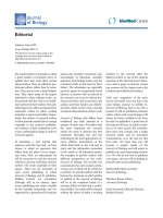
Figure 3
UNC-69 is homologous to mammalian SCOCO. (a) Sequence alignment of UNC-69/SCOCO proteins from S. cerevisiae, C. elegans, C. briggsae,
mosquito, Drosophila, Fugu, zebrafish, Xenopus, mouse and human. Residues identical in all ten sequences are shaded black; similar residues are
shaded gray. The underlined region is predicted in all cases to form a coiled-coil domain. The region boxed in green is acidic, and the region boxed in
red is serine/threonine-rich. The bracket indicates the carboxy-terminal basic region. Asterisks mark mutations in unc-69. (b) mRNA of the human
unc-69 homolog SCOCO is enriched in fetal brain and is also present in fetal kidney, liver and lung. (c) Expression of human SCOCO rescues the
locomotion defect of unc-69 mutant. Movement of the wild type (WT), mutants, and transgenic L4-stage hermaphrodites was scored as complete
sine waves per minute. For each genotype n = 10. Error bars represent the standard error of the mean.
1 MSAENISTGSPTGKQPSS
1 MSQKTEQDDIPLADDDDTVTIISGGKTPRAAQP
1 MSQKTEQDDIPLADDDDTVTIISGGKTPRAAQP
1 MSLKSQDD-IPLADDDLEVIINDDESSKYMCNGR
1 MSLLNNDDSIPNMDEDPQVVIPDDEPPATGRMPS
1 MVEREE-TPGMEAEVNEEDGTFINVSLADDPGQHISKLGRQQILQAVS
1 MNCEID
1 MDSDMD
1 MMNADMD
1 MMNADMD
19 EVNLGERE
34 LPKEEPPE
34 LPKEEPPE
34 SLDSIASSYTNGNSSPQQFLENESPDAD
35 GRSMDSLRSSFTNRSSTPDSSHNSLEAMEMAQD
48 NRGEPARHHELRPRRFARRRPPTFVSVRSIMERERDWTSVCLTGDVENQV
7 GDMENQV
7 ALDLENQI
8 AVDAENQV
8 AVDAENQV
27 AGTKNERMMRQTKLLKDTLDLLWNKTLEQQEVCEQLKQENDYLEDYIGNL
42
DPEEKARLITQVLELQNTLDDLSQRVESVKEESLKLRSENQVLGQYIQNL
42 DPEEKARMITQVLELQNTLDDLSQRVESVKEESLKLRSENQVLGQYIQNL
62 EQEEKARLIAQVLELQNTLDDLSQRVDSVKEENLKLRSENQVLGQYIENL
68 DREEKARLITQVLELQNTLDDLSQRVDSVKEENLKLRSENQVLGQYIENL
98 ELEEKTRLINQVLELQHTLEDLSARVDAVKEENLKLKSENQVLGQYIENL
14 EQEEKTRLINQVLELQHTLEDLSARVDAVKEENLKLKSENQVLGQYIENL
15 ELEEKTRLINQVLELQHTLEDLSARVDAVKEENLKLKSENQVLGQYIENL
16 ELEEKTRLINQVLELQHTLEDLSARVDAVKEENLKLKSENQVLGQYIENL
16 ELEEKTRLINQVLELQHTLEDLSARVDAVKEENLKLKSENQVLGQYIENL
___________________________________________
S. cerevisiae Slo1p 77 MRSSNVLEK
C. briggsae UNC-69 92 MASSSVFQSSQ PPRPKQ-
C. elegans UNC-69 92 MSSSSVFQSSQ PSRPKQ-
A. gambiae 112 MSASSVFQSTTPNNVQNKKK
D. melanogaster 118 MSASSVFQSTS PSAAKKK
F. rubripes 148 MSASSVFQAT DTKAKRK
D. rerio 64 MSASSVFQTT DTKSKRK
X. laevis 65 MSASSVFQTT DTKSKRK
M. musculus SCOCO 66 MSASSVFQTT DTKSKRK
H. sapiens SCOCO 66 MSASSVFQTT DTKSKRK
___
unc-69(e587); opEx318 [Punc-69
::scoco]
unc-69(e6
02); opEx317
[P
unc-69
::scoco]
unc-69(e602); opEx319 [P
unc-69::scoco]
unc-69(e602); opEx320 [P
unc-69::scoco]
unc-69(e587)
unc-69(e602)
WT
9.5
kb
7.5
Kidney
Liver
Lung
Brain
4.4
2.4
1.35
Actin
40
30
20
10
0
Body bends/minute
SCOCO
*
**
Similar
Identical
(a) (b)
(c)
S. cerevisiae Slo1p
C. briggsae UNC-69
C. elegans UNC-69
A. gambiae
D. melanogaster
F. rubripes
D. rerio
X. laevis
M. musculus SCOCO
H. sapiens SCOCO
S. cerevisiae Slo1p
C. briggsae UNC-69
C. elegans UNC-69
A. gambiae
D. melanogaster
F. rubripes
D. rerio
X. laevis
M. musculus SCOCO
H. sapiens SCOCO
S. cerevisiae Slo1p
C. briggsae UNC-69
C. elegans UNC-69
A. gambiae
D. melanogaster
F. rubripes
D. rerio
X. laevis
M. musculus SCOCO
H. sapiens SCOCO
___________________________________________
UNC-69 is required for axonal outgrowth and
guidance
The ventral coiler phenotype of unc-69 mutants suggests a
defect in nervous system development. Indeed, previous
studies had reported axonal guidance defects of the D-type
GABAergic motor neurons, mechanosensory neurons and
the HSN neurons in unc-69 mutants [23,24]. We confirmed
these observations and extended them to other cell types
(see Tables 1,2 and Figures 2, 5a-f). Incorrect targeting of
the DD and VD motor axons is likely to contribute to the
Unc phenotype of unc-69 mutants. In addition to outgrowth
and guidance defects, we also observed ectopic branching of
the DD/VD neurons and mechanosensory neurons in
unc-69 mutants (Figure 5d,f). In a few cases the axons had
unusual large swellings and occasionally meandered along
the lateral body wall.
FMRF-amide (Phe-Met-Arg-Phe-NH
2
) is a neuropeptide that
serves as a neuromodulator, and is co-released together with
other neurotransmitters. In examining other neuronal
classes in unc-69(e587) mutants, we observed premature ter-
mination of axons of the FMRF-amide-positive neurons
ALA, RID and AVKR, but not RMG (data not shown, and see
Table 2). FMRF-amide-positive neurons are so-called neuro-
peptidergic neurons and could be sensory, motor or
interneurons. We observed that 67% (20/30) of ALA axons
Journal of Biology 2006, Volume 5, Article 9 Su and Tharin et al. 9.7
Journal of Biology 2006, 5:9
Figure 4
UNC-69::GFP is expressed in neurons. Confocal micrographs of mosaic animals expressing a rescuing carboxy-terminal UNC-69::GFP fusion. A
1 m optical section is shown in (a); all other panels are projections of optical series. (a) Late gastrula (large arrowhead) and early comma-stage
embryo (arrow) with widespread expression of UNC-69::GFP. Embryos were still inside the mother. Small arrowheads indicate the maternal VNC;
v indicates the maternal vulva. (b) A two-fold-stage embryo with strong UNC-69::GFP expression in VNC neurons (between arrowheads).
(c) A three-fold embryo expressing UNC-69::GFP in a growth cone (arrowhead). The arrow indicates a neuronal cell body. (d) An L1-stage larva
expressing UNC-69::GFP in neurons and axons in the head (arrow), VNC (small arrowheads) and tail (large arrowhead). The asterisk indicates
reporter expression in labial sensory neuronal processes of an adjoining adult animal. (e) An L3 larva expressing UNC-69::GFP in the CAN neuron
(large arrow), excretory canal (small arrowheads) and in commissural axons (small arrow). (f) An L4 larva expressing UNC-69::GFP in the CAN
(large arrow), HSN (large arrowhead) and ALM (small arrowhead) neurons. Small arrows indicate commissures. All scale bars represent 10 m. In
all cases, anterior is to the left and dorsal is up.
(a) (b) (c)
(d) (e) (f)
terminated prematurely, and ALA axons sometimes
branched before termination. AVKR had frequent axonal
outgrowth and guidance defects: 85% (17/20) of AVKR
axons terminated prematurely or crossed from the left VNC
(VNCL) to the right VNC (VNCR). Taken together, these
observations support a role for unc-69 in ventral and dorsal
axonal guidance as well as in axonal elongation within
the fascicles.
UNC-69 is required for fasciculation
As unc-69 mutants have midline crossover defects (see Table
2), it is likely that axons running in the same fascicle lose
cell-cell adhesion and fail to stay together. We constructed a
series of electron micrograph (EM) cross-sections through
the major nerve cords (DNC, VNCL and VNCR) that run
antero-posteriorly in adult hermaphrodites. In wild-type
animals, the composition of axons in any of these nerve
cords is highly stereotyped, with four axons fasciculated to
run in VNCL and the other ventral axons running within
VNCR (Figure 5g) [25]. In unc-69(e587) and unc-69(e602)
mutants, many fascicles split into two or more groups and
in some cases defasciculated axons could be seen running
alone along the hypodermal ridge. Moreover, some axons of
both the DNC and VNCL appeared to be mislocalized and
can be seen on the wrong side of the hypodermal ridge
(Figure 5h and data not shown). Anti-tubulin and anti-
GABA staining confirmed the observed fasciculation defects
in unc-69(e587) mutants (data not shown).
UNC-69 acts cell autonomously to control neurite
outgrowth
To determine whether unc-69 expression is required in the
growing neurites or in the surrounding tissues, we created
unc-69 transgenic lines expressing unc-69(+) specifically in
the six touch neurons under the control of a mec-7 pro-
moter. We compared outgrowth and guidance defects of the
ALM and AVM neurons in three such lines with those of
unc-69(lf) mutants (see Table 1, Figure 2). In all three trans-
genic lines, the percentage of ALM neurites that failed to
extend to full length or send a branch into the nerve ring
9.8 Journal of Biology 2006, Volume 5, Article 9 Su and Tharin et al. />Journal of Biology 2006, 5:9
Table 2
Axon outgrowth and guidance defects of HSN, DD/VD, ALA
and AVK neurons
Axon guidance phenotype Defect in unc-69(e587) n
mutants (%)
HSN
Ventral outgrowth 16 70
Midline crossover (HSNL) 38 40
Failure to reach nerve ring 99 59
DD/VD
Dorsal outgrowth 33 45
ALA
Premature termination 67 30
AVKR
Premature termination or crossover 85 20
The morphology of HSN neurons was visualized using antibodies
against serotonin; that of DD/VD neurons using antibodies against
GABA; and that of ALA and AVKR neurons using antibodies against
FMRF-amide. See Materials and methods for details. n, number of
animals scored.
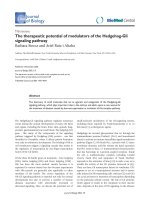
Figure 5 (see figure on the next page)
unc-69 is required for axonal outgrowth, guidance, branching and fasciculation in invertebrates and vertebrates. (a,b) Defect in the migration of the
HSN neuron in unc-69 mutant animals. (a) In wild-type animals, the HSN axons (HSNL and HSNR) migrate ventrally until they reach the VNC, which
they join and follow rostrally towards the head (arrow in (a)). (b) In unc-69 mutants, HSN axons occasionally fail to grow ventrally and instead project
laterally along the body wall (arrow in (b)). Animals were stained with anti-serotonin antibodies to visualize the HSN neurons. Arrowheads indicate
the vulva. Dotted lines mark the ventral margin of the body walls. (c,d) Commissures of D-type GABAergic neurons routinely reach the DNC in
wild-type animals (c), but often fail in unc-69(e587) animals (d) and prematurely bifurcate (arrow). D-type GABAergic neurons were visualized with the
unc-47::gfp transgene oxIs12. Asterisk in (d) marks a gap in the DNC. There are also often ectopic sprouts from the commissures (arrowheads in (d))
in unc-69(e587) mutants. (e,f) Images of the single ALM touch neuron in (e) wild-type and (f) unc-69(e602) animals. Many ectopic neurites branched
out from the soma and the axonal shaft of the ALM neuron in unc-69(e602) mutant (arrowheads). (g,h) Tracings of representative electron
micrographs of sections through the DNC and VNC. (g) In the wild type, the position and content of the three major fascicles are highly stereotyped
(black arrows). (h) In unc-69(e587) mutants, defasciculated axons can often be found migrating separately along the body wall (open arrows).
(i,j) Morphology of the bipolar AWC sensory neuron in (i) wild-type and (j) unc-69(e587) animals. Dendrites of AWC neurons in both animals reach
the nose (arrows). Axonal shape is normal in wild-type worms, but abnormal in unc-69(e587) mutants, with ectopic bulges occasionally extending from
the soma (arrowhead in (j)). (k,l) Expression pattern of SCOCO in stage 26 chick embryos. Sections were incubated with (k) antisense and (l) sense
RNA probes for chick SCOCO. SCOCO was highly expressed in neural tissue and was most prominent in DRGs and in motoneurons of both the lateral
motor column (LMC) and the medial motor column (MMC). Expression in the notochord (NC) and dermamyotome (DMT) was less pronounced.
(m,n) In ovo RNAi of chick SCOCO. Embryos injected and electroporated with double-stranded RNA corresponding to (m) a yfp-containing plasmid or
(n) chick SCOCO were immunostained with anti-neurofilament antibodies. (m) In control embryos, the epaxial nerves extending dorsally toward their
target, the epaxial muscle, were highly fasciculated. (n) RNAi of SCOCO led to defasciculation of epaxial nerve bundles and extensive branching
between muscle segments (arrows). In all panels dorsal is up. Scale bars represent: (a-j) 10 m, (k,l) 100 m and (m,n) 500 m.
Journal of Biology 2006, Volume 5, Article 9 Su and Tharin et al. 9.9
Journal of Biology 2006, 5:9
Figure 5 (see legend on the previous page)
(a)
(b)
(c)
(d)
(i) (j)
(e)
(f)
(g) (h)
(k) (l)
(m) (n)
dropped significantly. Similar observations were made for
AVM outgrowth and branching. Note that none of the trans-
genic lines completely rescued the ALM outgrowth and
branching defects. This could be due to loss or silencing of
the transgene carried on the extrachromosomal array or
could reflect a requirement for unc-69 in other neuronal
and/or non-neuronal cells. Nevertheless, we conclude that
UNC-69 promotes outgrowth and guidance largely, if not
completely, in a cell-autonomous manner.
UNC-69 is required for normal presynaptic
organization
The C. elegans synaptobrevin/vesicle-associated membrane
protein (VAMP) homolog SNB-1 is a vesicular soluble N-ethyl-
maleimide-sensitive factor attachment protein receptor
(v-SNARE) on synaptic vesicles (SVs). Tagged SNB-1 can be
used to follow SVs as they are transported to presynaptic
regions [26]. We isolated an allele of unc-69, ju69, in a visual
genetic screen for mislocalization of a SNB-1::GFP reporter in
D-type GABAergic motor neurons. In wild-type worms,
SNB-1::GFP expressed in the D neurons can be localized to dis-
crete puncta along the VNC and DNC, at sites of neuromuscu-
lar junctions (Figure 6a,c). In unc-69(ju69) mutant nerve cords,
SNB-1::GFP puncta were irregular in size and position, on
average larger than in wild type, and often completely missing
for extended stretches (Figure 6b,d,e). In addition, we occa-
sionally observed puncta that abnormally diffused from the
nerve cords into the commissures (Figure 6d). Despite the
abnormal shape and distribution of presynaptic regions, the
overall morphology of DD and VD neurons was grossly
normal (Figure 6f-i) and only occasionally (<10%; n = 50) did
one commissure fail to exit the VNC. We made similar obser-
vations in touch neurons using worms carrying the P
mec-4
::gfp
transgene zdIs5 (data not shown), a strain chosen for recon-
firming findings made on D-type GABAergic motor neurons.
Much more dramatic SNB-1::GFP distribution defects were
observed in the strong mutant unc-69(e587) (data not
shown). Because of the extensive pathfinding defects
observed in strong unc-69 mutants, however, which might
complicate interpretation of the SNB-1::GFP distribution
defect, we restricted our subsequent analysis to the
unc-69(ju69) background, in which axonal guidance is largely
normal. Indeed, although unc-69(ju69) mutant worms are
Unc, they move much better than strong unc-69 mutants.
Thus, the locomotion defect observed in unc-69(ju69)
mutants is probably a consequence of a defect in transport or
localization of axonal cargos rather than in axon guidance.
UNC-69 is not required for dendritic growth or for
targeting proteins into dendrites
To determine whether the outgrowth defects we observed in
unc-69 mutants are specific to axons, we examined the
morphology of the AWC class of sensory neurons using the
kyIs140 [P
str-2
::gfp] transcriptional reporter, which is normally
stochastically activated in either the right or left AWC neuron
[27]. The bilaterally symmetric AWC neurons have a distinct
bipolar structure, with a dendrite extending to the tip of the
nose and an axon extending into the nerve ring (Figure 5i).
In unc-69(e587) mutants, the axon of the AWC neuron often
stopped prematurely (Figure 5j), and str-2::gfp expression
was often silenced (see below). In contrast, the dendrite of
the AWC neuron had no outgrowth defect, as 100%
(136/136) of the AWC dendrites extended to their full
length. In unc-69(e587) mutants, 73% (99/136) of AWC
neurons had ectopic bulges or branches protruding from
either the cell body or the axon (similar to what we observed
in the mechanosensory neurons, Figure 5f,j). Ectopic
branches only rarely extended from dendrites, however (data
not shown). Dendritic morphology was also normal in the
ASI neurons (visualized by the str-3::gfp transgene), the
AWB, AWC, ASG, ASI, ASK, and ASJ neurons (visualized by
the tax-2
⌬
::gfp transgene) [28,29], and the sensory neurons
ASJ, ASH, ASI, ASK, ADL, and ADF (visualized by staining
with the lipophilic dye DiI; data not shown). Finally, an
odorant receptor was still properly localized to the cilia (see
below). From these observations, we conclude that UNC-69
is probably not required for either cilia formation or den-
dritic elongation within the amphid sensilla, a sensory organ
within the head of a worm.
In vesicle-trafficking mutants such as unc-16 and unc-116,
markers for synaptic vesicles are also mis-sorted into den-
drites [7]. We wondered whether unc-69 mutants also show
such a general sorting defect, or whether unc-69 might be
required more specifically for efficient trafficking within the
axons. At the L1 larval stage, the thirteen VD neurons are not
yet born, and the six DD neurons are the only D-type
GABAergic motor neurons present in the VNC. At this stage,
the DD neurons receive their synaptic inputs from the DNC
and output onto the ventral body-wall muscles. In wild-type
L1s, therefore, the SNB-1::GFP puncta can be seen only along
the VNC. In unc-69(ju69) mutants, the synaptic GFP was not
significantly mislocalized to the DNC (3.4%; n = 59; Figure
6k). In contrast, SNB-1::GFP puncta were frequently seen in
the DNC in unc-16(ju146) mutant L1s (90.6%; n = 32; Figure
6k). We also made similar observations in worms carrying a
snb-1::gfp transgene expressed in a pair of ASI sensory
neurons, in which SNB-1::GFP was not significantly mis-
localized to the ASI dendrites in unc-69(ju69) mutants
(C-W.S., Y.J. and M.O.H., unpublished data).
We next asked whether UNC-69 has any role in transporting
proteins within the dendrites. We used an odr-10::gfp trans-
gene that is expressed in the AWB neurons to answer this
question [30]. ODR-10 is an odorant receptor for diacetyl,
9.10 Journal of Biology 2006, Volume 5, Article 9 Su and Tharin et al. />Journal of Biology 2006, 5:9
and is actively transported in vesicles from the cell bodies to
the cilia at the end of the dendrites, where the GFP fusion is
deposited (Figure 6l). In dendritic targeting mutants, such
as unc-101 (which encodes the homolog of AP1 1 clathrin
adaptor protein), ODR-10::GFP is not targeted to the AWB
cilia [30] (Figure 6n); in contrast, in both unc-69(ju69) and
unc-69(e587) mutants, ODR-10::GFP was still properly tar-
geted (Figure 6m; data not shown). Taken together, our
results suggest that dendritic development and transport of
proteins into dendrites is not impaired in unc-69 mutants.
Thus, UNC-69 is possibly specifically required for axonal
transport and outgrowth.
UNC-69 interacts physically with UNC-76
To identify potential UNC-69 interactors, we screened three
C. elegans yeast two-hybrid libraries using full-length
UNC-69 as bait. From these screens, we isolated at least 34
independent clones of UNC-76, a 385-amino-acid protein
that was previously shown to be involved in axonal out-
growth and fasciculation in C. elegans [12-14]. The Drosophila
homolog of UNC-76 was identified as a KHC-binding
protein and shown to be a regulator of axonal transport [15].
A mammalian homolog of UNC-76, FEZ1, is a substrate for
PKC [16]. Worm, fly and mammalian UNC-76 proteins are
not only conserved in amino-acid sequence but also have
Journal of Biology 2006, Volume 5, Article 9 Su and Tharin et al. 9.11
Journal of Biology 2006, 5:9
Figure 6
unc-69 affects axonal but not dendritic trafficking. (a,c) SNB-1::GFP is seen as evenly spaced puncta along the (a) VNC and (c) DNC in wild-type
animals. (b,d,e) In unc-69(ju69) mutants, SNB-1::GFP puncta are on average bigger and often are absent from the VNC (arrowhead in (b)) and the
DNC (arrowheads in (d,e)). In addition, SNB-1::GFP sometimes diffuses into the commissure (arrow in (d)). (a,b,e) Lateral views; (c,d) dorsal views
of adult hermaphrodites. (f-i) As in (f,h) wild-type animals, neuronal morphology is grossly normal in (g,i) unc-69(ju69) mutants, and commissures still
routinely reach the DNC . D-type GABAergic neuron morphology is visualized with the P
unc-25
::gfp transgene juIs76. (f,g) Lateral views; (h,i) dorsal
views. (j) Distribution of SNB-1::GFP puncta in a stretch of axon labeled with P
unc-25
::DsRed monomer in the DNC in a unc-69(ju69) mutant
hermaphrodite. SNB-1::GFP puncta are unevenly distributed, even though the DNC anatomy is grossly normal. (k) SNB-1::GFP is not significantly
mislocalized into DD dendrites in unc-69(ju69) mutants. Animals carrying an snb-1::gfp transgene were scored at the L1 larval stage. Whereas 90% of
unc-16(ju146) L1 larvae (n = 32) show dorsal GFP, 0% of wild-type L1s (n = 47) and 3% unc-69(ju69) L1s (n = 59) show dorsal GFP. Error bars
represent the standard error of the mean. (l-n) The diacetyl odorant receptor ODR-10::GFP is targeted efficiently into AWB cilia both in
(l) wild-type worms and (m) in unc-69(ju69) mutants. (n) In contrast, ODR-10::GFP becomes diffused in the dendritic targeting mutant unc-101. The
arrow indicates the cilia; arrowheads indicate packets of ODR-10::GFP that shuttle in the dendrites. Anterior is to the left and dorsal is up.
100
80
60
%SNB-1::GFP in DNC
n = 47
n = 59
Wild type
unc-69(ju69)
unc-16(ju146)
n = 32
40
20
0
(a)
(b)
(f)
(g)
(h)
(i)
(l)
(k)
(m)
(n)
(j)
(c)
(d)
(e)
several conserved regions (Figure 7d) predicted to be capable
of forming coiled-coil domains [14,15]. UNC-76 localizes to
axons, and worms harboring mutations in unc-76 have a
severe Unc phenotype and coil ventrally, phenotypes very
similar to those observed in unc-69 mutants [14].
We used an in vitro glutathione S-transferase (GST) pull-down
assay to verify the physical interaction between UNC-69
and UNC-76. As shown in Figure 7a, in vitro translated full-
length UNC-76 (UNC-76FL) was pulled down efficiently
by GST-UNC-69 but only minimally by GST-CBP, a eukary-
otic transcription factor used as a negative control [31].
Conversely, in vitro translated adenoviral protein E1A effi-
ciently bound to its cognate partner GST-CBP but not to
GST-UNC-69. Therefore, the interaction between UNC-76
and UNC-69 is specific and most likely direct.
To narrow down the regions of interaction, we generated
truncated proteins lacking various parts of UNC-76 (Figure
7b,d) and tested for their interaction with GST-UNC-69. We
found that amino acids 281 to 299 of UNC-76 were neces-
sary to interact with UNC-69 in vitro. Interestingly, this 19-
amino-acid region overlaps with a region predicted to form
a coiled-coil structure (amino acids 265-292; purple region
9.12 Journal of Biology 2006, Volume 5, Article 9 Su and Tharin et al. />Journal of Biology 2006, 5:9
Figure 7
UNC-69 physically interacts with UNC-76, as shown by in vitro GST pull-down assays. (a) Full-length UNC-76 (UNC-76 FL) specifically binds to
full-length GST-UNC-69 but not GST-CBP. The E1A-CBP interaction was used as a positive control. (b) Serial deletions of UNC-76: a portion of
the carboxy-terminal region (deleted in UNC-76 ⌬␥ but contained within UNC-76 B3 and A3) is necessary for interaction with GST-UNC-69.
(c) Point mutation L287P or a small 19-amino-acid deletion (UNC-76 ⌬19), which deletes amino acids 281-299, totally abolishes the ability of
UNC-76 to bind GST-UNC-69. (d) Summary of the deletion analysis, as well as the results of rescuing experiments. Gray shading indicates
conserved regions. Note that UNC-76 ⌬19 not only loses its binding ability but also its rescuing activity for the unc-76(e911) mutants. The
19-amino-acid region (green) lies within a conserved region and overlaps with a region we predicted to form a coiled-coil domain (purple). A
previously described axonal targeting sequence [14] is in red. The positions of different unc-76 alleles are indicated.
kDa
UNC-76
E275A
A4
A3
B5
B4
FL
1 197 281 299 385
Interaction with
UNC-69 in vitro
Rescue of unc-76
(e911) in vivo
B3
B2
C3
C2
C1
∆βγ
∆γ
∆β
∆α
∆αγ
L281P
L287P
K291A
P2
12345
∆19
+
+
+
+
+
+
+
−
+
+
+
−
−
+/−
+
−
−
−
+
ND
−
+
ND
ND
ND
ND
+
ND
ND
ND
ND
ND
ND
−
ND
−
ND
ND
+
+
+/−
+/−
kDa
87
UNC-76 FLInput:
Input:
Input:
Input
UNC-76 FL
UNC-76 FL
UNC-76 ∆α
UNC-76 ∆19
UNC-76 FL(L287P)
UNC-76 ∆β
UNC-76
∆γ
UNC-76 B3
UNC-76 A3
Input
+ GST
Input
+ GST
+ GST
+ GST-UNC-69
+ GST-UNC-69
Input
+ GST
+ GST-UNC-69
+ GST-UNC-69
n2457
n2397
ev424
n2367
n2399
e911
n2398
+ GST-CBP
+ GST-CBP
E1A
62
kDa
49
38
28
49
38
28
17
14
6
40
31
Conserved regions
Axonal targeting sequence (aa 1-197)
Sequence predicted to form coiled coil (aa 265-292)
Minimal interacting peptide (aa 281-299)
(a) (d)
(b)
(c)
in Figure 7d) and lies within a region conserved from
worms to humans (gray-shaded region in Figure 7d).
UNC-76 may require interaction with UNC-69 to
function in vivo
To corroborate the in vitro interactions with the in vivo func-
tion of UNC-76, we expressed truncated UNC-76 proteins
tagged with yellow or cyan fluorescent protein (YFP or CFP)
in unc-76(e911) mutant worms (Figure 7d) and assayed for
rescue of the Unc phenotype. Both amino-terminally and
carboxy-terminally tagged full-length UNC-76::YFP or
CFP::UNC-76 fusion proteins were functional and rescued
unc-76(e911) mutants (Figure 7d). The CFP::UNC-76 ⌬␣
fusion protein (which lacked the amino terminus of
UNC-76) failed to rescue unc-76(e911) mutants, suggesting
that the amino-terminal region of UNC-76 is required for
its function in vivo. Bloom and Horvitz reported that amino
acids 1-197 of UNC-76 are sufficient to direct proteins into
the axons in C. elegans [14]. As the axonal targeting
sequence of UNC-76 includes the region deleted in UNC-76
⌬␣, we speculated that CFP::UNC-76 ⌬␣ fusion proteins
were not transported to axons. Indeed, the CFP signal was
weak and seemed to congregate more around the soma
(data not shown). In contrast, the CFP::UNC-76 ⌬␥ fusion
protein was both strongly expressed in soma and axons, but
failed to rescue unc-76(e911) mutants, consistent with the
hypothesis that binding to UNC-69 is critical for UNC-76 to
function in vivo.
If coiled-coil structures are important for the UNC-76-
UNC-69 interaction, any mutation that abolishes the coiled-
coil structure would possibly also abolish physical
interaction between the two proteins. To test this idea, we
mutagenized four conserved residues in UNC-76: Glu275,
Leu281, Leu287, and Lys291. Both UNC-76(E275A) and
UNC-76(K291A) mutant proteins still bound UNC-69 in
vitro (Figure 7d). Likewise, YFP fusions of these mutant pro-
teins rescued unc-76(e911) mutants. In contrast, both
UNC-76(L281P) and UNC-76(L287P) mutant proteins
failed to bind UNC-69 in vitro. Surprisingly, UNC-76(L287P)
was still able to rescue unc-76(e911) in vivo (Figure 7c,d; we
did not test UNC-76(L281P) for rescue). These data suggest
that a single -amino-acid substitution might not be potent
enough to destroy the coiled-coil structure when UNC-76
protein is folded in its native state. Finally, we created a
mutant protein carrying both L281P and L287P mutations
(P2), as well as an internal deletion mutant, ⌬19, which
deletes amino acids 281-299 of UNC-76. Both P2 and ⌬19
mutants largely failed to rescue unc-76(e911) in vivo (Figure
7d; occasionally, mutant hermaphrodites carrying the
unc-76 P2::yfp or the unc-76
⌬
19::yfp transgenes were slightly
rescued as young adults). In summary, amino acids 281-299
of UNC-76 probably contain or overlap with an
UNC-69-binding site, and UNC-76 may require interaction
with UNC-69 to function in vivo.
UNC-69 and UNC-76 act in the same pathway to
control axon extension
As both UNC-69 and UNC-76 are required for axon out-
growth and fasciculation, we asked whether they function in
the same genetic pathway to regulate axon extension. We
first tested whether overexpression of UNC-69 in unc-76(lf)
mutants could bypass the unc-76 mutant phenotype. We
overexpressed a functional unc-69::gfp transgene as an extra-
chromosomal array in unc-76(e911) mutants but did not
see any rescue in locomotion (three independent lines, data
not shown). Likewise, overexpression of a functional
unc-76::yfp transgene failed to rescue the locomotion defect
of unc-69(e587) mutants (data not shown).
We also performed a double-mutant analysis to further
address the question of whether unc-69 and unc-76 act in
the same pathway. In C. elegans, expression of the odorant
receptor gene str-2 is randomly turned on in either the left
or the right AWC sensory neuron (AWCL/R), but never in
both [27]. In wild-type worms, this ‘1 AWC
ON
’ phenotype is
determined by axonal contact and calcium signaling
between AWCL and AWCR. In axonal guidance mutants
such as unc-76, sax-3 and vab-3, the two AWC axons often
fail to meet, and P
str-2
::gfp expression is consequently
silenced in both AWCs, giving rise to a ‘2 AWC
OFF
’ pheno-
type [27]. We used this system to quantitatively score axon
extension defects in the nerve ring in different unc-69(lf)
and unc-76(lf) mutants as well as in unc-69(lf); unc-76(lf)
double mutants.
In both strong loss-of-function mutants, unc-69(e602) and
unc-69(e587), 30-34% of animals showed a 2 AWC
OFF
phenotype. In contrast, the hypomorphic allele unc-69(ju69)
resulted in only 1% of mutant worms (n = 190) having
P
str-2
::gfp expression silenced in both AWCs (Table 3). This
result was consistent with our previous observation that neu-
ronal morphology is largely normal in unc-69(ju69)
mutants. In agreement with previous studies [27], 47% of
unc-76(e911) mutants (n = 101) had the 2 AWC
OFF
pheno-
type; e911 was the strongest allele among all the nine alleles
that we tested. For the other unc-76 alleles, the 2 AWC
OFF
phenotype varied from 6% to 30%. Interestingly, the
strength of the AWC expression defect (which is an indica-
tion of axon extension defects) showed an inverse colinear
relationship with the position of each mutation in the open
reading frame: the most 5’ mutation, unc-76(n2457),
showed the least defect in axon extension, whereas alleles
located most carboxy-terminally showed greater defects than
alleles located close to the amino terminus (Table 3). Inter-
estingly, we did not observe enhancement of axon extension
Journal of Biology 2006, Volume 5, Article 9 Su and Tharin et al. 9.13
Journal of Biology 2006, 5:9
defects in unc-69; unc-76 double mutants: in all cases, the
defect in the double mutant was no stronger than in the
stronger of the single mutants (Table 3). In contrast, axon
extension defects were greatly enhanced in unc-76(e911);
sax-3(ky123), unc-76(e911); unc-6(n102) and unc-33(e204);
unc-76(e911), and slightly enhanced in unc-76(e911);
vab-3(e648) and unc-119(ed3); unc-76(e911) double mutants
(Table 3). Because unc-76 alleles failed to show any add-
itivity with the candidate null alleles unc-69(e587) and
unc-69(e602), we conclude that UNC-69 and UNC-76 prob-
ably act in the same pathway to control axon extension, at
least in the case of the AWC sensory neurons.
UNC-69 and UNC-76 regulate presynaptic
organization cooperatively
We showed above that UNC-69 is required for localization
of synaptic vesicles in axons. Does UNC-76 also have a role
in this process, and if so, does UNC-76 control presynaptic
organization together with UNC-69? Unfortunately, all
existing unc-76 alleles have severe axonal outgrowth defects,
making interpretations of defect in synaptic vesicle localiza-
tion difficult. To bypass this problem and to reveal possible
genetic interactions between unc-69 and unc-76, we looked
at the localization of the synaptobrevin SNB-1::GFP puncta
in unc-69(lf)/+; unc-76(lf)/+ double heterozygotes (Figure 8).
9.14 Journal of Biology 2006, Volume 5, Article 9 Su and Tharin et al. />Journal of Biology 2006, 5:9
Table 3
Quantitative analysis of axon extension defects in unc-69(lf), unc-76(lf) and other mutants
Genotype 2 AWC
OFF
(%) 1 AWC
ON
(%) 2 AWC
ON
(%) n
Wild type 1 99 0 442
unc-69
unc-69(ju69) 1 99 0 190
unc-69(e602) 34 66 0 119
unc-69(e587) 30 70 0 194
unc-76
unc-76(rh116) 11 89 0 83
unc-76(n2457) 6 94 0 102
unc-76(n2397) 892064
unc-76(ev424) 10 90 0 68
unc-76(n2367) 30 70 0 84
unc-76(n2399) 25 75 0 67
unc-76(e911) 47 53 0 101
unc-76(e911); lon-2(e678) 31 69 0 91
unc-76(n2398) 28 72 0 184
Double mutants
unc-69(e602); unc-76(n2457) 35 65 0 106
unc-69(e602); unc-76(e911) 48 52 0 118
unc-69(e587); unc-76(n2457) 33 67 0 108
unc-69(e587); unc-76(e911) 31 69 0 143
Other axonal guidance mutants
sax-3(ky123) 64 33 3 112
lon-2(e678) unc-6(n102) 38 62 0 65
vab-3(e648) 54 40 6 68
unc-33(e204) 7 73 20 135
unc-119(ed3) 12 48 39 99
Other double mutants
unc-76(e911); sax-3(ky123) 95 5 0 22
unc-76(e911); lon-2(e678) unc-6(n102) 73 27 0 152
unc-76(e911); vab-3(e648) 63 27 10 62
unc-33(e204); unc-76(e911) 80 18 1 291
unc-119(ed3); unc-76(e911) 65 32 3 167
All animals scored had kyIs140 (P
str-2
::gfp) in the background, which turns on its expression in only one of the two AWC neurons (1 AWC
ON
) in
wild-type animals. In axon guidance mutants, P
str-2
::gfp expression is silenced in both AWCs (2 AWC
OFF
) owing to failure of axonal contact. All unc-69
and unc-76 alleles except unc-76(rh116) are arranged in order according to their physical position (5’ to 3’) in the open reading frame. n, number of
animals scored.
In wild-type adult hermaphrodites, SNB-1::GFP can be seen
as evenly distributed puncta along the DNC [7] (Figure
8a,e). The distribution pattern of GFP puncta in DNC was
not significantly different in unc-69(e587)/+ heterozygotes
(Figure 8b) as compared with wild-type animals. However,
in both unc-69(e587)/+; unc-76(e911)/+ and unc-69(e587)/+;
unc-76(n2457)/+ double heterozygous hermaphrodites,
SNB-1::GFP puncta were occasionally more diffused, larger,
or completely absent within a stretch of DNC (Figure 8c,d,f);
the absence of SNB-1::GFP puncta may be due to either trans-
port or axon extension defects. In addition, unc-69(e587)/+;
unc-76(e911)/+ and unc-69(e587)/+; unc-76(n2457)/+ double
Journal of Biology 2006, Volume 5, Article 9 Su and Tharin et al. 9.15
Journal of Biology 2006, 5:9
Figure 8
UNC-69 and UNC-76 cooperate to regulate the size and position of synaptic vesicles. (a-d) Lateral view of adult hermaphrodites 52-54 h after
hatching, single section. (e,f) Lateral view of the DNC of adult hermaphrodites 52-54 h after hatching, flattened images of confocal z-stack. Anterior
is to the left and dorsal is up. (a,e) SNB-1::GFP is evenly distributed along the DNC in wild-type animals. (b) Removing one copy of unc-69 does not
affect SNB-1::GFP distribution. (c,d,f) SNB-1::GFP becomes diffused and the puncta becomes larger (arrows) in unc-69(e587)/+; unc-76(e911)/+ and
(unc-69(e587)/+; unc-76(n2457)/+ double heterozygotes. Occasionally, SNB-1::GFP is missing altogether from a stretch of the DNC (bracket in (d)).
The genotypes are as follows: (a,e) juIs1 [P
unc-25
::snb-1::gfp], (b) qC1/unc-69(e587); juIs1, (c) qC1/unc-69(e587); nT1[qIs51]/juIs1;
nT1[qIs51]/unc-76(e911), (d,f) qC1/unc-69(e587); nT1[qIs51]/juIs1; nT1[qIs51]/unc-76(n2457). Scale bars represent 10 m.
(a) WT (b) unc-69(e587)/+
(c) unc-69(e587)/+; unc76(e911)/+ (d) unc-69(e587)/+; unc76(n2457)/+
(e) WT (f) unc-69(e587)/+; unc76(n2457)/+
heterozygotes occasionally had a slight Unc phenotype in
locomotion, resembling weak synaptic transmission
mutants. The weak locomotion defect could be a direct or
indirect effect of the synaptic vesicle mislocalization defect.
In summary, the unc-69/+; unc-76/+ double heterozygotes
show phenotypes that are similar, albeit significantly weaker,
to those observed in unc-69(ju69) homozygotes. Haplo-
insufficient genetic interactions of this type, commonly
known as nonallelic (or unlinked) noncomplementation,
are often observed with proteins that form heterodimers or
function in a common protein complex (such as ␣- and
-tubulin; [32]). Several other explanations are also possible,
however (discussed in [33]). Thus, our observations are
compatible with, but do not definitively prove, the hypothe-
sis that UNC-69 and UNC-76 act in a common pathway
required for proper synaptic-vesicle localization.
UNC-69 and UNC-76 colocalize in punctate
structures in axons and cell bodies
To determine the subcellular localization of UNC-69 and
UNC-76, we coinjected P
unc-69
::cfp::unc-69 and
P
unc-76
::unc-76::yfp constructs at low concentration (5 ng/l)
into unc-76(e911) mutant hermaphrodites, and selected
rescued transgenic animals for examination. At low concen-
tration, both CFP::UNC-69 and UNC-76::YFP often
appeared as puncta along the DNC, in CAN neurons, as
well as in other neuronal processes that run along the sub-
dorsal and subventral tracts (Figure 9a-f). Less frequently,
these puncta could also be found in commissures that
connect the DNC to the VNC. The punctate pattern of
UNC-76 can also be observed when worms are stained
with anti-UNC-76 antisera [14], consistent with this being
the endogenous expression pattern of UNC-76. Both
CFP::UNC-69 and UNC-76::YFP puncta were of variable
size but were usually large and immobile, even in the com-
missures. Interestingly, CFP::UNC-69 and UNC-76::YFP
proteins also colocalized in round, perinuclear dots in the
soma (Figure 9j-l). These observations strengthen our belief
that UNC-69 and UNC-76 coexist in a protein complex.
The molecular nature of the observed UNC-69-UNC-76
puncta (multiprotein complexes or vesicles, perhaps)
remains to be determined.
UNC-116/kinesin heavy chain is required for proper
subcellular distribution of both UNC-69 and UNC-76
In Drosophila, Unc-76 associates and copurifies with KHC,
which is the major component of the conventional kinesin
motor Kinesin-1 required for axonal transport towards the
plus ends of microtubules [15]. A similar biochemical inter-
action between UNC-76 and the C. elegans KHC ortholog
UNC-116 [34] has not been reported so far. To determine
whether the UNC-69-UNC-76 complex is transported to
axons by UNC-116, or by another kinesin, the KIF1A
homolog UNC-104 [35], we compared the subcellular
localization of both CFP::UNC-69 and UNC-76::YFP in
wild-type and in different kinesin mutant backgrounds.
In unc-116(rh24) mutants, UNC-76::YFP puncta were occa-
sionally diffuse and sometimes failed to be accompanied by
CFP::UNC-69 puncta in a stretch of axon (Figure 9g-i). In
addition, both CFP::UNC-69 and UNC-76::YFP proteins
9.16 Journal of Biology 2006, Volume 5, Article 9 Su and Tharin et al. />Journal of Biology 2006, 5:9
Figure 9 (see figure on the following page)
UNC-69 and UNC-76 colocalize as puncta in neuronal processes. (a-o) Functional P
unc-69
::cfp::unc-69 and P
unc-76
::unc-76::yfp constructs were coinjected
at 5 ng/l each into unc-76(e911) mutants, and worms rescued for locomotion were selected. Note that the unc-76(e911) mutation was removed from
the background in (g-o). (d-o) are deconvoluted single-layer images. (a-c) Lateral view of an adult hermaphrodite from one line of transgenic animals
with a wild-type phenotype. Both CFP::UNC-69 and UNC-76::YFP form discrete, large puncta in the DNC, as well as in the commissure (arrow).
Vignette in (c) shows an enlarged image of colocalized puncta in the DNC from the rectangle. (d-f) Lateral view of an adult hermaphrodite from a
second line of transgenic animals with a wild-type phenotype. Note that CFP::UNC-69 and UNC-76::YFP are both cytoplasmic and punctate, and the
puncta are present in lateral and sublateral processes. (g-i) In unc-116(rh24) mutants, UNC-76::YFP puncta became diffuse in a stretch of axon in the
VNC, and failed to colocalize with CFP::UNC-69 (arrows in (h,i)). (j-l) CFP::UNC-69 and UNC-76::YFP colocalize in perinuclear structures in the soma
of a neuron in the tail ganglia. (m-o) In unc-116(rh24) mutants, both UNC-76::YFP and CFP::UNC-69 often appear as partially overlapping or non-
overlapping aggregates in the soma of (i) a preanal and (ii-iii) two tail ganglion neurons. (p-u) Expression pattern of (p-r) opIs124 (P
unc-69
::unc-69::gfp)
and (s-u) opIs130 (P
unc-76
::unc-76::yfp). Both transgenes were integrated into the genome to ensure stable gene expression. All pictures show the CAN
neuron soma (arrowhead) and its vicinity. (p,s) In wild-type worms, the CAN neuron extended its bipolar processes along the excretory canal, and the
CAN neurites were filled with UNC-69::GFP and UNC-76::YFP. Note that puncta cannot be seen in these integrants owing to overexpression of the
transgenes. (q) In unc-104(e1265) mutants, UNC-69::GFP accumulated near the CAN soma as well as in its neuronal processes (asterisks), giving it a
notched appearance. (t) UNC-76::YFP localization appeared to be grossly normal in unc-104(e1265) mutants. (r,u) In unc-116(rh24) mutants, both
UNC-69::GFP and UNC-76::YFP accumulated in CAN neurites (asterisk). UNC-69::GFP accumulation was prominent near the CAN soma and was
accompanied by ectopic branches. In contrast, UNC-76::YFP aggregated and was evenly distributed along the CAN processes. The scale bar
represents 20 m. (v,w) The CAN neuron visualized by the integrated transgene kyIs4 (P
ceh-23
::ceh-23::unc-76
1-197
::gfp). (v) In wild-type worms, GFP
appeared as string of dots, reminiscent of endogenous UNC-76 expression pattern. (w) In unc-116(rh24) mutants, GFP dots became larger and more
dispersed. (x,y) CAN neuron visualized by an extrachromosomal array opEx901 (P
unc-69
::gfp). Unlike the UNC-69::GFP fusion (r), GFP itself did not
accumulate significantly around CAN soma in unc-116(rh24) mutants (y), although ectopic branches were frequently observed. (p-y) are confocal
z-stack images. Anterior is to the left in (a-i) and (p-y); anterior is up and ventral is to the right in (j-o). All scale bars except in (p-u) represent 10 m.
Journal of Biology 2006, Volume 5, Article 9 Su and Tharin et al. 9.17
Journal of Biology 2006, 5:9
Figure 9 (see legend on the previous page)
often occupied distinct but partially overlapping perinuclear
territories in the soma in unc-116(rh24) mutants (Figure
9m-o). Whereas perinuclear CFP::UNC-69 dots increased in
size in unc-116(rh24) mutants, perinuclear UNC-76::YFP
either split into several smaller dots (as in Figure 9n(i)) or
formed an irregular reticular structure (as in Figure 9n(iii))
in unc-116(rh24) mutants. The unc-116(rh24) mutants carry
two missense mutations (I304M and E338K) at the end of
the motor domain of KHC (amino acids 1-358) [34]. Thus,
these mutations are likely to affect the processivity of KHC
and cargo transport along the microtubules.
We also generated functional integrated UNC-69::GFP and
UNC-76::YFP transgenes that were stably overexpressed in the
nervous system and studied their subcellular localization in
different kinesin mutant backgrounds. The CAN neurons are
a pair of bilaterally symmetric neurons that send processes
antero-posteriorly along the excretory canal (Figure 9p) [36].
In wild-type animals, UNC-69::GFP and UNC-76::YFP could
be observed both in the CAN soma and throughout the
processes (Figure 9p,s). In worms mutant for unc-104(e1265),
the C. elegans KIF1A homolog [35], subcellular distribution of
UNC-69::GFP and UNC-76::YFP was not significantly altered
(Figure 9q,t). In unc-116(rh24) mutants, overexpression
pattern of UNC-69::GFP and UNC-76::YFP were both signifi-
cantly different from wild-type animals. The CAN neuron
accumulated UNC-69::GFP in the vicinity of its cell body,
which was swollen and deformed. In addition, there were
ectopic branches near the cell body, and UNC-69::GFP also
accumulated in these processes (Figure 9r). Unlike
UNC-69::GFP, UNC-76::YFP appeared as giant dots along the
CAN processes in unc-116(rh24) mutant, as if UNC-76::YFP
was removed from the cytoplasm and concentrated in certain
subcellular compartments (Figure 9u). Moreover, a
CEH-23::UNC-76
1-197
::GFP fusion protein [37] also appeared
as large aggregates along CAN processes in unc-116(rh24)
mutants (Figure 9w).
In summary, our data show that the subcellular distribution
of both UNC-69 and UNC-76 is altered in unc-116(rh24)
mutants. It is striking that the nearly perfect co-localization
of UNC-69 and UNC-76 is disrupted in unc-116 mutants.
We are still at a loss to explain the molecular basis of this
unexpected finding. What is clear, however, is that axonal
transport of UNC-69 and UNC-76 is still occurring in
unc-116(rh24) mutants. Thus, other kinesin motors and/or
additional factors probably contribute to transport of
UNC-69 and UNC-76 along the axons.
UNC-69 does not interact with ARL-1, ARL-3, or
ARFRP
UNC-69 homologs in S. cerevisiae and mammals have been
reported to interact physically with members of the family
of ARF-like small GTPases. To investigate whether a similar
interaction occurs in C. elegans, we first used yeast two-
hybrid assays to study protein-protein interactions between
UNC-69 and three closely related but distinct ARF-like small
GTPases, ARL-1 (F54C9.10), ARL-3 (F19H8.3), and ARFRP
(Y54E10BR.2) [38]. Whereas UNC-69 readily interacted
with the carboxyl terminus of UNC-76 (UNC-76␥), it did
not interact with any of the three ARF-like proteins
(Figure 10a). As human SCOCO was isolated as an effector
for GTP-bound ARL1 [20], we also tested the ability of
UNC-69 to interact with GTPase-defective forms of ARL-1
and ARFRP. UNC-69 did not interact with either
ARL-1(Q70L) or ARFRP(Q79L) (Figure 10a). Deletion of
the amino-terminal myristoylation site [39] also had no
effect: UNC-69 did not interact with the amino-terminal
deletion ARL mutants, ARL-1⌬16 (with or without the
GTPase-defective mutation) or or ARL-3⌬17 (data not
shown). In contrast, we readily detected the previously
reported interaction between ARL-3 and UNC-119 [40], a
homolog of human retinal gene 4 (HRG4) [41-43] (Figure
10b). Thus, the failure to detect any interaction between
UNC-69 and the three ARF-like proteins might not have
been due to inappropriate protein folding or subcellular
compartmentalization in yeast.
UNC-69 and mannosidase II occupy partially
overlapping subcellular regions
To address directly the question of whether UNC-69 is a
Golgi-associating protein, we coexpressed CFP::UNC-69 and
a YFP-tagged fragment of the C. elegans Golgi protein man-
nosidase II F58H1.1 (mansII::YFP) [44]. Unlike the colocal-
ization pattern we observed previously for UNC-69 and
UNC-76, UNC-69 and mansII only occasionally colocalized
(Figure 11). Moreover, we clearly observed regions in which
both UNC-69 and mansII occupied non-overlapping sub-
cellular territories, even under overexpression conditions
(arrows in Figure 11c,f,i). This mutual exclusion could not
simply be explained by the squeezing out of UNC-69 from
the mansII-containing territories as a result of spatial con-
straint, as UNC-69 and mansII territories did sometimes
overlap (arrowheads in Figure 11c,i).
Taken together, our results suggest that any interaction
between UNC-69 and Golgi is at best transient, and that the
UNC-69 puncta probably represent a structure distinct from
the Golgi.
UNC-69/SCOCO is required for axon pathfinding
and fasciculation in chicken embryos
Although we failed to find any clear link between UNC-69
and Golgi-associated transport in C. elegans, two lines of evi-
dence do suggest that the molecular function of
UNC-69/SCOCO is conserved through evolution. First, the
9.18 Journal of Biology 2006, Volume 5, Article 9 Su and Tharin et al. />Journal of Biology 2006, 5:9
level of conservation between family members is extremely
high in all the metazoans analyzed (see Figure 3a). Second,
overexpression of human SCOCO is sufficient to rescue the
uncoordinated phenotype (and hence the axon guidance
defects) of unc-69 mutants, suggesting that human SCOCO
can substitute for UNC-69 (see Figure 3c). There are,
however, no reports so far on a possible role for
UNC-69/SCOCO in vertebrate development. To address
this issue, we studied the function of UNC-69/SCOCO in
nervous system development of chicken embryos.
Expression of the chick homolog of unc-69/SCOCO was
detected by in situ hybridization in the spinal cord of stage
22 embryos. Expression increased with time, peaking at
around stage 26 (Figure 5k). In chicken embryos, SCOCO
was expressed in motor neurons of both the lateral motor
column (LMC) and the medial motor column (MMC). In
addition to neural tissues, staining was also present in the
dermamyotome (Figure 5k). Blocking the function of
SCOCO with in ovo RNA interference (RNAi) [45] resulted
in aberrant pathfinding of the epaxial nerve fibers (Figure
5n). The epaxial nerve is formed by axons of motoneurons
of the MMC. These axons leave the spinal cord together
with the neurons of the LMC to form the ventral root.
Instead of growing into the developing limb, however,
they leave the ventral root by a sharp dorsal turn. In
control embryos, epaxial axons grew dorsally in a fascicu-
lated manner and started branching only after reaching the
territory of the prospective epaxial muscle (Figure 5m). In
contrast, axons of epaxial motoneurons lacking UNC-
69/SCOCO were strongly defasciculated and started to
extend along the longitudinal axis of the embryo before
reaching their dorsal destination (81% of unc-69/SCOCO
RNAi treated embryos (n = 26) showed defects, versus 10%
of control embryos (n = 20); Figure 5n and Additional data
file 1). Because our in ovo RNAi approach selectively knocks
down UNC-69/SCOCO expression in the spinal cord
neurons, we conclude that the chick homolog of UNC-
69/SCOCO is likely to function autonomously in epaxial
nerve cells to control axon pathfinding, consistent with our
observations in worms.
From the above analysis, we conclude that the function of
UNC-69/SCOCO in axon guidance and nervous-system
development is probably conserved through evolution. On
the basis of its high degree of sequence conservation and
its expression pattern, we predict that SCOCO is also
required for nervous system development in mammals,
including humans.
Discussion
UNC-69 is required for normal presynaptic
organization and axonal outgrowth
In this work we show that mutations that affect the small
108-amino-acid protein UNC-69 abrogated a spectrum of
processes, including synaptic-vesicle targeting, axonal out-
growth, pathfinding, and fasciculation. Although a weak
reduction-of-function allele of unc-69 results in a selective
Journal of Biology 2006, Volume 5, Article 9 Su and Tharin et al. 9.19
Journal of Biology 2006, 5:9
Figure 10
UNC-69 does not interact with ARL-1, ARL-3 or ARFRP. (a) Plasmids
containing LexA-unc-69 or LexA-human SCOCO were cotransformed into
yeast cells with vector alone or vectors containing GAD-unc-76
␥
,
GAD-arl-1, GAD-arl-3, or GAD-arfrp. Protein-protein interactions were
measured as -galactosidase activity by using ONPG liquid assays.
UNC-69 did not interaction with any of the three ARL proteins.
SCOCO did not interact with any of the three ARL proteins either
(data not shown). (b) Auxotrophic growth assays for interactions
between LexA-UNC-119 and GAD-ARL-3 or GAD-UNC-76␥. Cells
(3×10
4
) were plated onto +His or -His plates, and serial tenfold
dilutions of cells were then subsequently plated. Cells were grown at
30°C for 48 h before images were taken. Note that the -His plate did
not contain 3-amino triazol. The strength of interaction between
UNC-119 and ARL-3⌬17 was only a fifth of that between UNC-119
and ARL-3 FL, as assayed by -galactosidase activity (data not shown).
Vector + vector
SCOCO + vector
UNC-69 + vector
UNC-69 + UNC-76γ
SCOCO + UNC-76γ
UNC-69 + ARL-1(wt)
UNC-69 + ARL-1(Q70L)
UNC-69 + ARL-3(wt)
UNC-69 + ARFRP(wt)
UNC-69 + ARFRP(Q79L)
60
50
40
β-galactosidase units
30
20
10
0
Vector Vector
Bait Prey +His −His
UNC-119 Vector
UNC-119 ARL-3 FL
UNC-119 ARL-3 ∆17
UNC-119 UNC-76γ
(a)
(b)
defect in synaptic vesicle localization, strong unc-69
mutants have extensive axonal outgrowth, fasciculation and
guidance defects. Both of the strong unc-69(lf) alleles,
unc-69(e602) and unc-69(e587), truncate the coiled-coil
domain of UNC-69. In contrast, the hypomorphic allele
unc-69(ju69) results in a missense mutation of the start
codon and presumably interferes with translation initiation.
The lack of extensive axonal outgrowth defects in the
unc-69(ju69) mutants suggests that UNC-69 protein trans-
lation might not be totally abolished and enough UNC-69
protein is still being produced to meet the requirement for
growth-cone extension. In contrast, the process of proper
localization of synaptic vesicles appears to be more sensitive
to reduction in levels of UNC-69 protein. It is possible that
unc-69 mediates different cellular processes in parallel:
axonal outgrowth, fasciculation and guidance on one hand,
synaptic vesicle localization on the other.
UNC-69 is clearly different from most other molecules that
have been implicated in axon outgrowth and guidance,
which are either guidance cues, membrane receptors, or cell-
adhesion proteins [46,47]. It is tempting to place UNC-69
downstream of these molecules, acting possibly as an inte-
grator or transducer of extracellular guiding signals. Alterna-
tively, and perhaps more likely, the axonal-outgrowth and
fasciculation defects observed in strong unc-69(lf) mutants
could be secondary to a more general transport defect, as
loss of UNC-69 function might interfere not only with the
transport of synaptic cargos but also with the transport of
axonogenic vesicles to growth cones.
9.20 Journal of Biology 2006, Volume 5, Article 9 Su and Tharin et al. />Journal of Biology 2006, 5:9
Figure 11
UNC-69 does not colocalize with the Golgi marker mansII. P
unc-69
::cfp::unc-69 and P
unc-69
::mansII::yfp plasmids were coinjected at 5 ng/l each into
unc-69(e587) mutant hermaphrodites, and worms rescued for locomotion were selected for analysis. (a-c) Subcellular localization of CFP::UNC-69
and mansII::YFP in a stretch of axon in the DNC in an adult hermaphrodite animal. The scale bar represents 10 m. (d-i) Subcellular localization of
CFP::UNC-69 and mansII::YFP in the cell bodies of a tail neuron (d-f) and a neuron in the VNC (g-i) in adult hermaphrodite animals. UNC-69 and
mansII usually occupied distinct subcellular regions (arrows in (c,f,i)); only occasionally did the expression patterns of the two proteins overlap
(arrowheads in (c,i)). Anterior is to the left and dorsal is up in all pictures. All pictures are deconvoluted single focal plane images. (j) Mean
fluorescence intensity of CFP::UNC-69 and mansII::YFP along a 17 m distance inside the bracketed region in (c).
UNC-69 and UNC-76 interact in vivo
We identified UNC-76 as an UNC-69-interacting protein.
UNC-76 is required for axonal outgrowth and fasciculation
in worms and its homolog in Drosophila is an axonal trans-
port protein [14,15]. We identified a 19-amino-acid segment
(amino acids 281-299) in UNC-76 that is necessary for its
interaction with UNC-69, and possibly for its in vivo func-
tion. Our genetic experiments suggest that both UNC-69 and
UNC-76 act in the same pathway to regulate axon extension.
In addition, we found that UNC-76 and UNC-69 cooperate
to regulate the size and position of SNB-1::GFP puncta, a
marker of presynaptic regions. Finally, we showed that
UNC-69 and UNC-76 colocalize in neurons as puncta and
that their normal subcellular distribution requires
UNC-116/KHC. The physical interaction, subcellular co-
localization, and similar mutant phenotypes all suggest that
the UNC-69-UNC-76 protein complex acts as a functional
unit that promotes transport of vesicles along axons.
A possible role for UNC-69 and UNC-76 in axonal
transport?
Proper SNB-1::GFP localization in C. elegans requires many
proteins, including UNC-116/KHC, KLC-2, UNC-14,
UNC-51, as well as UNC-16, the worm homolog of
Drosophila Sunday Driver, which serves as a scaffold protein
receiving regulatory signals from the JNK pathway [7,9,48].
Both UNC-16 and UNC-14 are recruited to Kinesin-1
through their interaction with KLC-2 [9]. In UNC-116,
KLC-2, UNC-14, and UNC-16 mutants, SNB-1::GFP is mis-
localized from axons to dendrites. In contrast, SNB-1::GFP
puncta were largely excluded from dendrites in unc-69(ju69)
mutants, suggesting that UNC-69 is intrinsically different
from these proteins. UNC-69 may function in a distinct step
of axonal transport, and is possibly not involved in polarized
sorting. Moreover, UNC-69 is unlikely to control general
protein trafficking in neurons, as it was not required for den-
dritic transport of the transmembrane receptor ODR-10.
Kinesin-1 transports various cargos, including Golgi, endo-
plasmic reticulum, mitochondria and synaptic membrane
proteins, but not synaptic vesicles, in the nervous system
[34,49,50]. In contrast, UNC-104/KIF1A preferentially
transports synaptic vesicle precursors [35,51]. Thus, the
synaptic -vesicle marker mislocalization defects in various
Kinesin-1 mutants [7,9,52,53] are probably secondary to a
general defect of Kinesin-1-dependent cargo transport or
other intracellular trafficking events within the axons. As
subcellular localization of UNC-69 and UNC-76 is altered
in the unc-116(rh24) mutants, but not in the
unc-104(e1268) or unc-104(rh43) mutants (our unpub-
lished observations), our data imply that UNC-69 and
UNC-76 are possibly required for events other than trans-
porting synaptic vesicles.
Indeed, the observation that Drosophila UNC-76 is a KHC-
associating protein further suggests that the UNC-69-
UNC-76 protein complex might constitute an alternative
pathway for Kinesin-1-mediated transport. Association of
the UNC-69-UNC-76 protein complex with the tail of KHC
could either provide additional levels of regulation, or allow
intracellular trafficking of a different repertoire of cargos. So
far, however, we could not verify a direct physical interac-
tion between C. elegans UNC-76 and the tail region (amino
acids 696-815) of UNC-116 in vitro (C-W.S. and M.O.H.,
unpublished results). Further analysis will be required to
definitively address this issue.
Implication of UNC-69 in mediating post-Golgi
transport
UNC-69 is not predicted to have enzymatic activity and pos-
sibly functions only by interacting with other coiled-coil
domain-containing proteins. The budding yeast and mam-
malian homologs of UNC-69, Slo1p and SCOCO, have been
shown to interact, respectively, with Arl3p and ARL1 - two
related, Golgi-associated, GTP-binding ADP-ribosylation
factor (ARF)-like proteins. Mammalian ARL1 is involved in
post-Golgi transport [20]. Yeast Arl3p could target Arl1p to
the Golgi, where it then tethers vesicles derived from endo-
somes to the Golgi [21,54]. The fact that expression of
human SCOCO rescues unc-69 mutants suggests that a
similar interaction occurs in C. elegans. We failed to detect
any physical interaction between UNC-69 and either ARL-1
or ARFRP, however. Moreover, UNC-69 often appears to
occupy subcellular regions distinct from those of the Golgi
marker mansII. Thus, the exact step at which UNC-69 acts in
vesicular transport (if at all) is still obscure.
Model for UNC-69-UNC-76 protein complex in
vesicular trafficking
We propose that UNC-69 and UNC-76 participate in a
protein complex that is localized to certain subcellular com-
partments in the cytoplasm to control vesicle transport
between the Golgi and the plasma membrane. The whole
UNC-69-UNC-76 protein complex might be recruited and
targeted by Kinesin-1 to its final destination in axons. As
multiple ectopic branches were consistently observed in
both unc-69 and unc-76 mutants, the UNC-69-UNC-76
protein complex might also help restrict membrane addi-
tion to growth cones during neuronal development, thereby
preventing unwanted membrane extension elsewhere along
the axons. Similar biochemical properties of the UNC-69-
UNC-76 protein complex might be used to regulate synaptic
vesicle clustering or maintenance in the presynaptic regions
in the adult nervous system. UNC-69-UNC-76 puncta could
reflect certain post-Golgi compartments that shuttle
between their budding sites on the trans-Golgi and their
docking sites near the plasma membrane. As such, an
Journal of Biology 2006, Volume 5, Article 9 Su and Tharin et al. 9.21
Journal of Biology 2006, 5:9
UNC-69-UNC-76 protein complex might function at an
intermediate step before vesicle maturation. Indeed,
UNC-69-UNC-76 puncta were present along the commis-
sures in addition to axons. As commissures do not form
synapses [36], these puncta could define a sorting compart-
ment from which functional vesicles are formed.
Functional conservation of UNC-69 and SCOCO
through evolution
Several lines of evidence suggest that UNC-69/SCOCO has
an evolutionarily conserved role in nervous-system develop-
ment in different animal species. First, human SCOCO
rescues the Unc defect of C. elegans unc-69 mutants (see
Figure 3c). Second, human SCOCO interacts with worm
UNC-76␥ in our yeast two-hybrid assays (Figure 10a), sug-
gesting that the rescuing activity of human SCOCO is due at
least in part to its ability to associate with UNC-76 in
C. elegans. Third, human SCOCO and its chicken homolog
are highly expressed in developing CNS neurons (see
Figures 3b and 5k). Fourth, RNAi-mediated knockdown of
chicken UNC-69/SCOCO results in guidance and fascicu-
lation defects of the epaxial nerves (Figure 5m,n). It seems
plausible from these observations that SCOCO also has an
important role in promoting proper development (and pos-
sibly function) of the nervous system in mammals.
Conclusions
Our studies reveal an important role for the UNC-69-
UNC-76 protein complex in axonal outgrowth, fascicu-
lation and synapse formation. Our results suggest that
UNC-69 and UNC-76 act as a functional unit to regulate
one or multiple steps of vesicle dynamics in the C. elegans
nervous system. On the basis of our transgenic rescue and
RNAi experiments, we suggest that vertebrates also use the
UNC-69-UNC-76 complex in a similar fashion to control
synapse formation and axonal outgrowth. We expect
further studies to shed light on this hitherto less noticed
branch of axonal guidance.
Materials and methods
C. elegans strains and genetics
C. elegans strains were maintained as described [18]. All
strains were grown at 20°C, except dpy-20(e1282ts) and
lin-15(n765ts) mutants, which were grown at 15°C before
injection to improve viability and at 25°C following injec-
tion to enhance selection of transgenic F1 animals. Wild-
type worms were of the Bristol N2 strain.
Cloning of unc-69
All genetic mapping data were deposited into WormBase
[55].The unc-69 gene is tightly linked to RFLP nP55, which
is recognized by cosmid C15B3. The three overlapping
cosmids C15B3, C41B4 and F11D2, but not the flanking
cosmids C30B11 and F46H1, rescued the Unc phenotype of
unc-69(e587) mutant. Subsequent subclonings identified a
1.2-kb EcoRI-SacI rescuing genomic fragment, which con-
tained a single gene composed of three exons. A frameshift
mutation was introduced into the unc-69 open reading
frame of the rescuing EcoRI-SacI fragment by cutting and
filling the unique MluI restriction site, followed by re-
ligation of the blunt ends. The frameshifted construct failed
to rescue unc-69 mutant worms. To identify the molecular
lesion(s) present in unc-69 mutants, the unc-69 locus from
wild-type and unc-69 mutants was amplified using primers
flanking the gene (5’-GCTCCGCAGTACGTCTTCTAAGCCC-3’
and 5’-GCGAGAATGGAACAATCAATGGACG-3’) and
sequenced. In addition to the stop codon, e602 also con-
tains a silent (third base) G-to-A transition in Lys107.
Egg-laying assay
Assays of egg-laying behavior were performed either in M9
buffer [56] or on plates. For M9 assays, gravid hermaphro-
dites were individually transferred to microtiter wells con-
taining either M9 or a 5 mg/ml solution of serotonin (5-HT,
Sigma, St. Louis, USA) in M9 and the number of eggs laid
after 60 min was determined. For plate assays, five gravid
hermaphrodites were transferred onto fresh plates with or
without food, and the total number of eggs laid after 90
min was determined.
Immunocytochemistry and fluorescence microscopy
Indirect immunofluorescence staining for serotonin and
GABA were performed as previously described [57-59].
Anti-serotonin and anti-GABA antisera were generously pro-
vided by H. Steinbusch (Free University, Amsterdam, The
Netherlands) and used at 1%. Neuronal morphology was
observed on a Zeiss Axioplan microscope equipped for epi-
fluorescence, using the Zeiss filter set 488005 (excitation:
395-440 nm band-pass filter; emission: 470 nm long-pass
filter). For colocalization studies, animals were anesthetized
with 10 mM levamisole and mounted on 4% agarose pads
in M9. A Leica DMRA2 microscope equipped with a Hama-
matsu ORCA-ER CCD camera, a Leica Fluotar 40X oil objec-
tive, and appropriate filter sets was used to visualize YFP
and CFP. Images were taken and deconvoluted using the
Openlab software (Improvision, Coventry, UK), and ana-
lyzed using ImageJ. For confocal microscopy, a Zeiss 510 or
a Leica DMRE confocal laser-scanning microscope equipped
with TCS SP2 AOBS and PL APO objectives was used.
Electron microscopy
Adult hermaphrodites were fixed in 0.8% glutaraldehyde,
0.7% OsO
4
and 0.1 M cacodylate buffer for 1 h on ice. Sub-
sequently, samples were cut and postfixed in 2% OsO
4
and
9.22 Journal of Biology 2006, Volume 5, Article 9 Su and Tharin et al. />Journal of Biology 2006, 5:9
0.1 M cacodylate buffer, mounted into an agar block, dehy-
drated in series of alcohols, and embedded in a mixture of
epon-araldite. Thin sections (50 nm) were cut on an Ultra-
cut E and pictures were taken with a JEOL 1200X at 80 kV.
Dye filling
Adult worms were soaked in 10 g/ml DiI (Molecular
Probes, Eugene, USA) in M9, and incubated in the dark for
2 h at 20°C, followed by several washes in M9 buffer. Stain-
ing was analyzed using appropriate filters.
cDNA screening and northern blot
A rescuing 2.8 kb EcoRI genomic fragment was used to
probe a C. elegans lambdaZAP cDNA library (gift of
R. Barstead). From approximately 300,000 plaques we iso-
lated four cDNAs. The sequences present at the ends of the
inserts were determined for all four clones, and the DNA
sequence of the longest intact clone was determined (one
strand only). A random-primed,
32
P-labeled unc-69 cDNA
was used to probe a northern blot of wild-type poly(A)
+
RNA. The blot was visualized and band intensities quanti-
fied using a Fuji Film phosphor imager. Poly(A)
+
RNA was
isolated from purified embryos or mixed stages of wild-type
strain N2 as described by Sambrook et al. [60], except that
FastTrack columns (Invitrogen, San Diego, USA) were used
(according to the manufacturer’s protocols) instead of stan-
dard oligo(dT) columns. Likewise, a human fetal multiple
tissue northern blot was probed with a portion of the
SCOCO cDNA.
Molecular biology
All manipulations were done following standard protocols
[60]. A 1.5 kb unc-69 genomic fragment containing 700 bp
upstream and 300 bp downstream of the coding region was
engineered through site-directed mutagenesis to construct
various amino- and carboxy-terminal CFP or GFP fusions
(pgfpu69, pu69gfp and pSU083). The rescuing ability of the
constructs was tested for both amino- and carboxy-terminal
GFP fusions. A full-length unc-69 cDNA was cloned into
pGEX-4T1 to generate a prokaryotic expression unc-69-gst
construct. To generate a full-length unc-76 major splicing
form cDNA (pSU001), the EcoRI-XbaI fragment of p76-c4
[14] was replaced by the EcoRI-BamHI fragment of yk784h09
and the XbaI-BamHI fragment of p76-c7, using three-way
ligation. Progressive unc-76 deletions were made either by a
PCR-based method, or using ExoIII and S1 nucleases fol-
lowing Erase-a-Base (Promega, Madison, USA) and
ExoIII/S1 Deletion Kit (Fermentas, Hanover, USA) proto-
cols. To generate genomic unc-76 constructs, an AscI site was
engineered 5’ to the start ATG and was used to insert a 1 kb
KpnI-AscI unc-76 promoter region up to the start codon.
Subsequently a BglII-SphI genomic fragment encompassing
exons 1 to 3 was used to replace the corresponding region
of the cDNA. An AscI CFP cassette was inserted in-frame to
create the amino-terminal CFP fusion plasmid (pSU065).
Likewise, a NotI site was engineered immediately 5’ to the
stop codon to allow creation of the carboxy-terminal YFP
fusion plasmid (pSU026). A genomic fragment containing
the first 82 amino acids of mannosidase II was placed 3’ to
the unc-69 promoter and was carboxy-terminally fused with
YFP to create pSU137. Full-length and truncated cDNAs of
unc-69, scoco, unc-76, arl-1, arl-3, arfrp, and unc-119 were
amplified from mixed-stage N2 mRNA pool or existing
cDNA clones and subsequently subcloned into pRE192 and
pRH143 to create LexA-bait and GAD-prey plasmids.
pRE192 and pRH143 are derivatives of pBTM116 and
pGAD424, respectively, and are gifts of K. Basler (University
of Zurich).
Single-amino-acid changes were made following
QuikChange Site-Directed Mutagenesis Kit protocols (Strat-
agene, La Jolla, USA). Sequences for each of the mutations
are as follows: UNC-76(E275A): GAGǞGCG;
UNC-76(L281P): CTGǞCCA; UNC-76(L287P):
CTGǞCCG; UNC-76(K291A): AAAǞGCC; ARL-1(Q70L):
CAAǞCTA; ARFRP(Q79L): CAGǞCTG. Mutations were
confirmed by sequencing and swapped back into the origi-
nal plasmids before further subcloning. All plasmids and
construct sequences are available upon request.
Protein sequence analysis
Prediction of coiled-coil structures was carried out using the
COILS version 2.1 program [61]. The coiled-coil regions of
UNC-69, UNC-76 and their homologs were assigned on the
basis of a greater than 80% probability of forming coiled
coils according to this program. We used ClustalW or
T-Coffee for multiple sequence alignment and shaded the
ClustalW alignment using BOXSHADE.
Yeast two-hybrid
Random- and dT-primed yeast two-hybrid phage libraries
were generous gifts from R. Barstead (Oklahoma Medical
Research Foundation, Oklahoma City, USA). A further
random-primed library was provided by M. Vidal (Harvard
Medical School, Boston, USA). The unc-69 cDNA was sub-
cloned into pDBTrp and pDBLeu vectors (PROQUEST Two-
Hybrid System, GibcoBRL, Carlsbad, USA) and cotransformed
with the AD plasmids. Transformed yeast reporter strain
Mav203 (GibcoBRL) was patched onto plates lacking Leu
and Trp (LW plates), and then replica plated onto plates
lacking Leu, Trp and His with 75 mM 3-amino triazol
(LWH/3AT plates), onto plates lacking uracil (URA plates)
and onto filters for -galactosidase assays. Only clones that
activated all three (His, Ura, LacZ) reporter genes were kept
for further analysis. Plasmid DNAs were purified from posi-
tive clones and retested for interaction with the bait plasmid.
Journal of Biology 2006, Volume 5, Article 9 Su and Tharin et al. 9.23
Journal of Biology 2006, 5:9
To study the interactions between UNC-69 and various ARL
proteins, we used the yeast reporter strain L40, and followed
Clontech’s Yeast Protocols Handbook for o-nitrophenyl
-
D-galactopyranoside (ONPG) and auxotrophic assays.
In vitro translation and GST pull-down assay
In vitro transcription and translation of unc-76 was performed
following protocols of the TNT-coupled reticulocyte lysate
system (Promega) in the presence of
35
S-labeled methionine
(Amersham, Little Chalfont, UK). For pull-down experi-
ments, purified recombinant GST and UNC-69-GST proteins
were first immobilized on glutathione beads (Amersham).
Immobilized proteins (2-10 g) were incubated with 1-20 l
in vitro translated UNC-76 (depending on binding efficiency)
in 1x interaction buffer (20 mM HEPES pH 7.9, 5 mM MgCl
2
,
0.2% NP40, 0.2% BSA, 7.5% glycerol and protease inhibitor
cocktail (Complete Mini, Roche, Indianapolis, USA)) at 4°C
for 1.5-2 h. The beads were washed three times in washing
buffer (100 mM KCl, 20 mM HEPES pH 7.9, 5 mM MgCl
2
,
0.2% NP40 and protease inhibitor cocktail (Roche)), resus-
pended in 20 l 2x SDS sample buffer, boiled and analyzed
by SDS-PAGE and autoradiography.
Germline transformations and array integration
For high-copy overexpression, plasmids were injected into the
germline of adult hermaphrodites [62] at 50 ng/l together
with 150 ng/l rescuing dpy-20(+) or pL15-EK lin-15(+)
genomic fragments. To generate low-copy extrachromosomal
arrays, plasmids were individually or co-injected each at 5
ng/l together with 195 ng/l pL15-EK. Transgenic progeny
of injected animals were selected as non-Dpy or non-Muv
animals at 25°C. Stably transmitting extrachromosomal
arrays were integrated by ␥-irradiation at 120 kV for 4.2 min.
In ovo RNAi in chicken embryos
We obtained a cDNA clone of chick SCOCO, ChEST376p13,
from the HGMP Research Center [63]. Double-stranded RNA
for in ovo RNAi was transcribed in vitro as described [45].
Embryos were injected at stage 17-18 with 0.1 l dsRNA
(500 ng/l) or a plasmid encoding YFP in PBS followed by
electroporation. Stage 25-26 embryos [64] were stained and
examined under a dissecting microscope with epifluores-
cence. With the site of injection chosen, we knocked down
SCOCO only in MMC/LMC motor neurons, not in cells of
the dermamyotome and/or the epaxial mesenchyme.
Immunostaining of chicken embryos
For whole-mount immunostaining, staged embryos were
dissected and fixed as described [45]. Embryos were perme-
abilized with 1% Triton X-100 in PBS, incubated in 20 mM
lysine and 0.1 M sodium phosphate (pH 7.4), and in block-
ing solution (10% FCS in PBS) to reduce nonspecific stain-
ing. Mouse anti-neurofilament antibodies (RM0270, Zymed,
San Francisco, USA) were used at 1:1,500 and applied for
48 h at 4°C. Embryos were washed in PBS at 4°C, and incu-
bated in blocking solution before addition of secondary anti-
body (goat anti-mouse Cy3, 1:250; Jackson Laboratories,
West Grove, USA). Embryos were dehydrated by sequential
incubation in 25%, 50%, 75%, and 100% methanol. The
tissue was cleared by transferring the embryos to BBBA
(benzyl-benzoate:benzyl-alcohol = 2:1). The embryos were
gently agitated until translucent, and were analyzed by fluo-
rescent microscopy.
In situ hybridization of chicken embryos
Embryos were sacrificed at different developmental stages
(from stage 17 to 33) and fixed in 4% paraformaldehyde.
The tissue was cryoprotected in 25% sucrose, and 20-m-
thick transverse sections of the lumbosacral spinal cord
were used for the analysis of SCOCO expression as
described previously [65].
Additional data files
The following files are available: Supplementary results and
tables (Additional data file 1); a figure showing that the
overexpression of a full-length UNC-69(M1I)::GFP protein
rescues locomotion defects of the unc-69(e587) mutants
(Additional data file 2); a figure showing the extent of the
ok339 deletion (Additional data file 3); and a figure showing
that RNAi knockdown of chicken UNC-69/SCOCO results in
epaxial nerve pathfinding defects (Additional data file 4).
Acknowledgements
We thank A. Hajnal and J. M. Kinchen for critical reading of the manu-
script, B. Dickson for thoughtful comments, and W-C. Chou, R. Staedeli,
H-H. Chen, L. Martin, P. Gisler, E. Horvath and G. Stergiou for other
help. We thank E. Hartwieg for assistance with the EM sections;
D. Baillie for the C. briggsae genomic library; S. Clark for the mec-4::gfp
plasmid; R. Barstead, M. Vidal and K. Basler for Y2H libraries and plas-
mids; H. Steinbusch for antisera; R. Eckner for the E1A and CBP plas-
mids; Y. Kohara for cDNAs; and C. Bargmann for the kyIs4 strain. Some
strains were contributed by the Caenorhabditis Genetics Center (CGC),
which is funded by the National Institutes of Health (NIH) Center for
Research Resources. We would also like to thank G. Moulder at the
C. elegans Gene Knockout Consortium for providing us with the
unc-69(ok339) deletion strain. This work was funded by grants from the
Rita Allen Foundation, March of Dimes, Ernst Hadorn Foundation, and
Swiss National Science Foundation to M.O.H. The work at MIT was
supported by NIH Grant GM24663 to H.R.H. H.R.H. and Y.J. are inves-
tigators of the Howard Hughes Medical Institute. C.R. is supported by
the research fund of the University of Zurich. C-W.S. is supported by
the Ernst Hadorn Foundation and a Zentrum für Neurowissenschaften
Zürich (ZNZ) Ph.D. fellowship.
References
1. Martinez-Arca S, Coco S, Mainguy G, Schenk U, Alberts P, Bouille P,
Mezzina M, Prochiantz A, Matteoli M, Louvard D and Gallie T:
A common exocytotic mechanism mediates axonal and
dendritic outgrowth. J Neurosci 2001, 21:3830-3838.
9.24 Journal of Biology 2006, Volume 5, Article 9 Su and Tharin et al. />Journal of Biology 2006, 5:9
2. Washbourne P, Bennett JE, McAllister AK: Rapid recruitment
of NMDA receptor transport packets to nascent synapses.
Nat Neurosci 2002, 5:751-759.
3. Ahmari SE, Buchanan J, Smith SJ: Assembly of presynaptic active
zones from cytoplasmic transport packets. Nat Neurosci 2000,
3:445-451.
4. Burack MA, Silverman MA, Banker G: The role of selective trans-
port in neuronal protein sorting. Neuron 2000, 26:465-472.
5. Horton AC, Ehlers MD: Neuronal polarity and trafficking.
Neuron 2003, 40:277-295.
6. Vale RD: The molecular motor toolbox for intracellular
transport. Cell 2003, 112:467-480.
7. Byrd DT, Kawasaki M, Walcoff M, Hisamoto N, Matsumoto K, Jin Y:
UNC-16, a JNK-signaling scaffold protein, regulates
vesicle transport in C. elegans. Neuron 2001, 32:787-800.
8. Koushika SP, Schaefer AM, Vincent R, Willis JH, Bowerman B,
Nonet ML: Mutations in Caenorhabditis elegans cytoplasmic
dynein components reveal specificity of neuronal retro-
grade cargo. J Neurosci 2004, 24:3907-3916.
9. Sakamoto R, Byrd DT, Brown HM, Hisamoto N, Matsumoto K, Jin Y:
The Caenorhabditis elegans UNC-14 RUN domain protein
binds to the Kinesin-1/UNC-16 complex and regulates
synaptic vesicle localization. Mol Biol Cell 2005, 16:483-496.
10. Lai T, Garriga G: The conserved kinase UNC-51 acts with
VAB-8 and UNC-14 to regulate axon outgrowth in
C. elegans. Development 2004, 131:5991-6000.
11. Ogura K, Shirakawa M, Barnes TM, Hekimi S, Ohshima Y: The
UNC-14 protein required for axonal elongation and guid-
ance in Caenorhabditis elegans interacts with the serine/thre-
onine kinase UNC- 51. Genes Dev 1997, 11:1801-1811.
12. Hedgecock EM, Culotti JG, Thomson JN, Perkins LA: Axonal
guidance mutants of Caenorhabditis elegans identified by
filling sensory neurons with fluorescein dyes. Dev Biol 1985,
111:158-170.
13. McIntire SL, Garriga G, White J, Jacobson D, Horvitz HR: Genes
necessary for directed axonal elongation or fasciculation
in C. elegans. Neuron 1992, 8:307-322.
14. Bloom L, Horvitz HR: The Caenorhabditis elegans gene unc-76
and its human homologs define a new gene family
involved in axonal outgrowth and fasciculation. Proc Natl
Acad Sci USA 1997, 94:3414-3419.
15. Gindhart JG, Chen J, Faulkner M, Gandhi R, Doerner K, Wisniewski T,
Nandlestadt A: The Kinesin-associated protein UNC-76 is
required for axonal transport in the Drosophila nervous
system. Mol Biol Cell 2003, 14:3356-3365.
16. Kuroda S, Nakagawa N, Tokunaga C, Tatematsu K, Tanizawa K:
Mammalian homologue of the Caenorhabditis elegans
UNC-76 protein involved in axonal outgrowth is a protein
kinase C zeta-interacting protein. J Cell Biol 1999, 144:403-411.
17. Okumura F, Hatakeyama S, Matsumoto M, Kamura T, Nakayama
KI: Functional regulation of FEZ1 by the U-box-type ubiq-
uitin ligase E4B contributes to neuritogenesis. J Biol Chem
2004, 279:53533-53543.
18. Brenner S: The genetics of Caenorhabditis elegans. Genetics
1974, 77:71-94.
19. Hengartner MO, Horvitz HR: C. elegans cell survival gene
ced-9 encodes a functional homolog of the mammalian
proto-oncogene bcl-2. Cell 1994, 76:665-676.
20. Van Valkenburgh H, Shern JF, Sharer JD, Zhu X, Kahn RA: ADP-
ribosylation factors (ARFs) and ARF-like 1 (ARL1) have
both specific and shared effectors: characterizing ARL1-
binding proteins. J Biol Chem 2001, 276:22826-22837.
21. Panic B, Whyte JR, Munro S: The ARF-like GTPases Arl1p
and Arl3p act in a pathway that interacts with vesicle-
tethering factors at the Golgi apparatus. Curr Biol 2003,
13:405-410.
22. Huang CF, Buu LM, Yu WL, Lee FJ: Characterization of a novel
ADP-ribosylation factor-like protein (yARL3) in Saccha-
romyces cerevisiae. J Biol Chem 1999, 274:3819-3827.
23. Siddiqui SS: Mutations affecting axonal growth and guidance
of motor neurons and mechanosensory neurons in the
nematode Caenorhabditis elegans. Neurosci Res Suppl 1990,
13:S171-S190.
24. Wightman B, Baran R, Garriga G: Genes that guide growth
cones along the C. elegans ventral nerve cord. Development
1997, 124:2571-2580.
25. White JG, Southgate E, Thomson JN, Brenner S: The structure
of the ventral nerve cord of Caenorhabditis elegans. Philos
Trans R Soc Lond B Biol Sci 1976, 275:327-348.
26. Nonet ML: Visualization of synaptic specializations in live
C. elegans with synaptic vesicle protein-GFP fusions. J Neu-
rosci Methods 1999, 89:33-40.
27. Troemel ER, Sagasti A, Bargmann CI: Lateral signaling medi-
ated by axon contact and calcium entry regulates asym-
metric odorant receptor expression in C. elegans. Cell 1999,
99:387-398.
28. Peckol EL, Zallen JA, Yarrow JC, Bargmann CI: Sensory activity
affects sensory axon development in C. elegans. Development
1999, 126:1891-1902.
29. Coburn CM, Bargmann CI: A putative cyclic nucleotide-gated
channel is required for sensory development and function
in C. elegans. Neuron 1996, 17:695-706.
30. Dwyer ND, Adler CE, Crump JG, L’Etoile ND, Bargmann CI:
Polarized dendritic transport and the AP-1 mu1 clathrin
adaptor UNC-101 localize odorant receptors to olfactory
cilia. Neuron 2001, 31:277-287.
31. Eckner R, Yao TP, Oldread E, Livingston DM: Interaction and
functional collaboration of p300/CBP and bHLH proteins
in muscle and B-cell differentiation. Genes Dev 1996,
10:2478-2490.
32. Stearns T, Botstein D: Unlinked noncomplementation: iso-
lation of new conditional-lethal mutations in each of the
tubulin genes of Saccharomyces cerevisiae. Genetics 1988,
119:249-260.
33. Belanger KD, Kenna MA, Wei S, Davis LI: Genetic and physical
interactions between Srp1p and nuclear pore complex
proteins Nup1p and Nup2p. J Cell Biol 1994, 126:619-630.
34. Patel N, Thierry-Mieg D, Mancillas JR: Cloning by insertional
mutagenesis of a cDNA encoding Caenorhabditis elegans
kinesin heavy chain. Proc Natl Acad Sci USA 1993, 90:9181-9185.
35. Hall DH, Hedgecock EM: Kinesin-related gene unc-104 is
required for axonal transport of synaptic vesicles in
C. elegans. Cell 1991, 65:837-847.
36. White JG, Southgate E, Thomson JN, Brenner S: The structure
of the nervous system of the nematode Caenorhabditis
elegans. Philos Trans R Soc Lond B Biol Sci 1986, 314:1-340.
37. Zallen JA, Kirch SA, Bargmann CI: Genes required for axon
pathfinding and extension in the C. elegans nerve ring.
Development 1999, 126:3679-3692.
38. Li Y, Kelly WG, Logsdon JM Jr, Schurko AM, Harfe BD,
Hill-Harfe KL, Kahn RA: Functional genomic analysis of the
ADP-ribosylation factor family of GTPases: phylogeny
among diverse eukaryotes and function in C. elegans.
FASEB J 2004, 18:1834-1850.
39. Pasqualato S, Renault L, Cherfils J: Arf, Arl, Arp and Sar pro-
teins: a family of GTP-binding proteins with a structural
device for ‘front-back’ communication. EMBO Rep 2002,
3:1035-1041.
40. Li S, Armstrong CM, Bertin N, Ge H, Milstein S, Boxem M,
Vidalain PO, Han JD, Chesneau A, Hao T, et al.: A map of the
interactome network of the metazoan C. elegans. Science
2004, 303:540-543.
41. Maduro M, Pilgrim D: Identification and cloning of unc-119, a
gene expressed in the Caenorhabditis elegans nervous
system. Genetics 1995, 141:977-988.
42. Higashide T, Inana G: Characterization of the gene for HRG4
(UNC119), a novel photoreceptor synaptic protein
homologous to Unc-119. Genomics 1999, 57:446-450.
43. Swanson DA, Chang JT, Campochiaro PA, Zack DJ, Valle D:
Mammalian orthologs of C. elegans unc-119 highly
expressed in photoreceptors. Invest Ophthalmol Vis Sci 1998,
39:2085-2094.
44. Rolls MM, Hall DH, Victor M, Stelzer EH, Rapoport TA: Target-
ing of rough endoplasmic reticulum membrane proteins
and ribosomes in invertebrate neurons. Mol Biol Cell 2002,
13:1778-1791.
Journal of Biology 2006, Volume 5, Article 9 Su and Tharin et al. 9.25
Journal of Biology 2006, 5:9