Báo cáo sinh học: "Systemic 5-fluorouracil treatment causes a syndrome of delayed myelin destruction in the central nervous system" doc
Bạn đang xem bản rút gọn của tài liệu. Xem và tải ngay bản đầy đủ của tài liệu tại đây (1.36 MB, 22 trang )
Research article
SSyysstteemmiicc 55 fflluuoorroouurraacciill ttrreeaattmmeenntt ccaauusseess aa ssyynnddrroommee ooff ddeellaayyeedd mmyyeelliinn
ddeessttrruuccttiioonn iinn tthhee cceennttrraall nneer
rvvoouuss ssyysstteemm
Ruolan Han*, Yin M Yang*, Joerg Dietrich
†
, Anne Luebke
‡
,
Margot Mayer-Pröschel* and Mark Noble*
Addresses: *Department of Biomedical Genetics and University of Rochester Stem Cell and Regenerative Medicine Institute, University of
Rochester Medical Center, Elmwood Avenue, Rochester, NY 14642, USA.
†
Department of Neurology, Massachusetts General Hospital,
Harvard Medical School, Fruit Street, Wang 835, Boston, MA 02114, USA.
‡
Department of Neurobiology and Anatomy, University of
Rochester Medical Center, Elmwood Avenue, Rochester, NY 14642, USA.
Correspondence: Mark Noble. Email:
AAbbssttrraacctt
BBaacckkggrroouunndd::
Cancer treatment with a variety of chemotherapeutic agents often is associated
with delayed adverse neurological consequences. Despite their clinical importance, almost
nothing is known about the basis for such effects. It is not even known whether the occurrence
of delayed adverse effects requires exposure to multiple chemotherapeutic agents, the presence
of both chemotherapeutic agents and the body’s own response to cancer, prolonged damage to
the blood-brain barrier, inflammation or other such changes. Nor are there any animal models
that could enable the study of this important problem.
RReessuullttss::
We found that clinically relevant concentrations of 5-fluorouracil (5-FU; a widely used
chemotherapeutic agent) were toxic for both central nervous system (CNS) progenitor cells and
non-dividing oligodendrocytes
in vitro
and
in vivo
. Short-term systemic administration of 5-FU
caused both acute CNS damage and a syndrome of progressively worsening delayed damage to
myelinated tracts of the CNS associated with altered transcriptional regulation in oligodendrocytes
and extensive myelin pathology. Functional analysis also provided the first demonstration of
delayed effects of chemotherapy on the latency of impulse conduction in the auditory system,
offering the possibility of non-invasive analysis of myelin damage associated with cancer treatment.
CCoonncclluussiioonnss::
Our studies demonstrate that systemic treatment with a single chemo-
therapeutic agent, 5-FU, is sufficient to cause a syndrome of delayed CNS damage and provide
the first animal model of delayed damage to white-matter tracts of individuals treated with
systemic chemotherapy. Unlike that caused by local irradiation, the degeneration caused by 5-FU
treatment did not correlate with either chronic inflammation or extensive vascular damage
and appears to represent a new class of delayed degenerative damage in the CNS.
BioMed Central
Journal of Biology
2008,
77::
12
Open Access
Published: 22 April 2008
Journal of Biology
2008,
77::
12 (doi:10.1186/jbiol69)
The electronic version of this article is the complete one and can be
found online at />Received: 19 June 2007
Revised: 3 January 2008
Accepted: 19 February 2008
© 2008 Han
et al.
; licensee BioMed Central Ltd.
This is an Open Access article distributed under the terms of the Creative Commons Attribution License ( />which permits unrestricted use, distribution, and reproduction in any medium, provided the original work is properly cited.
BBaacckkggrroouunndd
Most treatments used to kill cancer cells also kill a diverse
range of normal cell types, leading to a broad range of
adverse side effects in multiple organ systems. In the
hematopoietic system, the tissue in which such adverse
effects have been most extensively studied, their detailed
analysis has led to the discoveries that bone marrow
transplants and cytokine therapies can improve the out-
come of many forms of cancer treatment. In contrast, there
has been no comparable level of analysis for most other
organ systems compromised by cancer treatments.
One of the tissues for which adverse side effects of cancer
treatment are clinically important is the central nervous
system (CNS). Although it has long been appreciated that
targeted irradiation of the CNS may be associated with
neurological damage, it has become increasingly clear that
systemic chemotherapy for non-CNS cancers also can have a
wide range of undesirable effects. This has been perhaps
most extensively studied in the context of breast cancer (for
examples, see [1-13]). For example, it has been reported
that 18% of all breast cancer patients receiving standard-
dose chemotherapy show cognitive defects after treatment
[9], with such problems reported in over 30% of patients
examined two years after treatment with high-dose
chemotherapy [10]; this is a greater than eightfold increase
over the frequency of such changes in control patients.
Adverse neurological sequelae include such complications
as leukoencephalopathy, seizures and cerebral infarctions,
as well as cognitive impairment [14-18]. Adverse neuro-
logical effects have been observed with almost all categories
of chemotherapeutic agents [19-22], including antimetabo-
lites (such as cytosine arabinoside (Ara-C) [23], 5-fluorouracil
(5-FU) [24,25], methotrexate [26-28], DNA cross-linking
agents (such as BCNU [29] and cisplatin [30]) and even
anti-hormonal agents [31-37]. Given the large number of
individuals treated for cancer, these adverse neurological
changes easily may affect as many people as some of the
more extensively studied neurological syndromes.
One of the most puzzling aspects of chemotherapy-induced
damage to the CNS is the occurrence of toxicity reactions
with a delayed onset. Although this has been particularly
well documented in children exposed to both chemo-
therapy and cranial irradiation [15,38-47], delayed toxicity
reactions also occur in individuals treated only with
systemic chemotherapy. For example, white matter changes
induced by high-dose chemotherapy for breast cancer, and
detected in up to 70% of treated individuals, usually arise
only several months after treatment is completed [48,49].
One widely used chemotherapeutic agent associated with
both acute and delayed CNS toxicities is 5-FU. Acute CNS
toxicities associated with systemically administered 5-FU
(most frequently in combination with other chemothera-
peutic agents) include a pancerebellar syndrome and sub-
acute encephalopathy with severe cognitive dysfunction,
such as confusion, disorientation, headache, lethargy and
seizures. With high-dose treatment, as many as 40% of
patients show severe neurological impairments that may
progress to coma [50-52]. In addition, a delayed cerebral
demyelinating syndrome reminiscent of multifocal leuko-
encephalopathy has been increasingly identified following
treatment with drug regimens that include 5-FU, with
diagnostic findings obtained by both magnetic resonance
imaging (MRI) and analysis of tissue pathology [24,53-78].
Despite the existence of multiple clinical studies describing
delayed CNS damage associated with systemic exposure to
chemotherapy, almost nothing is known about the basis for
these effects. For example, because of the multi-drug
regimens most frequently used in cancer treatment, it is not
even known whether delayed toxicities require exposure to
multiple drugs. Nor is it known whether such delayed
changes can be caused solely by exposure to chemotherapy
or if they represent a combination of the response to
chemotherapy and, for example, physiological changes
caused by the body’s reaction to the presence of a tumor. In
addition, the roles of ongoing inflammation or damage to
the vasculature in inducing such delayed CNS damage are
wholly unknown. Moreover, the absence of animal models
for the study of delayed damage makes progress in the
biological analysis of this important problem difficult.
Here, we demonstrate that delayed CNS damage in mice is
caused by short-term systemic treatment with 5-FU. Our
experiments demonstrate that CNS progenitor cells and
oligodendrocytes are vulnerable to clinically relevant
concentrations of 5-FU in vitro and in vivo. More impor-
tantly, 5-FU exposure in vivo was followed by degenerative
changes that were markedly worse than those observed
shortly after completion of chemotherapy and that grew still
worse with time. Systemic application of 5-FU in vivo (three
injections interperitoneally (i.p.) over 5 days) was sufficient
to induce delayed degeneration of CNS white-matter tracts.
We observed this degeneration using functional, cytological
and ultrastructural analysis and by altered expression of the
transcriptional regulator Olig2, which is essential for
generation of functional oligodendrocytes. The degeneration
was not associated with either the prolonged inflammation
or the extensive vascular damage to the CNS caused by local
irradiation. This study provides the first animal model of
delayed damage to white-matter tracts of individuals treated
with systemic chemotherapy and suggests that this impor-
tant clinical problem might represent a new class of damage,
different from that induced by local CNS irradiation.
12.2
Journal of Biology
2008, Volume 7, Article 12 Han
et al.
/>Journal of Biology
2008,
77::
12
RReessuullttss
NNeeuurraall pprrooggeenniittoorr cceellllss aanndd oolliiggooddeennddrrooccyytteess aarree vvuullnneerraabbllee
ttoo cclliinniiccaallllyy rreelleevvaanntt lleevveellss ooff 55 FFUU
iinn vviittrroo
We first examined the effects of exposure to clinically
relevant concentrations of 5-FU in vitro, as in our previous
studies on the chemotherapeutic agents cisplatin, BCNU
(carmustine) and cytarabine [79]. To estimate clinically
relevant concentrations, we used the following information:
routinely used continuous intravenous infusions of 5-FU
can result in steady-state plasma and cerebrospinal fluid
(CSF) concentrations in the range 0.3-71.0 µM, and
continuous pump infusions result in 3- to 25-fold higher
levels of exposure [80]. High-dose (bolus) injections of
5-FU can even expose brain tissue to peak concentrations in
the millimolar range [80,81], with tri-exponential elimina-
tion half-time values of 2, 12 and 124 minutes [82], and
CSF elimination half-times can be greatly extended after
localized application to brain tissue using slowly bio-
degradable polymer microspheres [83,84].
To identify potential targets of 5-FU toxicity, we first
examined the effects of clinically relevant concentrations of
5-FU on purified populations of CNS stem cells, lineage-
restricted progenitor cells and differentiated cell types. The
cells examined were: neuroepithelial stem cells (NSCs) [85];
neuron-restricted precursor (NRP) cells [86]; glial-restricted
precursor (GRP) cells [87,88]; and oligodendrocyte-type-2
astrocyte progenitor cells (O-2A/OPCs), the direct ancestors
of oligodendrocytes [89], astrocytes and oligodendrocytes
(the myelin-forming cells of the CNS). This is summarized
in Figure 1a. For comparison, we also analyzed human
umbilical vein endothelial cells (HUVECs) and cell lines
from human breast cancer (MCF-7, MB-MDA-231), ovarian
cancer (ES-2), meningioma and glioma (T98, UT-12, UT-4),
and murine lymphoma (EL-4) and murine lymphocytic
leukemia (L1210).
We found that progenitor cells and oligodendrocytes were
vulnerable to clinically relevant levels of 5-FU. Exposure to
1 µM 5-FU for 24 hours (which is at the low end of the
range of concentrations observed in the CSF of individuals
treated with 5-FU by intravenous infusion [80]) caused a
55-70% reduction in viability of dividing O-2A/OPCs and
also of non-dividing oligodendrocytes (Figure 1b). Exposure
for 24 hours to 5 µM 5-FU killed about 80% of O-2A/OPCs
and oligodendrocytes and more than 50% of GRP cells and
HUVECs. Even at concentrations as low as 0.5 µM, 5-FU
reduced the survival of O-2A/OPCs and oligodendrocytes
by approximately 45%. Exposure to 5 µM 5-FU for 5 days
killed almost all the oligodendrocytes (Figure 1c), and
exposure to 1 mM 5-FU for just 1 hour reduced the number
of viable oligodendrocytes by more than 55% (Figure 1d).
In marked contrast, these doses of 5-FU had no effect on
any of a variety of cancer cell lines, in agreement with
previous studies on the breast cancer lines examined
[90,91]. Thus, cell division was not sufficient to confer
vulnerability to 5-FU, and a lack of division by oligo-
dendrocytes was not sufficient to make them resistant.
Purified astrocytes and rapidly dividing NSCs were less
vulnerable to 5-FU than progenitor cells and oligodendro-
cytes (Figure 1b-d), although even these populations showed
some evidence of vulnerability when exposure time was
extended to 120 hours (as is often associated with continuous
intravenous infusion; Figure 1c). The relative resistance of
NSCs to 5-FU (as compared with O-2A/OPCs, GRP cells
and oligodendrocytes) demonstrates that, even in primary
cell populations, cell division is not by itself sufficient to
confer vulnerability to 5-FU.
We next investigated whether exposure to sublethal concen-
trations of 5-FU would disrupt normal progenitor cell
function by suppressing cell division, as we have seen with
BCNU, cisplatin and cytarabine [79]. Analysis of clonal
growth in these experiments was used as it provides more
detailed information on both cell division and progenitor
cell differentiation than does analysis in mass culture.
Progenitors, grown at cell densities that allow the study of
single clonally derived families of cells (as in, for example,
[92-94]), were exposed for 24 hours to 0.05 µM 5-FU (a
concentration equivalent to less than 10% of that found in
the CSF in standard-dose applications [81]), followed by
5 days of clonal growth.
Analysis of O-2A/OPC function at the clonal level indicated
that transient exposure to 0.05 µM 5-FU caused suppression
of O-2A/OPC division. Examination of the composition of
100 randomly selected clones showed that, at 5 days, the
control cultures and the cultures exposed to 0.05 µM 5-FU
contained similar numbers of oligodendrocytes (154 in
control cultures (Figure 2a), and 175 in 5-FU cultures
(Figure 2b)) but less than half as many O-2A/OPCs (336 in
control cultures versus 151 in 5-FU cultures). There was a
>85% reduction in the number of clones containing 8 or
more progenitors (these clones comprised 13% of control
cultures versus only 2% of 5-FU-treated cultures), along
with a more general shift towards clones with fewer
progenitors (Figure 2). There was also a greater than two-
fold increase in the number of clones consisting of just one
or two oligodendrocytes and no progenitors. In control
cultures, 16% of clones had such a composition, compared
with 35% in cultures transiently exposed to 0.05 µM 5-FU.
As clones were all initiated from single purified O-2A/OPCs,
these results demonstrate that transient exposure of these
progenitor cells to sublethal concentrations of 5-FU did not
prevent the subsequent generation of oligodendrocytes,
/>Journal of Biology
2008, Volume 7, Article 12 Han
et al.
12.3
Journal of Biology
2008,
77::
12
despite the adverse effects of even low-dose 5-FU on these
cells (Figure 1b-d). As these cultures do not contain
macrophages (which would ingest dead cells), cell death is
easily observed by visual inspection and was found to be a
relatively rare event, affecting ≤10% of total cells. Thus, it
appears that the major cause of the lower cell numbers in 5-
FU-treated cultures was a reduction in progenitor cell
division, an interpretation consistent with the outcomes of
the in vivo analyses discussed below.
SSyysstteemmiicc ttrreeaattmmeenntt wwiitthh 55 FFUU ccaauusseess iinnccrreeaasseess iinn aappooppttoossiiss
aanndd pprroolloonnggeedd rreedduuccttiioonnss iinn cceellll ddiivviissiioonn
iinn tthhee aadduulltt CCNNSS
In vivo treatment of mice with 5-FU (40 mg kg
-1
, 3 injections
i.p. on days -4, -2 and 0 from the end of treatment; exposure
determined as discussed in Materials and methods) caused
significant induction of apoptosis in the multiple CNS
regions examined (Figure 3a-c). For example, at day 1 after
treatment, there was a 2.5-fold increase in apoptosis in the
subventricular zone (SVZ) and a 4-fold increase in the
dentate gyrus of the hippocampus (DG). The increased cell
death persisted in the SVZ and DG for at least 14 days, but
was at near normal values at 56 days and 6 months after
treatment (Figure 3a,c). In the corpus callosum (CC) there
was also a significant increase in apoptosis at day 1 to approxi-
mately 70% above control values (Figure 3b; p < 0.05).
Confocal microscopic analysis of immunolabeling and
terminal deoxynucleotidyltransferase-mediated dUTP nick-
end labeling (TUNEL) staining confirmed that the
vulnerability of cells in vivo was similar to that observed in
vitro (Figure 3d). In untreated animals, TUNEL
+
cells (which
are apoptotic cells) were very rare, but such cells were
frequently found in the SVZ, DG and CC of animals
receiving chemotherapy. In the SVZ and DG, the majority of
TUNEL
+
cells observed after 5-FU treatment were double-
cortin
+
(DCX
+
) neuronal progenitors [95], followed by
12.4
Journal of Biology
2008, Volume 7, Article 12 Han
et al.
/>Journal of Biology
2008,
77::
12
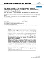
FFiigguurree 11
CNS progenitor cells are vulnerable to clinically relevant levels of 5-FU exposure.
((aa))
A summary of the putative relationships between the different
cell types under study (for discussion of this and alternative views on lineage relationships in the CNS, see [199,200]). Pluripotent neuroepithelial
stem cells (NSC) give rise to glial restricted precursor (GRP) cells and neuron restricted precursor (NRP) cells. GRP cells in turn give rise to
astrocytes and oligodendrocyte-type-2 astrocyte progenitor/oligodendrocyte precursor cells (O-2A/OPCs), the ancestors of oligodendrocytes.
((bb,,cc))
Primary CNS cells (b) or various cancer cell lines (c) were grown on coverslips and exposed to 5-FU for 24 h before analysis of cell viability as
described in Materials and methods. 5-FU concentrations were chosen on the basis of drug concentrations reached in humans after conventional
5-FU treatment. None of the tumor lines tested were sensitive to 5-FU treatment in this dose range, whereas O-2A/OPCs, oligodendrocytes, GRP
cells and human umbilical vein endothelial cells (HUVECs) were sensitive.
((dd,,ee))
Exposure conditions designed to mimic the exposure levels
associated with long-term infusion (d) or high-dose bolus administration (e) yielded similar results, with vulnerability of O-2A/OPCs and non-dividing
oligodendrocytes to 5-FU exceeding the vulnerability of rapidly dividing cancer cells. As shown in (b,d), the vulnerability of HUVECs also exceeds the
vulnerability of cancer cells. Each experiment was carried out in quadruplicate and was repeated at least twice in independent experiments. Data
represent mean of survival ± s.e.m, normalized to control values.
5-FU 5
µ
M 120 h
0 25 50 75 100
MDA-MB-231 (breast cancer)
Astrocytes
UT-4 glioma
GRP
HUVEC
O-2A/OPC
Oligodendrocytes
Survival (%)
MCF-7 (breast cancer)
ES-2 (ovarian cancer)
T98 glioma
meningioma
Cancer cellsNormal neural cells (rat) Normal cells (human)
5-FU 1 mM 1 h
0 25 50 75 100
T98 glioma
MDA-MB-231 (breast cancer)
meningioma
UT-4 glioma
HUVEC
GRP
Oligodendrocytes
O-2A/OPC
Survival (%)
MCF-7 (breast cancer)
Astrocytes
ES-2 (ovarian cancer)
0
20
40
60
80
100
120
0 0.01 0.1 1 10
5-FU [µM]
Survival (%)
O-2A/OPC
NSC
GRP
Oligodendrocytes
Astrocytes
HUVEC
(a)
(b)
(d) (e)
0
20
40
60
80
100
120
0 0.01 0.1 1 10
5-FU [µM]
Survival (%)
T98 glioma
Meningioma
MDA-MB-231 (breast cancer)
MCF-7 (breast cancer)
ES-2 (ovarian cancer)
(c)
NSC
GRP cells
NRP cells
O-2A/OPC
Oligodendrocytes
Neurons
Astrocytes
GFAP
+
cells (a subset of which may be stem cells in the SVZ
[96]). The SVZ also contained a smaller number of
TUNEL
+
Olig2
+
cells, which could be ancestors of oligo-
dendrocytes [97,98]. In the DG, there was also a very small
amount of NeuN
+
mature neurons that were TUNEL
+
. In
the CC, approximately 70% of the TUNEL
+
cells were
Olig2
+
, and thus would be either oligodendrocyte
progenitors or oligodendrocytes. Most of the remaining
TUNEL
+
cells in the CC were GFAP
+
, which in this tissue
would mean they are astrocytes. The specificity of TUNEL
labeling is demonstrated by representative images of
TUNEL
+
cells that were DCX
+
, Olig2
+
or GFAP
+
(Additional
data file 1) .
Analysis of cell division (as detected by incorporation of 5-
bromo-2-deoxyuridine (BrdU)) revealed that 5-FU caused
long-lasting suppression of proliferation in the SVZ and the
DG [99,100] (in which such proliferation is thought to be a
critical component of normal tissue function) as well as in
the CC (Figure 4a-c). Exposure to 5-FU caused reductions of
cell proliferation in all three regions. In contrast with the
return of levels of cell death to control levels (at least as
detected by TUNEL staining), cell division was suppressed for
long periods of time following completion of 5-FU treatment.
In the SVZ, 5-FU exposure was associated with a 40.9 ± 2.6%
decrease in numbers of BrdU
+
cells on day 1, with a
transient re-population of BrdU
+
cells at days 7 and 14,
followed by a subsequent decrease in animals examined at
day 56 and 6 months after completion of treatment. It was
striking that the most significant inhibition of DNA
synthesis in the SVZ was seen at 6 months post-treatment,
when there was a 67.7 ± 3.0% decrease in the number of
BrdU
+
cells compared with control animals (Figure 4a). In
the DG, suppression of DNA synthesis started on day 14
after treatment, and the greatest inhibition (60.7 ± 7.8%)
was also seen at 6 months (Figure 4c). In the CC, in
contrast, cell proliferation was significantly suppressed at all
time points examined (Figure 4b).
To determine whether exposure to 5-FU preferentially
reduced DNA synthesis in any particular cell population(s)
in vivo, we combined BrdU labeling with cell-type-specific
antibodies and analyzed individual BrdU
+
cells by confocal
microscopy (see Materials and methods). We analyzed the
CNS of animals sacrificed 1 day and 56 days after the
completion of 5-FU treatment in order to examine the acute
and long-term effects of treatment.
We found that neuronal precursors and oligodendrocyte
precursors were both affected in vivo. In the CC, where there
was a 42.6 ± 2.7% reduction in the number of BrdU
+
cells in
tissue sections from animals sacrificed 1 day after the
completion of treatment (Figure 4b), the proportion of
BrdU
+
cells that were Olig2
+
was similar between controls
and treated animals (Figure 5a,b). This result also held true
at day 56, when the proportion of Olig2
+
cells among the
BrdU
+
population was unchanged in untreated and treated
animals, despite a continued 53.2 ± 12.4% reduction in the
total number of BrdU
+
cells observed (Figure 5c,d). As
>90% of the BrdU
+
cells in the CC were Olig2
+
, these results
indicate that the reduction in DNA synthesis observed in
this tissue predominantly affected O-2A/OPCs [97,98,101].
In contrast with effects on putative O-2A/OPCs, there was a
somewhat enhanced loss of DCX
+
cells (which would have
been neuronal progenitors [95]) from among the BrdU
+
population in both the SVZ and the DG (Figure 5a-d). In
the SVZ, at 1 day after treatment, there was a dispropor-
tionate and significant reduction in the percentage of DCX
+
BrdU
+
cells, which represented 50.2 ± 1.9% of the cells incor-
porating BrdU in control animals and only 30.7 ± 3.9% in
animals treated with three injections of 5-FU (p < 0.01). At
day 56 the proportion of BrdU
+
cells that were DCX
+
was
not different between controls and treated animals,
although the total number of BrdU
+
cells in the SVZ of
treated animals continued to be significantly lower than
that of the control group (only 67.7 ± 4.9% compared with
/>Journal of Biology
2008, Volume 7, Article 12 Han
et al.
12.5
Journal of Biology
2008,
77::
12
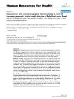
FFiigguurree 22
Sublethal doses of 5-FU inhibit division of O-2A/OPCs. Clonal analysis was
used to study the effects of low-dose 5-FU (0.05 µM for 24 h) on the
division and differentiation of freshly isolated progenitor cells. O-2A/OPCs
were grown at clonal density and exposed one day after plating to
((aa))
vehicle alone or
((bb))
0.05 µM 5-FU for 24 h, doses that killed less than 5%
of O-2A/OPCs in mass culture. The number of undifferentiated
O-2A/OPCs and differentiated cells (oligodendrocytes) was determined in
each individual clone from a total number of 100 clones in each condition
by morphological examination and by immunostaining with A2B5 and anti-
GalC antibodies (to label O-2A/OPCs and oligodendrocytes, respectively).
Results are presented as three-dimensional graphs. The number of
progenitors per clone is shown on the
x
(horizontal) axis, the number of
oligodendrocytes on the
z
(orthogonal) axis and the number of clones
with any particular composition on the
y
(vertical) axis. In 5-FU-treated
cultures analyzed five days after initiating 5-FU exposure, there was an
increase in the representation of small clones consisting wholly of
oligodendrocytes and clones containing large numbers of
oligodendrocytes, a reduction in the representation of large clones, a
general shift of clone size towards smaller values, and a clear reduction in
the total number of progenitor cells (see text for details). Experiments
were performed in triplicate in at least two independent experiments.
1
3
5
7
9
11
13
15
17
19
8
3
0
5
10
15
20
25
30
Number of clones
O-2A/OPC
Oligos
Control
Oligos
1
3
5
7
9
11
13
15
17
19
8
3
0
5
10
15
20
25
30
O-2A/OPC
5-FU
(a)
Number of clones
(b)
control animals at the same time point; p < 0.01). In
contrast, in the DG, a reduction in the number of DCX
+
cells was also seen, both at day 1 (with DCX
+
cells
comprising only 34.3 ± 4.4% of the BrdU
+
population in 5-
FU-treated mice compared with 63.2 ± 3.4% in the control
mice; p < 0.01), and at day 56 (23.7 ± 3.9% in 5-FU-treated
12.6
Journal of Biology
2008, Volume 7, Article 12 Han
et al.
/>Journal of Biology
2008,
77::
12
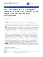
FFiigguurree 33
Systemic 5-FU treatment causes cell death in the adult CNS. Cell death was determined using the terminal deoxynucleotidyltransferase-mediated
dUTP nick-end labeling (TUNEL) assay. The number of TUNEL
+
cells was analyzed in control animals (that received 0.9% NaCl i.p.) and 5-FU-treated
animals and presented as percentage normalized values of controls at each time point. For ease of comparison, data presented in the figures show
the control value (mean set at 100% of the day 1 value) and normalized values of 5-FU treatment groups at all time points. Each treatment group and
the control group consisted of
n
= 5 animals at each time point. Figures show apoptosis in animals that received three bolus i.p. injections of 5-FU
(40 mg kg
-1
on days -4, -2 and 0 leading up to the analysis, where day 1 of analysis equals 1 day after the last treatment with 5-FU). There was
marked and prolonged increase of cell death in the 5-FU treatment group in
((aa))
the lateral subventricular zone (SVZ),
((bb))
the corpus callosum (CC)
and
((cc))
the dentate gyrus (DG) at 1, 7, 14 and 56 days and 6 months following treatment. Data are means ± s.e.m.; a two-way ANOVA test was
performed on the original un-normalized data set to test the statistical significance of treatment effect and time effect. Bonferroni post-tests were
performed to compare the 5-FU-treated group and the control group at each time point. The statistical significance of the Bonferroni post-tests is
labeled in the graphs where applicable: ***
p
< 0.001; **
p
< 0.01; and *
p
< 0.05. Two-way ANOVA test results indicate that, in the SVZ, the
treatment effect is extremely significant (
p
< 0.001), the time effect is very significant (
p
< 0.01); in the CC, the treatment effect is not quite
significant (
p
= 0.06), the time effect is not significant (
p
= 0.74); in the DG, the treatment effect is extremely significant (
p
< 0.001), the time effect is
significant (
p
< 0.05). The effect of the interaction between treatment and time is not significant for all three regions.
((dd))
To determine the
immediate cellular targets of 5-FU
in vivo
, we examined co-analysis of TUNEL labeling with antigen expression in animals sacrificed at day 1 after
completion of 5-FU treatment. The majority of TUNEL
+
cells in the SVZ and DG were doublecortin (DCX)
+
neuronal progenitors. Other TUNEL
+
cells in these two regions included GFAP
+
cells (which could be stem cells in the SVZ, or astrocytes in the DG) and Olig2
+
O-2A/OPCs. There was
also a small contribution of NeuN
+
mature neurons in the DG. In the CC, the majority of TUNEL
+
cells were Olig2
+
(which, in this white matter
tract, would be oligodendrocytes and O-2A/OPCs), with a small contribution of GFAP
+
astrocytes. Almost 100% of TUNEL
+
cells were accounted
for by known lineage markers. Each group consisted of
n
= 4 animals. Data are mean ± s.e.m.
SVZ
0
50
100
150
200
250
300
350
***
**
**
TUNEL
+
cells (% of controls)
CC
0
50
100
150
200
250
TUNEL
+
cells (% of controls)
DG
0
100
200
300
400
500
Control day 1
5-FU day 1
5-FU day 7
5-FU day 14
5-FU day 56
5-FU 6 months
*
TUNEL
+
cells (% of controls)
(a) (b) (c)
SVZ CC DG
0
25
50
75
100
NeuN
DCX
Olig2
GFAP
TUNEL
+
cells (% of controls)
(d)
mice versus 52.2 ± 2.8% in the control mice; p < 0.01). In
the CC, exposure to 5-FU was also associated with a small
increase in the proportion of GFAP
+
cells among the BrdU-
incorporating populations at both day 1 and day 56,
although such cells continued to represent a minority of the
BrdU
+
cells in this tissue. In addition, BrdU
+
cells that were
not labeled with any of the cell-type-specific antibodies
used in these studies were more prominent in treated
animals than in controls at day 1 (but not at day 56) in the
SVZ and were found in the DG at both time points (data
not shown). The DG was the only tissue in which these
unlabeled cells made up >10% of the entire BrdU
+
population. Such cells represented about 40% and 50% of
all BrdU-labeled cells in 5-FU-treated animals at days 1 and
56, respectively, compared with about 2% and 20%,
respectively, of all BrdU-labeled cells in control animals.
AAnnaallyyssiiss ooff aauuddiittoorryy ffuunnccttiioonn iinn 55 FFUU ttrreeaatteedd aanniimmaallss
ssuuggggeessttss ddeellaayyeedd ddiissrruuppttiioonn ooff mmyyeelliinnaattiioonn
To determine whether the exposure of experimental animals
to 5-FU was associated with functional impairment, we
investigated hearing function in treated animals at various
time points after treatment. Damage to the auditory system
is a well known correlate of treatments with cisplatin
[102,103]. This damage is associated with death of cochlear
outer hair cells, increases in the auditory brainstem response
(ABR) thresholds and decreases in transient evoked oto-
acoustic emissions (TEOAE) and distortion product oto-
acoustic emissions (DPOAE), all of which are indicators of
compromised cochlear function.
We examined the DPOAE as an indicator of cochlear function
and ABRs to provide information on changes in conduction
velocity from the ear to the brain, an indicator of myelination
status. Different peaks (called P1, P2, and so on) in the ABR
response are thought to correspond to different steps in the
transmission of information, and prior analysis of ABR inter-
peak latencies shows that loss of myelin (as in, for example,
CNS myelin-deficient mouse models [104,105]) causes
increases in specific ABR inter-peak latencies (P2-P1 and P3-
P1). Such measurements have been used by several investi-
gators to study myelination-associated problems in impulse
conduction in children with iron deficiency [106-109].
Our analysis of auditory function in 5-FU-treated animals
revealed what seems to be a previously unrecognized
consequence of chemotherapy exposure: increased latencies
of impulse transmission. Consistent with the absence from
/>Journal of Biology
2008, Volume 7, Article 12 Han
et al.
12.7
Journal of Biology
2008,
77::
12
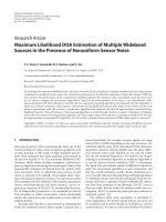
FFiigguurree 44
Systemic 5-FU exposure causes prolonged suppression of proliferation in the adult CNS
.
Animals were treated as described in Figure 3. The number
of BrdU
+
cells was analyzed in control animals and 5-FU-treated animals and presented as percentage normalized values of controls at each time
point. For ease of comparison, data presented in the figures show the control value (mean set at 100%) of day 1 and normalized values of 5-FU
treatment groups at all time points. Each group consisted of
n
= 5; a two-way ANOVA test was performed on the original un-normalized data set to
test the statistical significance of treatment effect and time effect. Bonferroni post-tests were performed to compare the 5-FU-treated group and the
control group at each time point. The statistical significance of the Bonferroni post-tests is labeled in the graphs where applicable: ***
p
< 0.001;
**
p
< 0.01; or *
p
< 0.05. Two-way ANOVA test results indicate that:
((aa))
in the SVZ, both the treatment effect and time effect are extremely
significant (
p
< 0.001), and the interaction of treatment and time is very significant (
p
< 0.01);
((bb))
in the CC, both the treatment effect and time effect
are extremely significant (
p
< 0.001), and the interaction of treatment and time is very significant (
p
< 0.01); and
((cc))
in the DG, the treatment effect
is very significant (
p
< 0.01), the time effect is extremely significant (
p
< 0.001), and the effect of the interaction between treatment and time is not
significant.
SVZ
0
25
50
75
100
125
***
***
***
BrdU
+
cells (% of controls)
CC
0
25
50
75
100
125
**
***
***
**
BrdU
+
cells (% of controls)
DG
0
25
50
75
100
125
Control day 1
5-FU day 1
5-FU day 7
5-FU day 14
5-FU day 56
5-FU 6 months
*
*
BrdU
+
cells (% of controls)
(a) (b) (c)
*
the literature of reported deficits in cochlear function
associated with 5-FU administration, DPOAEs in treated
animals were not significantly different from those in
untreated animals. In contrast, treated animals showed a
progressive alteration in ABRs when inter-peak latencies
were examined at days 1, 7, 14 and 56 after completion of
treatment and compared with baseline measurements of
each individual 1 day before 5-FU application.
In contrast with the lack of effect of 5-FU treatment on
DPOAEs, comparison of the changes in inter-peak latencies
P2-P1 and P3-P1 with those of a sham-treated control group
revealed that at the later time points of day 14 and day 56,
both inter-peak latency values of 5-FU-treated animals
showed marked increases (indicative of myelin damage or
loss), whereas those of sham-treated controls did not
(Figure 6). For example, at day 14, the P2-P1 and P3-P1
inter-peak latencies in 5-FU-treated animals increased by
0.179 ± 0.022 ms and 0.146 ± 0.050 ms, respectively, whereas
in control animals these latencies decreased by 0.037 ±
0.078 ms (p < 0.05 compared with 5-FU group) and 0.087 ±
0.123 ms (p < 0.01 compared with the 5-FU group). To
place these changes in context, a 0.1 ms delay in nerve
impulse transmission is considered to be a highly signifi-
cant functional change [104,110,111]. At day 56, the P2-P1
and P3-P1 inter-peak latencies in 5-FU-treated animals
increased by 0.191 ± 0.052 ms and 0.136 ± 0.088 ms, respec-
tively, whereas in control animals the P2-P1 inter-peak
12.8
Journal of Biology
2008, Volume 7, Article 12 Han
et al.
/>Journal of Biology
2008,
77::
12
FFiigguurree 55
Cell-type analyses of BrdU
+
cells in control and 5-FU-treated animals at early and late time points after completion of treatment. Co-analysis of BrdU
incorporation with antigen expression was conducted as described in Materials and methods. Both control and 5-FU-treated groups were analyzed
at (
aa,,bb
) day 1 and (
cc,,dd
) day 56 to evaluate the immediate and long-term effects of 5-FU treatment. Results indicate that division of both DCX
+
neuronal progenitors and Olig2
+
oligodendrocyte precursors was reduced by systemic exposure to 5-FU. In the CC, the reduction in apparent
division of Olig2
+
cells was proportionate to the overall reduction in all BrdU
+
cells. In the SVZ, there was an enhanced reduction of DCX
+
cells
from among the BrdU
+
population at day 1 but not at day 56. In the DG, there was an enhanced reduction in the dividing DCX
+
population at both
day 1 and day 56. In addition, the proportion of GFAP
+
cells in the CC was increased among the BrdU
+
population at both time points examined.
Data are mean ± s.e.m; *
p <
0.05, in comparisons with control animals (confidence interval = 95%, by unpaired, two-tailed Student’s
t
-test).
Control day 1
SVZ CC DG
0
25
50
75
100
DCX
Olig2
GFAP
BrdU
+
cells (% of controls)
5-FU day 1
SVZ CC DG
0
25
50
75
100
*
*
*
BrdU
+
cells (% of controls)
Control day 56
SVZ CC DG
0
25
50
75
100
BrdU
+
cells (% of controls)
5-FU day 56
SVZ CC DG
0
25
50
75
100
*
*
BrdU
+
cells (% of controls)
(a) (b)
(c) (d)
latency showed a small increase of 0.035 ± 0.075 ms
(p < 0.05 compared with the 5-FU group), and the P3-P1
inter-peak latency decreased by 0.002 ± 0.088 ms (p < 0.01
compared with the 5-FU group). At earlier time points,
there were no increases greater than 0.1 ms in these inter-
peak latencies in either the control or the treated groups.
55 FFUU ttrreeaattmmeenntt ccaauusseess ddeellaayyeedd cchhaannggeess iinn eexxpprreessssiioonn ooff
OOlliigg22 aanndd lloossss ooff mmyyeelliinn iinntteeggrriittyy
The results of our ABR analysis raised the possibility that 5-
FU-treated animals show a syndrome of delayed white
matter damage. Although our analysis of cell division and
cell death following systemic treatment with 5-FU revealed
a long-lasting suppression of cell division in the CC, we
observed only an increased level of apoptosis in this tissue
at one day after the cessation of treatment. We therefore
conducted a more detailed analysis of the CC, the major
myelinated tract in the rodent CNS.
Our further investigations revealed that systemic 5-FU
exposure was sufficient to cause substantial delayed
abnormalities in oligodendrocyte biology, in regard to both
transcriptional regulation and maintenance of myelin
integrity. Following treatment of six- to eight-week-old CBA
mice with three injections of 5-FU (40 mg kg
-1
, every other
day over 5 days), we first observed a slight increase in Olig2
+
cells in the CC at day 1 after completion of treatment.
Examination at later time points, in contrast, revealed a
substantial fall in the numbers of these cells. At day 56 after
treatment, the number of Olig2
+
cells was markedly
decreased, to 32.4 ± 9.7% (p < 0.001) of control levels at this
time point (Figure 7a-c). Immunofluorescence staining with
an anti-myelin basic protein (anti-MBP) antibody revealed
that there was also markedly decreased MBP staining in
animals treated with 5-FU examined 56 days after treatment
(data not shown). When we double-labeled sections with
the anti-CC1 antibody (to identify oligodendrocytes [112]),
however, we found that the reduction in the number of
Olig2
+
cells seen at day 56 was not matched by a similar fall
in the number of CC1
+
oligodendrocytes. Thus, whereas
almost all CC1
+
oligodendrocytes in the CC of the controls
were co-labeled with anti-Olig2 antibodies at day 56, in 5-
FU-treated animals many CC1
+
oligodendrocytes showed no
detectable expression of Olig2 (Figure 7d-i).
Ultrastructural analysis of the CC of animals 56 days after
treatment supported the interpretation of our immuno-
cytochemical analyses that many oligodendrocytes were
present at this time point, but also demonstrated the presence
of abundant myelin pathology. As shown in Figure 8, midline
/>Journal of Biology
2008, Volume 7, Article 12 Han
et al.
12.9
Journal of Biology
2008,
77::
12
FFiigguurree 66
Systemic 5-FU treatment caused delayed increases in auditory brainstem response (ABR) inter-peak latencies P2-P1 and P3-P1. Baseline ABR
hearing tests were performed on each animal one day before initiation of treatment with 5-FU (as for Figure 4). After treatment ended, follow-up
ABR tests were conducted on each animal at various points during a time course of 56 days. Control and treatment groups both consisted of
n
= 4
animals. ABR latencies were analyzed for each individual at each time point, and change of latency was calculated as L
t
- L
0
(L
t
, latency values at day
1, day 7, day 14, or day 56 post treatment; L
0
, baseline latency values 1 day before treatment initiation).
((aa))
The change of inter-peak P2-P1 latency
values;
((bb))
the change of inter-peak P3-P1 latency values. At the later time points day 14 and day 56, both P2-P1 and P3-P1 inter-peak latency values
of 5-FU-treated animals show average increases of more than 0.13 ms, whereas the same inter-peak latency values of sham-treated controls show
average decreases or an increase of less than 0.04 ms. Data are mean ± s.e.m. Statistical significance of the difference between the means of control
and treated groups was
p
< 0.05 in (a), and
p
< 0.01 in (b) (confidence interval = 95%; paired, one-tailed Student’s
t
-test).
Change of latency P2-P1
Day 1 Day 7 Day 14 Day 56
-0.3
-0.2
-0.1
-0.0
0.1
0.2
0.3
Ctrl
5-FU
Change of latency P2-P1 (ms)
Change of latency P3-P1
Day 1 Day 7 Day 14 Day 56
-0.5
-0.4
-0.3
-0.2
-0.1
-0.0
0.1
0.2
0.3
Ctrl
5-FU
Change of latency P3-P1 (ms)
Clicks at 80 dB
n = 4
(a) (b)
Clicks at 80 dB
n = 4
12.10
Journal of Biology
2008, Volume 7, Article 12 Han
et al.
/>Journal of Biology
2008,
77::
12
FFiigguurree 77
Systemic 5-FU treatment causes delayed dysregulation of Olig2 expression in oligodendrocytes in the CC. Animals were treated with 5-FU as in
Figure 3 and analyzed for expression of Olig2 in the CC at various time points. There was a marked reduction in the number of such cells at 56 days
(
((aa))
control;
((bb))
5-FU) after completion of treatment, but not at 1 or 14 days after treatment.
((cc))
Percent-corrected number of Olig2
+
cells in the
CC at day 1 and day 56 post-treatment with 5-FU, normalized to control values at each time point. Data represent averages from three animals in
each group, shown as mean ± s.e.m (
**
p
< 0.001, one-way ANOVA) in comparison with control values at each time point. The scale bar represents
150 µm.
((dd ii))
Representative confocal micrographs showing loss of Olig2 expression in a subset of CC1
+
oligodendrocytes in the CC of a 5-FU-
treated animal at day 56 in comparison with a sham-treated animal at the same time point. The reduction in numbers of Olig2
+
cells seen at day 56
after treatment was not associated with an equivalent fall in oligodendrocyte numbers, as determined by analysis of CC1
+
expression. (d-f) In control
animals, there is a close equivalence between CC1 expression (d) and Olig2 expression (e); a merged image is shown in (f). Three Olig2
+
CC1
-
cells
can be seen in (e,f) (arrowheads), which are probably O-2A/OPCs. (g-i) In contrast, in 5-FU-treated animals there is a reduction in the number of
Olig2
+
cells (h), but the CC of these animals contains many CC1
+
cells (g) that do not express Olig2 (i) (arrows, Olig2
+
CC1
+
; arrowheads, Olig2
+
CC1
-
cells). The scale bar represents 25 µm.
longitudinal sections of CC displayed scattered foci of
demyelinated axons, including partial or complete loss of
myelin sheaths and increases in inter-laminar splitting of the
myelin sheaths. Analysis of transverse sections (Figure 9)
provided further evidence of myelin vacuolization and
breakdown. It was also of interest to note the axonal
/>Journal of Biology
2008, Volume 7, Article 12 Han
et al.
12.11
Journal of Biology
2008,
77::
12
FFiigguurree 88
Delayed myelin and axonal degeneration in the CC caused by systemic
5-FU treatment (representative electron micrographs of longitudinal
sections of axons). Sections were taken from midline coronal sections
of the CC.
((aa))
A representative image from a sham-treated control
animal, showing normal myelinated axons and the normal axonal
cytoskeleton structures;
((bb ff))
representative images from a 5-FU-
treated animal, showing several pathological changes of both the myelin
and axonal structures. Asterisks, axonal abnormality; single arrows,
damaged myelin sheaths; double arrows, myelin debris; arrowheads,
engulfed myelin debris. (b) Several swollen axons with disrupted
cytoskeleton (asterisks), damaged myelin sheaths (single arrows) and
myelin debris (double arrows) can be seen. (c) Several swollen axons
(asterisks) with or without myelin can be seen, the axoplasm of which
show disruption of cytoskeleton and altered organelles. (d) Several
axons (asterisks) with absent or degenerating myelin (arrows) can be
seen; one axon shows a severely damaged axonal structure and myelin
on one side of a node of Ranvier (n) and partially disrupted myelin
sheath on the other side (arrow). (e) Several loci of myelin
degeneration can be seen (arrows); one axon seems to be transected
on one side of a node of Ranvier (n). An axon next to it shows partial
degeneration of the myelin sheath and disruption of the cytoskeleton
(asterisk). (f) Edema in what is likely to be a process of an astrocyte can
be seen, with some engulfed myelin debris (arrowhead) and the
adjacent axons are distorted; there are also swollen axons (asterisks)
with and without myelin (arrows).
FFiigguurree 99
Ultrastructural evidence of myelinopathy in 5-FU-treated animals.
Electron micrographs were taken from the midline transverse sections
of the CC (cross-sections of the axons).
((aa))
A representative image
from a sham-treated control animal, showing normal myelinated axons;
((bb ff))
representative images from a 5-FU-treated animal, showing
multiple pathological changes of both the myelin and axonal structures.
Single asterisks indicate demyelinated axons with rarefaction (that is,
decreased density of the axoplasm staining possibly due to disruptions
in cytoskeletal structures and organelles); double asterisks indicate an
abnormal axon with partially destructed myelin sheaths; single arrows
indicate inter-laminar splitting of the myelin sheaths; and double arrows
indicate myelin debris. (b) Two axons with damaged myelin sheaths
(asterisks), myelin debris (double arrows) and a smaller axon that
seems to be detaching from its myelin sheath (single arrow) can be
seen. (c) A large demyelinated axon with rarefaction of the axoplasm
(asterisk) and two axons with collapsed centers and inter-laminar
splitting of the myelin sheaths (arrows) can be seen, indicating on-going
myelin degeneration. (d) Two large axons with completely (asterisk) or
partially (double asterisks) damaged myelin can be seen, the axoplasm
of which shows altered cytoskeleton and organelles. One axon has a
collapsed center and inter-laminar splitting (arrow). (e) Myelin debris
can be seen, possibly from a degenerating axon (double arrows) and an
axon with inter-laminar splitting (arrow). (f) A demyelinated axon with
rarefaction of the axoplasm and possible axonal swelling (asterisk) and
two neighboring axons with inter-laminar splitting (arrows) can be seen.
pathology observed in these ultrastructural studies. Transverse
sections revealed degenerating axons with multi-laminated
structures and collapsed centers, swelling of axons and altered
axonal cytoskeleton and organelles. In the transverse sections,
pathological changes in axons were also readily apparent and
included axonal swelling and focal degeneration of the
axoplasmic cytoskeleton and microtubules.
Despite the presence of BrdU
+
Olig2
+
cells in day 56 CC,
raising the possibility of repair of the myelinopathy found
at this time point, examination of animals six months after
5-FU treatment revealed eventual loss of almost all cells and
myelin in this tissue. Hematoxylin and eosin staining
revealed markedly decreased cellularity in the CC in treated
animals at the 6 month time point, along with markedly
decreased levels of MBP in the CC and in the white-matter
tracts of the striatum of treated animals (Figure 10). In
agreement with the majority of the cell bodies in the mature
CC belonging to oligodendrocytes or glial progenitor cells,
the decrease in the number of CC1
+
cells at 6 months
matched the decrease of Olig2 labeling (data not shown),
confirming loss of oligodendrocytes at this time point.
55 FFUU ttrreeaattmmeenntt ccaauusseess oonnllyy ttrraannssiieenntt bbrraaiinn vvaassccuullaattuurree
eennddootthheelliiaall cceellll aappooppttoossiiss aanndd CCNNSS iinnffllaammmmaatti
ioonn iinn aa ssuubbsseett
ooff ttrreeaatteedd aanniimmaallss
The occurrence of delayed damage to the CNS following
irradiation has been a subject of interest for many years, and
both vascular damage and delayed inflammatory reactions
have been implicated as being important in the adverse
effects of this treatment on the CNS [113-116]. To begin to
determine whether similar mechanisms might be relevant
to analysis of the delayed effects of 5-FU administration, we
examined microglial activation and endothelial cell apop-
tosis in 5-FU-treated animals.
Unlike the consistent observations of microglial activation
in the irradiated CNS [116], such evidence of inflammation
following treatment with 5-FU was observed in only one of
ten treated animals and only at day 1 after the cessation of
treatment. Inflammatory reactions were examined in sections
labeled with antibody directed against the mouse antigen
F4/80, a 160 kDa glycoprotein expressed by activated murine
microglia and macrophages [117]. We found that F4/80
staining was markedly increased at day 1 in one of the ten
mice treated with 5-FU (Figure 11a,b). In this animal, there
was diffuse microglial activation throughout the brain,
including the primary motor cortex, CC, periventricular
striatum and hippocampus. The activation of microglia
seemed to be an acute inflammatory reaction, however,
since it was not found in any treated animals at later time
points. Thus, inflammation was not a frequent response to
treatment with 5-FU, and no prolonged inflammatory
reactions similar to those seen following irradiation were
observed in our experiments.
Damage to the vasculature following irradiation has also
been suggested as a possible contributor to delayed CNS
damage but, as for inflammation, it seems unlikely that such
damage contributed to the delayed effects of 5-FU
administration. Analysis of TUNEL labeling in 5-FU-treated
animals revealed a subset of animals (four out of ten total
treated animals from two independent experiments) that
showed markedly increased diffuse TUNEL
+
nuclei; the
distribution, morphology and size of these nuclei resembled
those of the microvasculature endothelial cells of the CNS
(Figure 11c-e). Double-labeling to visualize expression of
12.12
Journal of Biology
2008, Volume 7, Article 12 Han
et al.
/>Journal of Biology
2008,
77::
12
FFiigguurree 1100
5-FU treatment causes reduced cellularity and loss of myelin basic
protein (MBP) at 6 months after treatment. Representative images of
hematoxylin and eosin staining from the periventricular region of
((aa,,cc))
a control animal and
((bb,,dd))
a 5-FU-treated animal. (c) Partial
enlargement of the CC shown in (a); (d) partial enlargement of the CC
shown in (b). (a) Normal cellular density is seen in the CC of the
control; (b) in the CC from a 5-FU-treated animal, the cellular density
in the CC has decreased markedly.
((ee,,ff))
The expression of MBP seen in
control animals (e) (the fiber-like green fluorescence staining in the CC
and white-matter tracts in the peri-ventricular striatum) is greatly
reduced in treated animals (f). The bright green punctuated fluorescent
staining is BrdU
+
cells, which are present in control animals but greatly
depleted in treated animals. All sections were processed at the same
time and all images were taken under equal exposure times. The scale
bar represents 100 µm.
the vascular endothelial cell marker PECAM/CD31 [118]
confirmed that these apoptotic cells were vascular
endothelial cells (Figure 11c-e). However, these indications
of vascular damage were seen only in a subset of animals
examined 1 day after the cessation of treatment and were
not observed in animals examined at any later time points.
DDiissccuussssiioonn
Our studies demonstrate that systemic treatment with 5-FU
is associated with both acute and delayed toxicity reactions,
outcomes that are of particular concern because of the use
of this agent in the treatment of many cancers. As in our
recent studies on cisplatin, cytarabine and carmustine [79],
in vitro analysis of vulnerability to 5-FU revealed that
lineage-restricted progenitor cells of the CNS and non-
dividing oligodendrocytes were vulnerable to the effects of
5-FU at or below clinically relevant exposure levels. Thus,
toxicity of 5-FU was not limited to dividing cells. Toxicity of
5-FU was not limited to induction of cell death and was also
associated with suppression of O-2A/OPC division, even
when applied transiently at exposure levels that represent
small fractions of the CNS concentrations achieved during
cancer treatment. Although previous in vitro studies on
neurons and oligodendrocytes also observed vulnerability
of these cells [119,120], the effective concentrations used in
our present study are considerably lower than those used in
previous studies. Our in vitro analyses also predicted the
acute in vivo effects of 5-FU with considerable accuracy, just
as was the case with our previous studies on cisplatin,
BCNU and cytarabine [79]. 5-FU exposure transiently
increased apoptosis and suppressed proliferation for
extended periods of time in the SVZ, DG and CC. Cell-type-
specific analyses confirmed that the main populations
affected in vivo were also progenitor cells and oligo-
dendrocytes. Suppression of progenitor cell proliferation
was also seen in vitro in analyses of division and
differentiation in clonal families of cells.
This study is the first to demonstrate that delayed
degenerative damage can be caused by systemic application
of a single chemotherapeutic agent (5-FU) and does not
require the concurrent presence of cancer to manifest, as
well as the first to provide an animal model of delayed
/>Journal of Biology
2008, Volume 7, Article 12 Han
et al.
12.13
Journal of Biology
2008,
77::
12
FFiigguurree 1111
5-FU induces transient inflammation and apoptosis of microvasculature endothelial cells in a subset of treated animals. Representative photographs
showing the inflammatory response on day 1 after treatment with
((aa))
vehicle or
((bb))
5-FU treatment as indicated by immunostaining for the activated
microglia/macrophage marker F4/80. The basal level of F4/80 staining was very low in the controls but increased after treatment. These sections are
from the CC (the region between the two dotted lines), with similar evidence of inflammation seen in the DG and cortex of this same mouse. The
scale bar represents 50 µm.
((cc ee))
TUNEL/PECAM (CD31) double immunostaining was performed in 5-FU-treated animals, with representative
images taken from the DG showing double-labeling of the vascular endothelial cell marker PECAM (CD31) with TUNEL
+
nuclei. In the subset of
animals in which evidence of endothelial cell death was observed, similar TUNEL
+
profiles were also found in the cortex and CC.
damage to white matter associated with the systemic
administration of chemotherapy. These results are of
particular interest in the context of many clinical reports
that have identified neurotoxicity as a complication of
treatment regimens in which 5-FU is a component. Although
most reports of 5-FU-associated neurotoxicity indicate a
relatively acute onset, a delayed demyelinating cerebral
complication reminiscent of multifocal leukoencephalo-
pathy has also been increasingly reported in patients treated
with chemotherapy regimens that include 5-FU [24,53-78].
Although 5-FU is used most extensively in the treatment of
colorectal cancers, it is also an important component of
adjuvant therapies for the treatment of a variety of other
cancers, including breast [121-128], gastric [129-136], pan-
creatic [137-142] and lung [129,143,144], and is thus given
to large numbers of patients. Neurological symptoms may
occur in some patients several months after adjuvant
therapy with 5-FU and include declines in mental status,
ataxia and the appearance of prominent multifocal enhan-
cing white matter lesions detectable by MRI. In addition,
both acute and delayed neurological side effects have been
observed for many other chemotherapeutic agents
[9,14,23,26,27,29-31,33,145-154], and it will be of interest
to determine whether the pattern of degenerative changes
observed with 5-FU exposure is representative of delayed
changes associated with other chemotherapeutic agents.
We also have provided several novel findings regarding the
problem of delayed white matter damage caused by 5-FU
exposure. Our findings of aberrant regulation of Olig2
expression, with the presence of many Olig2-negative oligo-
dendrocytes at 56 days after treatment, provide the first
indication that chemotherapy alters the normal expression
of important transcriptional regulators in oligodendrocytes.
Our ultrastructural studies demonstrate extensive myelin
pathology at this time point, along with indications of
neuronal pathology. It is not yet known whether damage to
myelin precedes damage to neurons (as is thought to occur
in multiple sclerosis (see, for example [155-162]), or whether
neuronal damage occurs concurrent with or preceding myelin
pathology. The vulnerability of oligodendrocytes to 5-FU in
vitro and the increased apoptosis in these cells following 5-
FU exposure in vivo, however, suggests strongly that
oligodendrocytes are a direct target of this anti-metabolite.
Although this is a somewhat surprising result (in that 5-FU
has been thought to target dividing cells specifically, while
oligodendrocytes do not divide in the conditions used in
our experiments), previous studies have shown that
experimental derivatives of 5-FU, and its metabolites, also
cause myelin damage in vitro and in vivo [163,164]. Whether
5-FU derivatives such as capecitabine (an orally active form
of 5-FU) cause similar damage is not yet known, but the
presence of the activating enzyme for this drug (thymidine
phosphorylase) in white-matter tracts [165] makes this a
matter of concern.
Although the continuing presence of at least some BrdU
+
cells in the CC at 56 days offered the possibility that the
damage to myelin occurring at this time point might be
reversible, analysis at 6 months demonstrated a striking loss
of cells and of MBP. Thus, it appears that even a short-term
exposure to 5-FU can cause long-term and apparently
irreversible damage to white-matter tracts.
Analysis of alterations in myelination caused by chemo-
therapy would benefit enormously from the ability to
conduct functional analysis in a non-invasive manner, and
our analysis of alterations in inter-peak latencies in ABRs
provides a tool of particular potential interest in this regard,
as well as revealing a novel form of chemotherapy-induced
neurological damage. Despite extensive investigations of
ototoxicity induced by exposure to cisplatin (for reviews, see
[166-171]), such studies appear to have been focused
exclusively on the effects of chemotherapy on hair cells and
cochlear function and have not used ABR analysis of inter-
peak latencies to analyze changes that may be related to
white matter damage. Thus, our ABR analyses seem to
provide the first demonstration of adverse effects of
chemotherapy on a functional outcome related to CNS
myelination. ABR inter-peak latency analysis has been used,
however, to study myelination-related maturation and
function of the auditory pathway in normal infants in
conditions in which myelination is compromised (for
example, iron deficiency, fetal cocaine syndrome) and in
experimental animals [105,106,108,172-175]. Thus, this
approach provides a non-invasive functional analysis of a
myelination-related outcome measure that can be used in
both experimental animals and human populations.
The progressive alterations in ABR inter-peak latencies
observed in our studies also highlight the fact that at least
some of the delayed damage associated with 5-FU adminis-
tration is greater than the damage observed acutely. The
ability to study progressive deterioration in the same
animals over prolonged periods will make this approach of
particular value in further investigations of these changes.
Moreover, because of the ease of conducting such studies in
humans, such analysis may provide a simple, non-invasive
approach to the analysis of adverse effects on white matter
complementary to the imaging-based detection of leuko-
encephalopathy.
The underlying causes of delayed damage induced by
chemotherapy will be the subject of continued investigation,
but the observations that vascular damage and inflammatory
reactions were rare and were observed only at short intervals
12.14
Journal of Biology
2008, Volume 7, Article 12 Han
et al.
/>Journal of Biology
2008,
77::
12
after completion of treatment makes it seem unlikely that
these are causally important. This is in striking contrast to
the effects of irradiation, where inflammation is thought to
be essential in delayed suppression of hippocampal
neurogenesis [115,116]. It is possible that the appearance of
delayed damage following 5-FU treatment reflects the
combined effects of delayed oligodendrocyte death and a
loss of the progenitor cell populations required for
replacement. Recent findings that aging is associated with a
loss of expression of important transcriptional regulators,
including Olig2, in oligodendrocytes [176] and may be
associated with degenerative white matter changes [177-
185] also raises the possibility, however, that the effect of
5-FU results from an acceleration of the normal aging
processes.
Our findings also raise the question of whether multiple
pathological changes contribute to the effects of chemo-
therapy on cognition. The ability of irradiation to the CNS
to suppress the generation of new neurons in the hippo-
campus has been suggested to be relevant to the
understanding of cognitive impairment associated with this
particular form of cancer treatment [115]. Although reduced
numbers of dividing hippocampal neuronal progenitors are
also seen in association with exposure to 5-FU, BCNU or
cytarabine [79], the additional damage to white-matter
tracts caused by chemotherapy would be expected to impair
normal neuronal impulse conduction (in accordance with
the changes in ABR latency seen here) and thus might also
contribute to alterations in cognition. It is particularly
interesting in this regard that recent studies on breast cancer
patients treated with adjuvant chemotherapy have revealed
that, relative to controls, patients had slower speeded
processing and altered fractional anisotropy (a measure of
white matter integrity) in the corpus callosum. It has been
suggested that these white matter changes are related to the
cognitive deficits that may be associated with treatment
with systemic chemotherapy [186].
As adverse effects on several normal tissues have been
observed for almost all classes of chemotherapeutic agents
[19-22,187] (including alkylating agents [29,30], anti-
metabolites [23-26,57], methotrexate [27] and even anti-
hormonal agents [31-37]) and such treatments will clearly
remain the standard of care for cancer patients for many
years to come, the need to understand such damage better is
great. Indeed, some of the most important advances in the
treatment of cancer have emerged from the study of such
damage, the necessary first step in its prevention. Moreover,
evaluation of potential new therapeutics that does not
include adequate analysis of these potential toxicities may
lead to the approval of treatments that are no better than
existing treatments in avoiding serious damage to normal
tissue. The clinical study of such side effects does not
provide the experimental foundations required for the
analysis of such problems. Indeed, treatment for neuro-
logical complications of 5-FU treatment has largely been
ineffective so far, with some patients responding to
immediate discontinuance of chemotherapy and steroid
treatment [57,60], but with others continuing to deteriorate
and, in some severe cases, progressing to death [188]. In
contrast, recent studies on the toxicities in vitro and in vivo of
several chemotherapeutic agents [79,189], and our discovery
of an animal model for delayed damage to the CNS caused
by chemotherapy, provide experimental foundations that
should prove of great value in the discovery and evaluation
of therapies that either allow selective killing of cancer cells
or offer selective protection to the normal cells of the body.
MMaatteerriiaallss aanndd mmeetthhooddss
Most materials and methods are as described in [79] and are
presented here in brief.
PPrreeppaarraattiioonn ooff pprriimmaarryy cceellll ccuullttuurreess
In vitro studies were performed on purified cultures of
primary CNS cells isolated from the developing rat CNS.
Purified populations of neuroepithelial stem cells, neuron
restricted precursor cells, glial restricted precursor cells,
O-2A/OPCs, oligodendrocytes and astrocytes were all
prepared and grown as described previously [79]. HUVECs
(Cambrex) were cultured in endothelial growth medium
(EGM-2) and used within two passages after thawing.
Cancer cell lines used were established breast cancer cell
lines (MCF-7 and MDA-MB-231), ovarian cancer (ES-2) cells,
L1210 lymphocytic leukemia and EL-4 lymphoma cells, a
meningioma cell line and two cell lines isolated from
patients with glioblastoma multiforme (UT-4 and T98 cell
lines); these were grown as previously described [79].
IInn vviittrroo
ttooxxiicciittyy aanndd vviiaabbiilliittyy aassssaayy
In vitro toxicity studies involved microscopic analysis of
staining with the 3,(4,5-dimethylthiazol-2-yl) 2,5-diphenyl-
tetrazoliumbromide (MTT) assay in combination with 4’,6-
diamidino-2-phenylindole (DAPI) and staining with cell-
type-specific antibodies, as previously described [79]. Each
experiment was carried out in quadruplicate and was
repeated at least twice in independent experiments. Data
points represent means from single experiments and error
bars shown in figures represent ± standard error of the mean
(s.e.m).
CClloonnaall aannaallyyssiiss
Clonal analysis of O-2A/OPC division and differentiation
was carried out as described previously [92-94]. One day
after plating, cells were exposed for 24 h to low-dose 5-FU
/>Journal of Biology
2008, Volume 7, Article 12 Han
et al.
12.15
Journal of Biology
2008,
77::
12
(0.01 µM), a concentration that did not cause significant
killing of O-2A/OPCs in mass culture. The number of
undifferentiated progenitors and differentiated oligo-
dendrocytes was determined in each individual clone from
a total of 100 clones in each condition by morphological
examination and by immunostaining to confirm cell-type
identification. Experiments were performed in triplicate in
at least two independent experiments.
CChheemmootthheerraappyy aapppplliiccaattiioonn
iinn vviivvoo
For in vivo experiments, 6-8-week-old CBA mice were treated
with chemotherapy under approved protocols. 5-FU
(Sigma) was administered by i.p. injections. Animals
received 5-FU as three consecutive injections every other
day (40 mg kg
-1
body weight). Control animals received
equal amounts of 0.9% NaCl i.p Animals were sacrificed
on days 1, 7, 14 and 56 and 6 months after completion of
treatment with 5-FU (where day 0 is the time of the last
injection of the agent). For all in vivo experiments, animals
were perfused transcardially with 4% paraformaldehyde in
phosphate buffer (pH 7.4), under deep anesthesia using
Avertin (tribromoethanol; Sigma; 250 mg kg
-1
, 1.2%
solution).
We chose the in vivo dosage on the basis of conversion from
human treatment dosage to an equivalent mouse dosage
and previous animal studies of 5-FU effects in mice. As is
standard practice, we used a conversion factor of 3
[190-194] to calculate the equivalent mouse dose range
(20-1,167 mg kg
-1
) from the clinical human treatment dose
range (60-3,500 mg m
-2
). On the basis of animal studies in
mice (for example, [195-197]), in which doses of 5-FU used
ranged from 40-200 mg kg
-1
, we first used 60 mg kg
-1
every
other day for three doses as the initial trial treatment. As this
treatment caused death in half of treated CBA mice one
week after completion of treatment, we lowered the dosage
to 40 mg kg
-1
, which was tolerated well by the animals (it
caused less than a 10% increase in death over a six-month
period compared with sham-treated controls). The
differences in tolerance to 5-FU treatment between our
study and others may result from the different mouse
strains used. In clinical practice, patients are often given the
highest tolerated dosage of chemotherapy to achieve
maximum fractional kill of the malignant cells. Considering
this situation, we determined the appropriate in vivo dosage
to be 40 mg kg
-1
.
IImmmmuunnoofflluuoorreesscceennccee,, TTUUNNEELL ssttaaiinniinngg aanndd aannaallyyssiiss ooff BBrrddUU
iinnccoorrppoorraattiioonn
iinn vviivvoo
All in vivo analysis was carried out as described in [79].
Using free-floating sections (40 µm), detection of nuclear
profiles with DNA fragmentation, a hallmark of apoptosis,
was performed using a TUNEL assay combined with DAPI
counterstaining to visualize nuclear profiles. To combine
TUNEL staining with immunofluorescence staining for
different cell lineage markers, TUNEL staining was performed
first, followed by labeling with one of the following primary
antibodies for 24-48 h: mouse anti-NeuN (1:500,
Chemicon), goat anti-DCX (doublecortin; 1:500, Santa
Cruz), rat anti-S-100β (1:2,500, Swant), mouse anti-MBP
(1:1,000, Chemicon), mouse anti-GFAP (1:2,500, DAKO),
rabbit anti-Olig2 (1:1,000, a gift of David Rowitch), mouse
anti-CC1 (1:300, Calbiochem; which was used under
conditions [101] in which specificity for oligodendrocytes is
preserved and no double-labeling with GFAP
+
astrocytes
was observed), rat anti-CD31/PECAM (1:500, Chemicon)
and rat anti-F4/80 (Abcam). All secondary antibodies,
generated in donkey (anti-rat, anti-rabbit, anti-goat and
anti-mouse), were coupled to TritC, FitC or Cy5 (Jackson
ImmunoResearch) for in vivo staining and were used
according to the species of primary antibody. Fluorescent
signals were detected using a confocal laser scanning
microscope Leica TCS SP2 and a 40× oil immersion lens,
with pinhole settings corresponding to an optical thickness
of less than 2 µm used to avoid false positive signals from
adjacent cells.
To label the proportion of dividing cells engaged in DNA
synthesis in vivo, mice received a single injection of BrdU
(50 mg kg
-1
body weight, dissolved in 0.9% NaCl) given i.p.
4 h before perfusion. Anti-BrdU antibody was used to
identify BrdU
+
cells by standard techniques (as in [79]). A
minimum of 50 BrdU
+
cells was counted for each labeling
condition in each animal (n = 3 animals in each group
examined), with the sole exception of the DG of the
animals examined 56 days after cytarabine treatment, for
which an identical number of sections were examined as in
controls, but the frequency of labeled cells was not
sufficient to reveal 50 cells in these sections. Quantification
of BrdU
+
cells was accomplished with unbiased counting
methods. BrdU immunoreactive nuclei were counted in one
focal plane to avoid over-sampling. Brain structures were
sampled either by selecting predetermined areas on each
section (lateral SVZ) or by analyzing the entire structure on
each section (CC and DG). Differences were considered
significant when p < 0.01.
SVZ: BrdU
+
cells were counted in every sixth section (40
µm) from a coronal series between interaural anterior-
posterior (AP) +5.2 mm and AP +3.9 mm (the anterior
commissure crossing). BrdU
+
cells were counted along the
lateral ventricular wall up to 200 µm distance from the
lateral ventricle wall.
CC: BrdU
+
cells were counted in every sixth section (40 µm)
from a coronal series between interaural AP +5.2 mm and
12.16
Journal of Biology
2008, Volume 7, Article 12 Han
et al.
/>Journal of Biology
2008,
77::
12
AP +3.0 mm in the entire extension of the rostral and
medial part of the CC and analyzed as for the SVZ.
DG: BrdU
+
cells were counted in every sixth section (40
µm) from a coronal series between interaural AP +2.5 mm
and AP +1.1 mm. BrdU
+
cells were counted in the area of
the dentate gyrus, including the hilus, subgranular zone
and the granule cell layer and analyzed as for the SVZ.
Quantitative data in all figures are presented as mean
percentage normalized to control animals. Error bars
represent ± s.e.m.
To analyze BrdU incorporation in specific cell types, anti-
BrdU immunostaining was combined with immuno-
labeling to identify DCX
+
neuronal precursor cells [95],
Olig2
+
oligodendrocyte precursor cells (defined as cells
that were BrdU
+
and Olig2
+
, in order to discriminate these
cells from Olig2
+
non-dividing oligodendrocytes
[97,98,101]) and GFAP
+
cells; the latter would have been
astrocytes in the CC or DG or, in the SVZ, may also have
been stem cells [96]. Labeling and confocal analysis was
carried out as for the combination of immunolabeling
with TUNEL staining.
AAuuddiittoorryy bbrraaiinn sstteemm rreessppoonnsseess
Baseline ABRs were measured in each animal one day
before initiation of treatment. After treatment ended,
follow-up ABR tests were conducted on each animal at
various points during a time course of 56 days. For the
measurement, mice were anesthetized with xylazine
(20 mg kg
-1
i.p.) and ketamine (100 mg kg
-1
i.p.) [198].
Needle electrodes were inserted at the vertex and pinna of
the ear, with a ground near the tail. ABR potentials were
evoked with click stimuli at 80 dB SPL. The response was
amplified (10,000×) and 1,024 responses were averaged
with an analog-digital board in a LabVIEW-driven data-
acquisition system. For comparison of change of latencies,
the wave peaks P1, P2 and P3 were identified by visual
inspection at recorded wave forms, and the inter-peak
latencies of wave P2-P1 and P3-P1 computed. The change
of inter-peak latencies was calculated as L
t
- L
0
(where L
t
is
the inter-peak latency values at day 1, day 7, day 14, or day
56 post-treatment; and L
0
is the baseline inter-peak latency
values one day before treatment initiation).
IImmaaggeess aanndd ddaattaa pprroocceessssiinngg aanndd ssttaattiissttiiccss
Digital images were captured using a Nikon Eclipse E400
upright microscope with a spot camera (Diagnostic
Instruments) and the spot advanced software for Macintosh
(Diagnostic Instruments), or using the confocal laser
scanning microscope (Leica TCS SP2). Paired or unpaired
Student t-tests were used for statistical analyses where
applicable.
EElleeccttrroonn mmiiccrroossccooppyy
The animals were anesthetized and injected with heparin
and perfused with a 0.1 M phosphate buffered 4.0% para-
formaldehyde/2.5% glutaraldehyde fixative. The brain was
removed and allowed to fix overnight at 4°C and the CC
was then sectioned sagitally and coronally at approximately
1.0 mm. The sections were rinsed in 0.1 M sodium
phosphate buffer, post-fixed in buffered 1.0% osmium
tetroxide, dehydrated in a graded series of ethanol to 100%,
transitioned to propylene oxide and infiltrated with
EPON/Araldite epoxy resin overnight. They were embedded
into mold capsules (BEEM) and polymerized for 2 days at
70°C. Sections of 1 µm were cut onto glass slides and
stained with toluidine blue to determine areas to be thin
sectioned at 70 nm onto grids. The grids were stained
sequentially with uranyl acetate and lead citrate. A Hitachi
7100 transmission electron microscope was used to
examine and digitally capture images using a MegaView III
digital camera (Soft Imaging).
AAddddiittiioonnaall ddaattaa ffiilleess
An additional data file is available with this article online.
Additional data file 1 shows a representative confocal
micrograph of TUNEL
+
cells co-labeled with cell-type-
specific markers. (A-A”) show a TUNEL
+
DCX
+
cell in the
sub-ventricular zone; (B-B”) show a TUNEL
+
Olig2
+
cell in
the CC; (C-C”) show a TUNEL
+
GFAP
+
cell in the CC. The
scale bars represent 10 µm.
AAcckknnoowwlleeddggeemmeennttss
It is a pleasure to acknowledge the many helpful discussions with our
colleagues regarding this research, in particular Chris Pröschel and
Hartmut Land. This research was funded with generous support from
the National Institutes of Health (HD39702 and NS44701), from the
Susan B. Komen Foundation for the Cure and from the Wilmot Cancer
Center.
RReeffeerreenncceess
1. Stewart A, Collins B, Mackenzie J, Tomiak E, Verma S, Bielajew
C:
TThhee ccooggnniittiivvee eeffffeeccttss ooff aaddjjuuvvaanntt cchheemmootthheerraappyy iinn eeaarrllyy ssttaaggee
bbrreeaasstt ccaanncceerr:: aa pprroossppeeccttiivvee ssttuuddyy
Psychooncology
2008,
1177::
122-130.
2. Inagaki M, Yoshikawa E, Matsuoka Y, Sugawara Y, Nakano T,
Akechi T, Wada N, Imoto S, Murakami K, Uchitomi Y:
SSmmaalllleerr
rreeggiioonnaall vvoolluummeess ooff bbrraaiinn ggrraayy aanndd wwhhiittee mmaatttteerr ddeemmoonnssttrraatteedd iinn
bbrreeaasstt ccaanncceerr ssuurrvviiv
voorrss eexxppoosseedd ttoo aaddjjuuvvaanntt cchheemmootthheerraappyy
Cancer
2007,
110099::
146-156.
3. Silverman DH, Dy CJ, Castellon SA, Lai J, Pio BS, Abraham L,
Waddell K, Petersen L, Phelps ME, Ganz PA:
AAlltteerreedd ffrroonnttooccoorrttii
ccaall,, cceerreebbeellllaarr,, aanndd bbaassaall ggaanngglliiaa aaccttiivviittyy iinn aaddjjuuvvaanntt ttrreeaatteedd bbrreeaasst
t
ccaanncceerr ssuurrvviivvoorrss 55 1100 yyeeaarrss aafftteerr cchheemmootthheerraappyy
Breast Cancer
Res Treat
2007,
110033::
303-311.
4. Hurria A, Rosen C, Hudis C, Zuckerman E, Panageas KS, Lachs
MS, Witmer M, van Gorp WG, Fornier M, D'Andrea G, Moasser
M, Dang C, Van Poznak C, Hurria A, Holland J
:
CCooggnniittiivvee ffuunnccttiioonn
ooff oollddeerr ppaattiieennttss rreecceeiivviinngg aaddjjuuvvaanntt cchheemmootthheerraappyy ffoorr bbrreeaasstt
ccaanncceerr:: aa ppiilloott pprroos
sppeeccttiivvee lloonnggiittuuddiinnaall ssttuuddyy
J Am Geriatr Soc
2006,
5544::
925-931.
/>Journal of Biology
2008, Volume 7, Article 12 Han
et al.
12.17
Journal of Biology
2008,
77::
12
5. Downie FP, Mar Fan HG, Houede-Tchen N, Yi Q, Tannock IF:
CCooggnniittiivvee ffuunnccttiioonn,, ffaattiigguuee,, aanndd mmeennooppaauussaall ssyymmppttoommss iinn bbrreeaasstt
ccaanncceerr ppaattiieennttss rreecceeiivviinngg aaddjjuuvvaanntt c
chheemmootthheerraappyy:: eevvaalluuaattiioonn wwiitthh
ppaattiieenntt iinntteerrvviieeww aafftteerr ffoorrmmaall aasssseessssmmeenntt
Psychooncology
2006,
1155::
921-930.
6. Fan HG, Houede-Tchen N, Yi QL, Chemerynsky I, Downie FP,
Sabate K, Tannock IF:
FFaattiigguuee,, mmeennooppaauussaall ssyymmppttoommss,, aanndd ccooggnnii
ttiivvee ffuunnccttiioonn iinn wwoommeenn aafftteerr aaddjjuuvvaanntt cchheemmootthheerraappyy
ffoorr bbrreeaasstt
ccaanncceerr:: 11 aanndd 22 yyeeaarr ffoollllooww uupp ooff aa pprroossppeeccttiivvee ccoonnttrroolllleedd ssttuuddyy
J
Clin Oncol
2005,
2233::
8025-8032.
7. Bender CM, Sereika SM, Berga SL, Vogel VG, Brufsky AM, Paraska
KK, Ryan CM:
CCooggnniittiivvee iimmppaaiirrmmeenntt aassssoocciiaatteedd wwiitthh aaddjjuuvvaanntt
tthheerraappyy iinn bbrreeaasstt ccaanncceerr
Psychooncology
2006,
1155::
422-430.
8. Falleti MG, Sanfilippo A, Maruff P, Weih L, Phillips KA:
TThhee nnaattuurree
aanndd sseevveerriittyy ooff ccooggnniittiivvee iimmppaaiirrmmeenntt aassssoocciiaatteedd wwiitthh aaddjjuuvvaanntt
cchheemmootthheerraappyy iinn wwoomme
enn wwiitthh bbrreeaasstt ccaanncceerr:: aa mmeettaa aannaallyyssiiss ooff tthhee
ccuurrrreenntt lliitteerraattuurree
Brain Cogn
2005,
5599::
60-70.
9. Meyers CA, Abbruzzese JL:
CCooggnniittiivvee ffuunnccttiioonniinngg iinn ccaanncceerr
ppaattiieennttss:: eeffffeecctt ooff pprreevviioouuss ttrreeaattmmeenntt
Neurology
1992,
4422::
434-436.
10. Schagen SB, van Dam FS, Muller MJ, Boogerd W, Lindeboom J,
Bruning PF:
CCooggnniittiivvee ddeeffiicciittss aafftteerr ppoossttooppeerraattiivvee aaddjjuuvvaanntt
cchheemmootthheerraappyy ffoorr bbrreeaasstt ccaarrcciinnoommaa
Cancer
1999,
8855::
640-
650.
11. Jenkins V, Shilling V, Deutsch G, Bloomfield D, Morris R, Allan S,
Bishop H, Hodson N, Mitra S, Sadler G, Shah E, Stein R, White-
head S, Winstanley J:
AA 33 yyeeaarr pprroossppeeccttiivvee ssttuuddyy ooff tthhee eeffffeeccttss ooff
aaddjjuuvvaanntt ttrreeaattmmeennttss oonn ccooggnniittiioonn iinn wwoommeenn wwiitthh eeaarrllyy ssttaaggee bbrreeaasstt
ccaanncceerr
Br J Cancer
2006,
9944::
828-834.
12. Jenkins V, Beveridge H, Low R, Mitra S:
AAttyyppiiccaall hheeaarriinngg lloossss iinn
wwoommeenn wwiitthh bbrreeaasstt ccaanncceerr rreecceeiivviinngg aaddjjuuvvaanntt ttrreeaattmmeenntt
Breast
2006,
1155::
448-451.
13. Kreukels BP, Schagen SB, Ridderinkhof KR, Boogerd W, Ham-
burger HL, van Dam FS:
EElleeccttrroopphhyyssiioollooggiiccaall ccoorrrreellaatteess ooff iinnffoorrmmaa
ttiioonn pprroocceessssiinngg iinn bbrreeaasstt ccaanncceerr ppaattiieennttss ttrreeaatteedd wwiitthh aaddjjuuvvaanntt
cchheemmootthheerraappyy
Breast Cancer Res Treat
2005,
9944::
53-61.
14. Brezden CB, Phillips KA, Abdolell M, Bunston T, Tannock IF:
CCoogg
nniittiivvee ffuunnccttiioonn iinn bbrreeaasstt ccaanncceerr ppaattiieennttss rreecceeiivviinngg aaddjjuuvvaanntt
cchheemmootthheerraappyy
J Clin Oncol
2000,
1188::
2695-2701.
15. Duffner PK:
LLoonngg tteerrmm eeffffeeccttss ooff rraaddiiaattiioonn tthheerraappyy oonn ccooggnniittiivvee
aanndd eennddooccrriinnee ffuunnccttiioonn iinn cchhiillddrreenn wwiitthh lleeuukkeemmiiaa aanndd bbrraaiinn
ttuummoorrss
Neurologist
2004,
1100::
293-310.
16. Reddy AT, Witek K:
NNeeuurroollooggiicc ccoommpplliiccaattiioonnss ooff cchheemmootthheerraappyy
ffoorr cchhiillddrreenn wwiitthh ccaanncceerr
Curr Neurol Neurosci Rep
2003,
33::
137-
142.
17. Schagen SB, Hamburger HL, Muller MJ, Boogerd W, van Dam FS:
NNeeuurroopphhyyssiioollooggiiccaall eevvaalluuaattiioonn ooff llaattee eeffffeeccttss ooff aaddjjuuvvaanntt hhiigghh ddoossee
cchheemmootthheerraappyy oonn ccooggnniittiivvee ffuunnccttiioonn
J Neurooncol
2001,
5511::
159-165.
18. Omuro AM, Ben-Porat LS, Panageas KS, Kim AK, Correa DD,
Yahalom J, DeAngelis LM, Abrey LE:
DDeellaayyeedd nneeuurroottooxxiicciittyy iinn
pprriimmaarryy cceennttrraall nneerrvvoouuss ssyysstteemm llyymmpphhoommaa
Arch Neurol
2005,
6622::
1595-1600.
19. Dropcho EJ:
NNeeuurroottooxxiicciittyy ooff ccaanncceerr cchheemmootthheerraappyy
Semin Neurol
2004,
2244::
419-426.
20. Keime-Guibert F, Napolitano M, Delattre JY:
NNeeuurroollooggiiccaall ccoommppllii
ccaattiioonnss ooff rraaddiiootthheerraappyy aanndd cchheemmootthheerraappyy
J Neurol
1998,
224455::
695-708.
21. Minisini A, Atalay G, Bottomley A, Puglisi F, Piccart M, Biganzoli L:
WWhhaatt iiss tthhee eeffffeecctt ooff ssyysstteemmiicc aannttiiccaanncceerr ttrreeaattmmeenntt oonn ccooggnniittiivvee
ffuunnccttiioonn??
Lancet Oncol
2004,
55::
273-282.
22. Wefel JS, Kayl AE, Meyers CA:
NNeeuurrooppssyycchhoollooggiiccaall ddyyssffuunnccttiioonn
aassssoocciiaatteedd wwiitthh ccaanncceerr aanndd ccaanncceerr tthheerraappiieess:: aa ccoonncceeppttuuaall rreevviieeww
ooff aann eemmeerrggiinngg ttaarrggeett
Br J Cancer
2004,
9900::
1691-1696.
23. Poppelreuter M, Weis J, Külz AK, Tucha O, Lange KW, Bartsch
HH:
CCooggnniittiivvee ddyyssffuunnccttiioonn aanndd ssuubbjjeeccttiivvee ccoommppllaaiinnttss ooff ccaanncceerr
ppaattiieennttss:: aa ccrroossss sseeccttiioonnaall ssttuuddyy iinn aa ccaanncceerr rreehhaabbiilliittaattiioonn cceennttrree
Eur J Cancer
2004,
4400::
43-49.
24. Choi SM, Lee SH, Yang YS, Kim BC, Kim MK, Cho KH:
55 fflluuoo
rroouurraacciill iinndduucceedd lleeuukkooeenncceepphhaallooppaatthhyy iinn ppaattiieennttss wwiitthh bbrreeaasstt
ccaanncceerr
J Korean Med Sci
2001,
1166::
328-334.
25. Johnson MR, Hageboutros A, Wang K, High L, Smith JB, Diasio RB:
LLiiffee tthhrreeaatteenniinngg ttooxxiicciittyy iinn aa ddiihhyyddrrooppyyrriimmiiddiinnee ddeehhyyddrrooggeennaassee
ddeeffiicciieenntt ppaattiieenntt aafftteerr ttrreeaattmmeenntt wwiitthh ttooppiiccaall 55 fflluuoorroouurraacciill
Clin
Cancer Res
1999,
55::
2006-2011.
26. Haykin ME, Gorman M, van Hoff J, Fulbright RK, Baehring JM:
DDiiff
ffuussiioonn wweeiigghhtteedd MMRRII ccoorrrreellaatteess ooff ssuubbaaccuuttee mmeetthhoottrreexxaattee rreellaatteedd
nneeuurroottooxxiicciittyy
J Neurooncol
2006,
7766::
153-157.
27. Shuper A, Stark B, Kornreich L, Cohen IJ, Aviner S, Steinmetz A,
Stein J, Goshen Y, Yaniv I:
MMeetthhoottrreexxaattee ttrreeaattmmeenntt pprroottooccoollss aanndd
tthhee cceennttrraall nneerrvvoouuss ssyysstteemm:: ssiiggnniiffiiccaanntt ccuurree wwiitthh ssiiggnniiffiiccaanntt nneeuurroo
ttooxxiicciittyy
J Child Neurol
2000,
1155::
573-580.
28. Shuper A, Stark B, Kornreich L, Cohen IJ, Avrahami G, Yaniv I:
MMeetthhoottrreexxaattee rreellaatteedd nneeuurroottooxxiicciittyy iinn tthhee ttrreeaattmmeenntt ooff cchhiillddhhoooodd
aaccuuttee llyymmpphhoobbllaassttiicc lleeuukkeemmiiaa
Isr Med Assoc J
2002,
44::
1050-1053.
29. Kleinschmidt-DeMasters BK:
IInnttrraaccaarroottiidd BBCCNNUU lleeuukkooeenn
cceepphhaallooppaatthhyy
Cancer
1986,
5577::
1276-1280.
30. Bruck W, Heise E, Friede RL:
LLeeuukkooeenncceepphhaallooppaatthhyy aafftteerr cciissppllaattiinn
tthheerraappyy
Clin Neuropathol
1989,
88::
263-265.
31. Castellon SA, Ganz PA, Bower JE, Petersen L, Abraham L, Green-
dale GA:
NNeeuurrooccooggnniittiivvee ppeerrffoorrmmaannccee iinn bbrreeaasstt ccaanncceerr ssuurrvviivvoorrss
eexxppoosseedd ttoo aaddjjuuvvaanntt cchheemmootthheerraappyy aanndd ttaammooxxiiffeenn
J Clin Exp Neu-
ropsychol
2004,
2266::
955-969.
32. Eberling JL, Wu C, Tong-Turnbeaugh R, Jagust WJ:
EEssttrrooggeenn aanndd
ttaammooxxiiffeenn aassssoocciiaatteedd eeffffeeccttss oonn bbrraaiinn ssttrruuccttuurree aanndd ffuunnccttiioonn
Neu-
roimage
2004,
2211::
364-371.
33. Silverman DH, Dy CJ, Castellon SA, Lai J, Pio BS, Abraham L,
Waddell K, Petersen L, Phelps ME, Ganz PA:
AAlltteerreedd ffrroonnttooccoorrttii
ccaall,, cceerreebbeellllaarr,, aanndd bbaassaall ggaanngglliiaa aaccttiivviittyy iinn aaddjjuuvvaanntt ttrreeaatteedd bbrreeaasstt
ccaanncceerr ssuurrvviivvoorrss 55 1100 yyeeaarrss aafftteerr cchheemmootthheerraappyy
Breast Cancer
Res Treat
2007,
110033::
303-311.
34. Jenkins V, Shilling V, Fallowfield L, Howell A, Hutton S:
DDooeess
hhoorrmmoonnee tthheerraappyy ffoorr tthhee ttrreeaattmmeenntt ooff bbrreeaasstt ccaanncceerr hhaavvee aa ddeettrrii
mmeennttaall eeffffeecctt oonn mmeemmoorryy aanndd ccooggnniittiioonn?? AA ppiilloott ssttuuddyy
Psychoon-
cology
2004,
1133::
61-66.
35. Paganini-Hill A, Clark LJ:
PPrreelliimmiinnaarryy aasssseessssmmeenntt ooff ccooggnniittiivvee ffuunncc
ttiioonn iinn bbrreeaasstt ccaanncceerr ppaattiieennttss ttrreeaatteedd wwiitthh ttaammooxxiiffeenn
Breast
Cancer Res Treat
2000,
22000000::
165-176.
36. Shilling V, Jenkins V, Fallowfield L, Howell T:
TThhee eeffffeeccttss ooff
hhoorrmmoonnee tthheerraappyy oonn ccooggnniittiioonn iinn bbrreeaasstt ccaanncceerr
J Steroid Biochem
Molec Biol
2003,
8866::
405-412.
37. Castellon SA, Silverman DH, Ganz PA:
BBrreeaasstt ccaanncceerr ttrreeaattmmeenntt
aanndd ccooggnniittiivvee ffuunnccttiioonniinngg:: ccuurrrreenntt ssttaattuuss aanndd ffuuttuurree cchhaalllleennggeess iinn
aasssseessssmmeenntt
Breast Cancer Res Treat
2005,
9922::
199-206.
38. Duffner PK, Cohen ME, Thomas PRM:
LLaattee eeffffeeccttss ooff ttrreeaattmmeenntt oonn
tthhee iinntteelllliiggeennccee ooff cchhiillddrreenn wwiitthh ppoosstteerriioorr ffoossssaa ttuummoorrss
Cancer
1983,
5511::
233-237.
39. Hirsch JF, Renier D, Czernichow P, Benveniste L, Pierre-Kahn A:
MMeedduulllloobbllaassttoommaa iinn cchhiillddhhoooodd SSuurrvviivvaall aanndd ffuunnccttiioonnaall rreessuullttss
Acta
Neurochirurgica
1979,
4488::
1-15.
40. Raimondi AJ, Tomita T:
AAddvvaannttaaggeess ooff ‘‘ttoottaall’’ rreesseeccttiioonn ooff mmeedduull
lloobbllaassttoommaa aanndd ddiissaaddvvaannttaaggeess ooff ffuullll hheeaadd ppoossttooppeerraattiivvee rraaddiiaattiioonn
tthheerraappyy
Childs Brain
1979,
55::
550-551.
41. Duffner PK, Cohen ME, Parker MS:
PPrroossppeeccttiivvee iinntteelllleeccttuuaall tteessttiinngg
iinn cchhiillddrreenn wwiitthh bbrraaiinn ttuummoorrss
Ann Neurol
1988,
2233::
575-579.
42. Packer RJ, Sutton LN, Atkins TE, Radcliffe J, Bunin GR, D’Angio G,
Siegel KR, Schut L:
AA pprroossppeeccttiivvee ssttuuddyy ooff ccooggnniittiivvee ffuunnccttiioonn iinn
cchhiillddrreenn rreecceeiivviinngg wwhhoollee bbrraaiinn rraaddiiootthheerraappyy aanndd cchheemmootthheerraappyy:: 22
yyeeaarr rreessuullttss
J Neurosurg
1989,
7700::
707-713.
43. Packer RJ, Goldwein J, Nicholson HS, Vezina LG, Allen JC, Ris MD,
Muraszko K, Rorke LB, Wara WM, Cohen BH, Boyett JM
:
TTrreeaatt
mmeenntt ooff cchhiillddrreenn wwiitthh mmeedduulllloobbllaassttoommaa wwiitthh rreedduucceedd ddoossee ccrraann
iioossppiinnaall rraaddiiaattiioonn tthheerraappyy aanndd aaddjjuuvvaanntt cchheemmootthheerraappyy:: aa CChhiillddrreenn’’ss
CCaanncceerr GGrroouupp ssttuuddyy
J Clin Oncol
1999,
1177::
2127-2136.
44. Rowland JH, Glidewell OJ, Sibley RF, Holland JC, Tull R,
Berman A, Brecher ML, Harris M, Glickman AS, Forman E
:
EEffffeeccttss ooff ddiiffffeerreenntt ffoorrmmss ooff cceennttrraall nneerrvvoouuss pprroopphhyyllaaxxiiss oonn
nneeuurrooppssyycchhoollooggiicc ffuunnccttiioonn iinn cchhiillddhhoooodd lleeuukkeemmiiaa
J Clin Oncol
1984,
22::
1327-1335.
45. Peylan-Ramu N, Poplack DG, Pizzo PA, Adornato BT, DiChiro G:
AAbbnnoorrmmaall CCTT ssccaannss ooff tthhee bbrraaiinn iinn aassyymmppttoommaattiicc cchhiillddrreenn wwiitthh
aaccuuttee llyymmpphhooccyyttiicc lleeuukkeemmiiaa aafftteerr pprroopphhyyllaaccttiicc ttrreeaattmmeenntt ooff tthhee
cceennttrraall nneerrvvoouuss ssyysstteemm wwiitthh rraaddiiaattiioonn aanndd iinnttrraatthheeccaall cchheemmootthheerr
aappyy
N Engl J Med
1978,
229988::
815-818.
46. Duffner PK, Cohen ME, Brecher ML, Berger P, Parthasarathy KL,
Bakshi S, Ettinger LJ, Freeman A:
CCTT aabbnnoorrmmaalliittiieess aanndd aalltteerreedd
mmeetthhoottrreexxaattee cclleeaarraannccee iinn cchhiillddrreenn wwiitthh CCNNSS lleeuukkeemmiiaa
Neurology
1984,
3344::
229-233.
12.18
Journal of Biology
2008, Volume 7, Article 12 Han
et al.
/>Journal of Biology
2008,
77::
12
47. Mulhern RK, Palmer SL, Reddick WE, Glass JO, Kun LE, Taylor J,
Langston J, Gajjar A:
RRiisskkss ooff yyoouunngg aaggee ffoorr sseelleecctteedd nneeuurrooccooggnnii
ttiivvee ddeeffiicciittss iinn mmeedduulllloobbllaassttoommaa aarree aassssoocciiaatteedd wwiitthh wwhhiittee mmaatttteerr
lloossss
J Clin Oncol
2001,
1199::
472-479.
48. Brown MS, Stemmer SM, Simon JH, Stears JC, Jones RB, Cagnoni
PJ, Sheeder JL:
WWhhiittee mmaatttteerr ddiisseeaassee iinndduucceedd bbyy hhiigghh ddoossee
cchheemmootthheerraappyy:: lloonnggiittuuddiinnaall ssttuuddyy wwiitthh MMRR iimmaaggiinngg aanndd pprroottoonn
ssppeeccttrroossccooppyy
Am J Neuroradiol
1998,
1199::
217-221.
49. Stemmer SM, Stears JC, Burton BS, Jones RB, Simon JH:
WWhhiittee
mmaatttteerr cchhaannggeess iinn ppaattiieennttss wwiitthh bbrreeaasstt ccaanncceerr ttrreeaatteedd wwiitthh hhiigghh
ddoossee cchheemmootthheerraappyy aanndd aauuttoollooggoouuss bboonnee mmaarrrrooww ssuuppppoorrtt
Am J
Neuroradiol
1994,
1155::
1267-1273.
50. Moore DH, Fowler WCJ, Crumpler LS:
55 FFlluuoorroouurraacciill nneeuurroottooxxiicc
iittyy
Gynecol Oncol
1990,
3366::
152-154.
51. Tuxen MK, Hansen SW:
NNeeuurroottooxxiicciittyy sseeccoonnddaarryy ttoo aannttiinneeooppllaassttiicc
ddrruuggss
Cancer Treat Rev
1994,
2200::
191-214.
52. Bygrave HA, Geh JI, Jani Y, Glynne-Jones R:
NNeeuurroollooggiiccaall ccoomm
pplliiccaattiioonnss ooff 55 fflluuoorroouurraacciill cchheemmootthheerraappyy:: ccaassee rreeppoorrtt aanndd
rreevviieeww ooff tthhee lliitteerraattuurree
Clin Oncol (R Coll Radiol)
1998,
1100::
334-336.
53. Chen TC, Hinton DR, Leichman L, Atkinson RD, Apuzzo ML,
Couldwell WT:
MMuullttiiffooccaall iinnffllaammmmaattoorryy lleeuukkooeenncceepphhaallooppaatthhyy
aassssoocciiaatteedd wwiitthh lleevvaammiissoollee aanndd 55 fflluuoorroouurraacciill:: ccaassee rreeppoorrtt
Neuro-
surgery
1994,
3355::
1138-1142.
54. Cossaart N, SantaCruz KS, Preston D, Johnson P, Skikne BS:
FFaattaall
cchheemmootthheerraappyy iinndduucceedd eenncceepphhaallooppaatthhyy ffoolllloowwiinngg hhiigghh ddoossee tthheerraappyy
ffoorr mmeettaassttaattiicc bbrreeaasstt ccaanncceerr:: aa ccaassee rreeppoorrtt aanndd rreevviieeww ooff tthhee lliitteerraa
ttuurree
Bone Marrow Transplant
2003,
3311::
57-60.
55. Fassas AB, Gattani AM, Morgello S:
CCeerreebbrraall ddeemmyyeelliinnaattiioonn wwiitthh 55
fflluuoorroouurraacciill aanndd lleevvaammiissoollee
Cancer Invest
1994,
1122::
379-383.
56. Fujimoto C, Ito K, Iwasaki S, Nakao K, Sugasawa M:
RReevveerrssiibbllee
iimmppaaiirrmmeenntt ooff aauuddiittoorryy ccaalllloossaall ppaatthhwwaayy iinn 55 fflluuoorroouurraacciill iinndduucceedd
lleeuukkooeenncceepphhaallooppaatthhyy:: ppaarraalllleell cchhaannggeess iinn ffuunnccttiioonn aanndd iimmaaggiinngg
Otol Neurotol
2006,
2277::
716-719.
57. Hook CC, Kimmel DW, Kvols LK, Scheithauer BW, Forsyth PA,
Rubin J, Moertel CG, Rodriguez M:
MMuullttiiffooccaall iinnffllaammmmaattoorryy
lleeuukkooeenncceepphhaallooppaatthhyy wwiitthh 55 fflluuoorroouurraacciill aanndd lleevvaammiissoollee
Ann
Neurol
1992,
3311::
262-267.
58. Ishibashi S, Nishimura H, Mizusawa H:
55 FFUU iinndduucceedd aaccuuttee
lleeuukkooeenncceepphhaallooppaatthhyy
Intern Med
2004,
4433::
1009-1010.
59. Israel ZH, Lossos A, Barak V, Soffer D, Siegal T:
MMuullttiiffooccaall
ddeemmyyeelliinnaattiivvee lleeuukkooeenncceepphhaallooppaatthhyy aassssoocciiaatteedd wwiitthh 55 fflluuoorroouurraacciill
aanndd lleevvaammiissoollee
Acta Oncol
2000,
3399::
117-120.
60. Kimmel DW, Schutt AJ:
MMuullttiiffooccaall lleeuukkooeenncceepphhaallooppaatthhyy:: ooccccuurr
rreennccee dduurriinngg 55 fflluuoorroouurraacciill aanndd lleevvaammiissoollee tthheerraappyy aanndd rreessoolluuttiioonn
aafftteerr ddiissccoonnttiinnuuaattiioonn ooff cchheemmootthheerraappyy
Mayo Clin Proc
1993,
6688::
363-365.
61. Bleeker WA, Mulder NH, Hermans J, Otter R, Plukker JT:
TThhee
aaddddiittiioonn ooff llooww ddoossee lleeuuccoovvoorriinn ttoo tthhee ccoommbbiinnaattiioonn ooff 55 fflluuoo
rroouurraacciill lleevvaammiissoollee ddooeess nnoott iimmpprroovvee ssuurrvviivvaall iinn tthhee aaddjjuuvvaanntt
ttrreeaattmmeenntt ooff DDuukkeess’’ CC ccoolloonn ccaanncceerr IIKKNN CCoolloonn TTrriiaall GGrroouupp
Ann
Oncol
2000,
1111::
547-552.
62. Critchley P, Abbott R, Madden FJ:
MMuullttiiffooccaall iinnffllaammmmaattoorryy lleeuukkoo
eenncceepphhaallooppaatthhyy ddeevveellooppiinngg iinn aa ppaattiieenntt rreecceeiivviinngg 55 fflluuoorroouurraacciill aanndd
lleevvaammiissoollee
Clin Oncol (R Coll Radiol)
1994,
66::
406.
63. Enterline DS, Davey NC, Tien RD:
NNeeuurroorraaddiioollooggyy ccaassee ooff tthhee
ddaayy MMuullttiiffooccaall iinnffllaammmmaattoorryy lleeuukkooeenncceepphhaallooppaatthhyy dduuee ttoo ttrreeaatt
mmeenntt wwiitthh 55 fflluuoorroouurraacciill aanndd lleevvaammiissoollee
Am J Roentgenol
1995,
116655::
214-215.
64. Figueredo AT, Fawcet SE, Molloy DW, Dobranowski J,
Paulseth JE:
DDiissaabblliinngg eenncceepphhaallooppaatthhyy dduurriinngg 55 fflluuoorroouurraacciill
aanndd lleevvaammiissoollee aaddjjuuvvaanntt tthheerraappyy ffoorr rreesseecctteedd ccoolloorreeccttaall
ccaanncceerr:: aa rreeppoorrtt ooff ttwwoo ccaasseess
Cancer Invest
1995,
1133::
608-
611.
65. Galassi G, Tassone G, Sintini M, Spagnoli M, Bertolani L, Mavilla L:
55 FFlluuoorroouurraacciill aanndd lleevvaammiissoollee aassssoocciiaatteedd mmuullttiiffooccaall lleeuukkoo
eenncceepphhaallooppaatthhyy
Eur Neurol
1996,
3366::
244-246.
66. Luppi G, Zoboli A, Barbieri F, Crisi G, Piccinini L, Silingardi V:
MMuullttiiffooccaall lleeuukkooeenncceepphhaallooppaatthhyy aassssoocciiaatteedd wwiitthh 55 fflluuoorroouurraacciill aanndd
lleevvaammiissoollee aaddjjuuvvaanntt tthheerraappyy ffoorr ccoolloonn ccaanncceerr AA rreeppoorrtt ooff ttwwoo
ccaasseess aanndd rreevviieeww ooff tthhee lliitteerraattuurree TThhee IINNTTAACCCC IInntteerrggrruuppppoo
NNaazziioonnaallee TTeerrppiiaa AAddiiuuvvaannttee CCoolloonn CCaarrcciinnoommaa
Ann Oncol
1996,
77::
412-415.
67. Murray CL, Ford WJ, Swenson KK, Heros D, Sperduto PW:
MMuullttii
ffooccaall iinnffllaammmmaattoorryy lleeuukkooeenncceepphhaallooppaatthhyy aafftteerr fflluuoorroouurraacciill aanndd lleevv
aammiissoollee tthheerraappyy ffoorr ccoolloonn ccaanncceerr
Am J Neuroradiol
1997,
1188::
1591-1592.
68. Neu IS:
MMuullttiiffooccaall iinnffllaammmmaattoorryy lleeuukkeenncceepphhaallooppaatthhyy ccaauusseedd bbyy
aaddjjuuvvaanntt tthheerraappyy wwiitthh 55 fflluuoorroouurraacciill aanndd lleevvaammiissoollee aafftteerr rreesseeccttiioonn
ffoorr aann aaddeennooccaarrcciinnoommaa ooff tthhee ccoolloonn
Acta Neurol Scand
1993,
8877::
70.
69. Savarese DM, Gordon J, Smith TW, Litofsky NS, Licho R, Ragland
R, Recht L:
CCeerreebbrraall ddeemmyyeelliinnaattiioonn ssyynnddrroommee iinn aa ppaattiieenntt ttrreeaatteedd
wwiitthh 55 fflluuoorroouurraacciill aanndd lleevvaammiissoollee TThhee uussee ooff tthhaalllliiuumm SSPPEECCTT
iimmaaggiinngg ttoo aassssiisstt iinn nnoonniinnvvaassiivvee ddiiaaggnnoossiiss —— aa ccaassee rreeppoorrtt
Cancer
1996,
7777::
387-394.
70. Scheithauer W, Kornek GV, Marczell A, Karner J, Salem G, Greiner
R, Burger D, Stöger F, Ritschel J, Kovats E, Vischer HM,
Schneeweiss B, Depisch D:
CCoommbbiinneedd iinnttrraavveennoouuss aanndd iinnttrraappeerrii
ttoonneeaall cchheemmootthheerraappyy wwiitthh fflluuoorroouurraacciill ++ lleeuuccoovvoorriinn vvss fflluuoorroouurraacciill
++ lleevvaammiissoollee ffoorr aaddjjuuvvaanntt tthheerraappyy ooff rreesseecctteedd ccoolloonn ccaarrcciinnoommaa
Br J
Cancer
1998,
7777::
1349-1354.
71. Tha KK, Terae S, Sugiura M, Nishioka T, Oka M, Kudoh K, Kaneko
K, Miyasaka K:
DDiiffffuussiioonn wweeiigghhtteedd mmaaggnneettiicc rreessoonnaannccee iimmaaggiinngg iinn
eeaarrllyy ssttaaggee ooff 55 fflluuoorroouurraacciill iinndduucceedd lleeuukkooeenncceepphhaallooppaatthhyy
Acta
Neurol Scand
2002,
110066::
379-386.
72. Wu VC, Huang JW, Lien HC, Hsieh ST, Liu HM, Yang CC, Lin YH,
Hwang JJ, Wu KD:
LLeevvaammiissoollee iinndduucceedd mmuullttiiffooccaall iinnffllaammmmaattoorryy
lleeuukkooeenncceepphhaallooppaatthhyy:: cclliinniiccaall cchhaarraacctteerriissttiiccss,, oouuttccoommee,, aanndd iimmppaacctt
ooff ttrreeaattmmeenntt iinn 3311 ppaattiieennttss
Medicine (Baltimore)
2006,
8855::
203-213.
73. Fujikawa A, Tsuchiya K, Katase S, Kurosaki Y, Hachiya J:
DDiiffffuussiioonn
wweeiigghhtteedd MMRR iimmaaggiinngg ooff CCaarrmmooffuurr iinndduucceedd lleeuukkooeenncceepphhaallooppaatthhyy
Eur Radiol
2001,
1111::
2602-2606.
74. Hwang YH, Suh CK, Park SP:
MMuullttiiffooccaall iinnffllaammmmaattoorryy lleeuukkooeenn
cceepphhaallooppaatthhyy:: uussee ooff tthhaalllliiuumm 220011 SSPPEECCTT aanndd pprroottoonn MMRRSS
J
Korean Med Sci
2003,
1188::
621-624.
75. Ki SS, Jeong JM, Kim SH, Jeong SH, Lee JH, Han CJ, Kim YC, Lee
JO, Hong YJ:
AA ccaassee ooff nneeuurroottooxxiicciittyy ffoolllloowwiinngg 55 fflluuoorroouurraacciill
bbaasseedd cchheemmootthheerraappyy
Korean J Intern Med
2002,
1177::
73-77.
76. Mak W, Cheng PW, Cheung RT:
LLeeuukkooeenncceepphhaallooppaatthhyy ffoolllloowwiinngg
cchheemmootthheerraappyy ffoorr ccoolloonniicc ccaarrcciinnoommaa
Clin Radiol
2001,
5566::
333-335.
77. Ohara S, Hayashi R, Hata S, Itoh N, Hanyu N, Yamamoto K:
LLeeuukkooeenncceepphhaallooppaatthhyy iinndduucceedd bbyy cchheemmootthheerraappyy wwiitthh tteeggaaffuurr,, aa 55
fflluuoorroouurraacciill ddeerriivvaattiivvee
Acta Neuropathol
1998,
9966::
527-531.
78. Pirzada NA, Ali, II, Dafer RM:
FFlluuoorroouurraacciill iinndduucceedd nneeuurroottooxxiicciittyy
Ann Pharmacother
2000,
3344::
35-38.
79. Dietrich J, Han R, Yang Y, Mayer-Pröschel M, Noble M:
CCNNSS pprroogg
eenniittoorr cceellllss aanndd oolliiggooddeennddrrooccyytteess aarree ttaarrggeettss ooff cchheemmootthheerraappeeuuttiicc
aaggeennttss
iinn vviittrroo
aanndd
iinn vviivvoo
J Biol
2006,
55::
22.
80. Bourke RS, West CR, Chheda G, Tower DB:
KKiinneettiiccss ooff eennttrryy aanndd
ddiissttrriibbuuttiioonn ooff 55 fflluuoorroouurraacciill iinn cceerreebbrroossppiinnaall fflluuiidd aanndd bbrraaiinn ffooll
lloowwiinngg iinnttrraavveennoouuss iinnjjeeccttiioonn iinn aa pprriimmaattee
Cancer Res
1973,
3333::
1735-1746.
81. Diasio RB, Harris BE:
CClliinniiccaall pphhaarrmmaaccoollooggyy ooff 55 fflluuoorroouurraacciill
Clin
Pharmacokinet
1989,
1166::
215-237.
82. McDermott BJ, van den Berg HW, Murphy RF:
NNoonnlliinneeaarr pphhaarrmmaa
ccookkiinneettiiccss ffoorr tthhee eelliimmiinnaattiioonn ooff 55 fflluuoorroouurraacciill aafftteerr iinnttrraavveennoouuss
aaddmmiinniissttrraattiioonn iinn ccaanncceerr ppaattiieennttss
Cancer Chemother Pharmacol
1982,
99::
173-178.
83. Fournier E, Passirani C, Vonarbourg A, Lemaire L, Colin N,
Sagodira S, Menei P, Benoit JP:
TThheerraappeeuuttiicc eeffffiiccaaccyy ssttuuddyy ooff nnoovveell
55 FFUU llooaaddeedd PPMMMM 22 11 22 bbaasseedd mmiiccrroosspphheerreess oonn CC66 gglliioommaa
Int J
Pharm
2003,
226688::
31-35.
84. Fournier E, Passirani C, Montero-Menei C, Colin N, Breton P,
Sagodira S, Menei P, Benoit JP:
TThheerraappeeuuttiicc eeffffeeccttiivveenneessss ooff nnoovveell
55 fflluuoorroouurraacciill llooaaddeedd ppoollyy((mmeetthhyylliiddeennee mmaalloonnaattee 22 11 22)) bbaasseedd
mmiiccrroosspphheerreess oonn FF9988 gglliioommaa bbeeaarriinngg rraattss
Cancer
2003,
9977::
2822-
2829.
85. Rao MS:
MMuullttiippootteenntt aanndd rreessttrriicctteedd pprreeccuurrssoorrss iinn tthhee cceennttrraall
nneerrvvoouuss ssyysstteemm
Anat Rec
1999,
225577::
137-148.
86. Mayer-Pröschel M, Kalyani A, Mujtaba T, Rao MS:
IIssoollaattiioonn ooff
lliinneeaaggee rreessttrriicctteedd nneeuurroonnaall pprreeccuurrssoorrss ffrroomm mmuullttiippootteenntt nneeuurrooeepp
iitthheelliiaall sstteemm cceellllss
Neuron
1997,
1199::
773-785.
87. Rao M, Mayer-Pröschel M:
GGlliiaall rreessttrriicctteedd pprreeccuurrssoorrss aarree ddeerriivveedd
ffrroomm mmuullttiippootteenntt nneeuurrooeeppiitthheelliiaall sstteemm cceellllss
Dev Biol
1997,
118888::
48-63.
/>Journal of Biology
2008, Volume 7, Article 12 Han
et al.
12.19
Journal of Biology
2008,
77::
12
88. Rao M, Noble M, Mayer-Pröschel M:
AA ttrriippootteennttiiaall gglliiaall pprreeccuurrssoorr
cceellll iiss pprreesseenntt iinn tthhee ddeevveellooppiinngg ssppiinnaall ccoorrdd
Proc Natl Acad Sci
USA
1998,
9955::
3996-4001.
89. Raff MC, Miller RH, Noble M:
AA gglliiaall pprrooggeenniittoorr cceellll tthhaatt ddeevveellooppss
iinn vviittrroo
iinnttoo aann aassttrrooccyyttee oorr aann oolliiggooddeennddrrooccyyttee ddeeppeennddiinngg oonn tthhee
ccuullttuurree mmeeddiiuumm
Nature
1983,
330033::
390-396.
90. Chow LW, Loo WT:
TThhee ddiiffffeerreennttiiaall eeffffeeccttss ooff ccyycclloopphhooss
pphhaammiiddee,, eeppiirruubbiicciinn aanndd 55 fflluuoorroouurraacciill oonn aappooppttoottiicc mmaarrkkeerr
((CCPPPP 3322)),, pprroo aappooppttoottiicc pprrootteeiinn ((pp2211
WWAAFF 11
)) aanndd aannttii aappooppttoottiicc
pprrootteeiinn ((bbccll 22)) iinn bbrreeaasstt ccaanncceerr cceellllss
Breast Cancer Res Treat
2003,
8800::
239-244.
91. Longley DB, Allen WL, McDermott U, Wilson TR, Latif T, Boyer J,
Lynch M, Johnston PG:
TThhee rroolleess ooff tthhyymmiiddyyllaattee ssyynntthhaassee aanndd pp5533
iinn rreegguullaattiinngg FFaass mmeeddiiaatteedd aappooppttoossiiss iinn rreessppoonnssee ttoo aannttiimmeettaabboo
lliitteess
Clin Cancer Res
2004,
1100::
3562-3571.
92. Ibarrola N, Mayer-Pröschel M, Rodriguez-Pena A, Noble M:
EEvvii
ddeennccee ffoorr tthhee eexxiisstteennccee ooff aatt lleeaasstt ttwwoo ttiimmiinngg mmeecchhaanniissmmss tthhaatt
ccoonnttrriibbuuttee ttoo oolliiggooddeennddrrooccyyttee ggeenneerraattiioonn
iinn vviittrroo
Dev Biol
1996,
118800::
1-21.
93. Power J, Mayer-Pröschel M, Smith J, Noble M:
OOlliiggooddeennddrrooccyyttee
pprreeccuurrssoorr cceellllss ffrroomm ddiiffffeerreenntt bbrraaiinn rreeggiioonnss eexxpprreessss ddiivveerrggeenntt
pprrooppeerrttiieess ccoonnssiisstteenntt wwiitthh tthhee ddiiffffeerriinngg ttiimmee ccoouurrsseess ooff mmyyeelliinnaa
ttiioonn iinn tthheessee rreeggiioonnss
Dev Biol
2002,
224455::
362-375.
94. Smith J, Ladi E, Mayer-Pröschel M, Noble M:
RReeddooxx ssttaattee iiss aa
cceennttrraall mmoodduullaattoorr ooff tthhee bbaallaannccee bbeettwweeeenn sseellff rreenneewwaall aanndd ddiiffffeerr
eennttiiaattiioonn iinn aa ddiivviiddiinngg gglliiaall pprreeccuurrssoorr cceellll
Proc Natl Acad Sci USA
2000,
9977::
10032-10037.
95. Brown JP, Couillard-Despres S, Cooper-Kuhn CM, Winkler J,
Aigner L, Kuhn HG:
TTrraannssiieenntt eexxpprreessssiioonn ooff ddoouubblleeccoorrttiinn dduurriinngg
aadduulltt nneeuurrooggeenneessiiss
J Comp Neurol
2003,
446677::
1-10.
96. Doetsch F, Caille I, Lim DA, Garcia-Verdugo JM, Alvarez-Buylla A:
SSuubbvveennttrriiccuullaarr zzoonnee aassttrrooccyytteess aarree nneeuurraall sstteemm cceellllss iinn tthhee aadduulltt
mmaammmmaalliiaann bbrraaiinn
Cell
1999,
9977::
703-716.
97. Fancy SP, Zhao C, Franklin RJ:
IInnccrreeaasseedd eexxpprreessssiioonn ooff NNkkxx22 22 aanndd
OOlliigg22 iiddeennttiiffiieess rreeaaccttiivvee oolliiggooddeennddrrooccyyttee pprrooggeenniittoorr cceellllss rreessppoonndd
iinngg ttoo ddeemmyyeelliinnaattiioonn iinn tthhee aadduulltt CCNNSS
Mol Cell Neurosci
2004,
2277::
247-254.
98. Talbott JF, Loy DN, Liu Y, Qiu MS, Bunge MB, Rao MS, Whitte-
more SR:
EEnnddooggeennoouuss NNkkxx22 22++//OOlliigg22++ oolliiggooddeennddrrooccyyttee pprreeccuurrssoorr
cceellllss ffaaiill ttoo rreemmyyeelliinnaattee tthhee ddeemmyyeelliinnaatteedd aadduulltt rraatt ssppiinnaall ccoorrdd iinn
tthhee aabbsseennccee ooff aassttrrooccyytteess
Exp Neurol
2005,
119922::
11-24.
99. Alvarez-Buylla A, Garcia-Verdugo JM:
NNeeuurrooggeenneessiiss iinn aadduulltt ssuubb
vveennttrriiccuullaarr zzoonnee
J Neurosci
2002,
2222::
629-634.
100. Romanko MJ, Rola R, Fike JR, Szele FG, Dizon ML, Felling RJ,
Brazel CY, Levison SW:
RRoolleess ooff tthhee mmaammmmaalliiaann ssuubbvveennttrriiccuullaarr
zzoonnee iinn cceellll rreeppllaacceemmeenntt aafftteerr bbrraaiinn iinnjjuurryy
Prog Neurobiol
2004,
7744::
77-99.
101. Kitada M, Rowitch DH:
TTrraannssccrriippttiioonn ffaaccttoorr ccoo eexxpprreessssiioonn ppaatt
tteerrnnss iinnddiiccaattee hheetteerrooggeenneeiittyy ooff oolliiggooddeennddrroogglliiaall ssuubbppooppuullaattiioonnss iinn
aadduulltt ssppiinnaall ccoorrdd
Glia
2006,
5544::
35-46.
102. Humes HD:
IInnssiigghhttss iinnttoo oottoottooxxiicciittyy AAnnaallooggiieess ttoo nneepphhrroottooxxiicciittyy
Ann NY Acad Sci
1999,
888844::
15-18.
103. Trimmer EE, Essigmann JM:
CCiissppllaattiinn
Essays Biochem
1999,
3344::
191-
121.
104. Kanzaki J, Mikoshiba K, Tsukada Y:
AAuuddiittoorryy bbrraaiinn sstteemm rreessppoonnssee
iinn nneeuurrooppaatthhoollooggiiccaall mmuuttaanntt mmiiccee ((sshhiivveerreerr aanndd rreeeelleerr))
ORL J
Otorhinolaryngol Relat Spec
1985,
4477::
294-298.
105. Ito T, Tokuriki M, Shibamori Y, Saito T, Nojyo Y:
CCoocchhlleeaarr nneerrvvee
ddeemmyyeelliinnaattiioonn ccaauusseess pprroolloonnggaattiioonn ooff wwaavvee II llaatteennccyy iinn AABBRR ooff tthhee
mmyyeelliinn ddeeffiicci
ieenntt ((mmdd)) rraatt
Hear Res
2004,
119911::
119-124.
106. Algarin C, Peirano P, Garrido M, Pizarro F, Lozoff B:
IIrroonn ddeeffii
cciieennccyy aanneemmiiaa iinn iinnffaannccyy:: lloonngg llaassttiinngg eeffffeeccttss oonn aauuddiittoorryy aanndd vviissuuaall
ssyysstteemm ffuunnccttiioonniinngg
Pediatr Res
2003,
5533::
217-223.
107. Cankaya H, Oner AF, Egeli E, Caksen H, Uner A, Akcay G:
AAuuddii
ttoorryy bbrraaiinnsstteemm rreessppoonnssee iinn cchhiillddrreenn wwiitthh iirroonn ddeeffiicciieennccyy aanneemmiiaa
Acta Paediatr Taiwan
2003,
4444::
21-24.
108. Roncagliolo M, Garrido M, Walter T, Peirano P, Lozoff B:
EEvviiddeennccee
ooff aalltteerreedd cceennttrraall nneerrvvoouuss ssyysstteemm ddeevveellooppmmeenntt iinn iinnffaannttss wwiitthh iirroonn
ddeeffiicciieennccyy aanneemmiiaa aatt 66 mmoo:: ddeellaayyeedd mmaattuurraattiioonn ooff aauuddiittoorryy bbrraaiinn
sstteemm rreessppoonnsseess
Am J Clin Nutr
1998,
6688::
683-690.
109. Shankar N, Tandon OP, Bandhu R, Madan N, Gomber S:
BBrraaiinn
sstteemm aauuddiittoorryy eevvookkeedd ppootteennttiiaall rreessppoonnsseess iinn iirroonn ddeeffiicciieenntt aanneemmiicc
cchhiillddrreenn
Indian J Physiol Pharmacol
2000,
4444::
297-303.
110. Fujiyoshi T, Hood L, Yoo TJ:
RReessttoorraattiioonn ooff bbrraaiinn sstteemm aauuddiittoorryy
eevvookkeedd ppootteennttiiaallss bbyy ggeennee ttrraannssffeerr iinn sshhiivveerreerr mmiiccee
Ann Otol
Rhinol Laryngol
1994,
110033::
449-456.
111. Roncagliolo M, Benitez J, Eguibar JR:
PPrrooggrreessssiivvee ddeetteerriioorraattiioonn ooff
cceennttrraall ccoommppoonneennttss ooff aauuddiittoorryy bbrraaiinnsstteemm rreessppoonnsseess dduurriinngg ppoosstt
nnaattaall ddeevveellooppmmeenntt ooff tthhee mmyyeelliinn mmuuttaanntt ttaaiieepp rraatt
Audiol Neu-
rootol
2000,
55::
267-275.
112. Murtie JC, Zhou YX, Le TQ, Armstrong RC:
IInn vviivvoo
aannaallyyssiiss ooff
oolliiggooddeennddrrooccyyttee lliinneeaaggee ddeevveellooppmmeenntt iinn ppoossttnnaattaall FFGGFF22 nnuullll mmiiccee
Glia
2005,
4499::
542-554.
113. Tofilon PJ, Fike JR:
TThhee rraaddiioorreessppoonnssee ooff tthhee cceennttrraall nneerrvvoouuss
ssyysstteemm:: aa ddyynnaammiicc pprroocceessss
Radiat Res
2000,
115533::
357-370.
114. Hopewell JW, Withers HR:
LLaattee eeffffeeccttss ooff rraaddiiaattiioonn aarree dduuee pprriinnccii
ppaallllyy ttoo vvaassccuullaarr ddaammaaggee iinn ttiissssuuee
Med Phys
1998,
2255::
2265-2269.
115. Monje ML, Mizumatsu S, Fike FR, Palmer TD:
IIrrrraaddiiaattiioonn iinndduucceess
nneeuurraall pprreeccuurrssoorr cceellll ddyyssffuunnccttiioonn
Nat Med
2002,
88::
955-962.
116. Monje ML, Toda H, Palmer TD:
IInnffllaammmmaattoorryy bblloocckkaaddee rreessttoorreess
aadduulltt hhiippppooccaammppaall nneeuurrooggeenneessiiss
Science
2003,
330022::
1760-1765.
117. Austyn JM, Gordon S:
FF44//8800,, aa mmoonnoocclloonnaall aannttiibbooddyy ddiirreecctteedd ssppeecciiffii
ccaallllyy aaggaaiinnsstt tthhee mmoouussee mmaaccrroopphhaaggee
Eur J Immunol
1981,
1111::
805-815.
118. Wong D, Dorovini-Zis K:
PPllaatteelleett//eennddootthheelliiaall cceellll aaddhheessiioonn mmoollee
ccuullee 11 ((PPEECCAAMM 11)) eexxpprreessssiioonn bbyy hhuummaann bbrraaiinn mmiiccrroovveesssseell
eennddootthheelliiaall cceellllss iinn pprriimmaarryy ccuullttuurree
Brain Res
1996,
773311::
217-220.
119. Cho KH, Choi SM, Kim BC, Lee SH, Park MS, Kim MK, Kim JK:
55
fflluuoorroouurraacciill iinndduucceedd oolliiggooddeennddrrooccyyttee ddeeaatthh aanndd iinnhhiibbiittoorryy eeffffeecctt ooff
ccyycclloohheexxiimmiiddee,, TTrroollooxx,, aanndd ZZ VVAADD FFMMKK iinn mmuurriinnee ccoorrttiiccaall
ccuullttuurree
Cancer
2004,
110000::
1484-1490.
120. Yamada M, Nakagawa H, Fukushima M, Shimizu K, Hayakawa T,
Ikenaka K:
IInn vviittrroo
ssttuuddyy oonn iinnttrraatthheeccaall uussee ooff 55 fflluuoorroo 22’’
ddeeooxxyyuurriiddiinnee ((FFddUUrrdd)) ffoorr mmeenniinnggeeaall ddiisssseemmiinnaattiioonn ooff mmaalliiggnnaanntt
bbrraaiinn ttuummoorrss
J Neurooncol
1998,
3377::
115-121.
121. Costa LG:
TThhee pphhoosspphhooiinnoossiittiiddee//pprrootteeiinn kkiinnaassee CC ssyysstteemm aass aa
ttaarrggeett ffoorr nneeuurroottooxxiicciittyy
Pharmacol Res
1990,
2222::
393-408.
122. Ohnoa S, Toi M, Kuroi K, Nakamura S, Iwata H, Kusama M,
Masuda N, Yamazaki K, Hisamatsu K, Sato Y, Takatsuka Y, Shin E,
Kaise H, Kurozumi M, Tsuda H, Akiyama F:
UUppddaattee rreessuullttss ooff FFEECC
ffoolllloowweedd bbyy ddoocceettaaxxeell nneeooaaddjjuuvvaanntt ttrriiaallss ffoorr pprriimmaarryy bbrreeaasstt ccaanncceerr
Biomed Pharmacother
2005,
5599((SSuuppppll 22))::
S323-S324.
123. Hackshaw A, Knight A, Barrett-Lee P, Leonard R:
SSuurrrrooggaattee
mmaarrkkeerrss aanndd ssuurrvviivvaall iinn wwoommeenn rreecceeiivviinngg ffiirrsstt lliinnee ccoommbbiinnaattiioonn
aanntthhrraaccyycclliinnee cchheemmootthheerraappyy ffoorr aaddvvaanncceedd bbrreeaasstt ccaanncceerr
Br J
Cancer
2005,
9933::
1215-1221.
124. Trudeau M, Charbonneau F, Gelmon K, Laing K, Latreille J, Mackey
J, McLeod D, Pritchard K, Provencher L, Verma S:
SSeelleeccttiioonn ooff
aaddjjuuvvaanntt cchheemmootthheerraappyy ffoorr ttrreeaattmmeenntt ooff nnooddee ppoossiittiivvee bbrreeaasstt
ccaanncceerr
Lancet Oncol
2005,
66::
886-898.
125. Martin M, Villar A, Sole-Calvo A, Gonzalez R, Massuti B, Lizon J,
Camps C, Carrato A, Casado A, Candel MT, Albanell J, Aranda J,
Munarriz B, Campbell J, Diaz-Rubio E; GEICAM Group (Spanish
Breast Cancer Research Group), Spain:
DDooxxoorruubbiicciinn iinn ccoommbbiinnaa
ttiioonn wwiitthh fflluuoorroouurraacciill aanndd ccyycclloopphhoosspphhaammiiddee ((ii vv FFAACC rreeggiimmeenn,,
ddaayy 11,, 2211)) vveerrssuuss mmeetthhoottrreexxaattee iinn ccoommbbiinnaattiioonn wwiitthh fflluuoorroouurraacciill
aanndd ccyycclloopphhoosspphhaammiiddee ((ii vv CCMMFF rreeggiimmeenn,, ddaayy 11,, 2211)) aass aaddjjuuvvaanntt
cchheemmootthheerraappyy ffoorr ooppeerraabbllee bbrreeaasstt ccaanncceerr:: aa ssttuuddyy bbyy tthhee GGEEIICCAAMM
ggrroouupp
Ann Oncol
2003,
1144::
833-842.
126. Therasse P, Mauriac L, Welnicka-Jaskiewicz M, Bruning P, Cufer T,
Bonnefoi H, Tomiak E, Pritchard KI, Hamilton A, Piccart MJ:
FFiinnaall
rreessuullttss ooff aa rraannddoommiizzeedd pphhaassee IIIIII ttrriiaall ccoommppaarriinngg ccyycclloopphhooss
pphhaammiiddee,, eeppiirruubbiicciinn,, aanndd fflluuoorroouurraacciill wwiitthh aa ddoossee iinntteennssiiffiieedd eeppiirruu
bbiicciinn aanndd ccyycclloopphhoosspphhaammiiddee ++ ffiillggrraassttiimm aass nneeooaaddjjuuvvaanntt t
trreeaattmmeenntt
iinn llooccaallllyy aaddvvaanncceedd bbrreeaasstt ccaanncceerr:: aann EEOORRTTCC NNCCIICC SSAAKKKK mmuullttii
cceenntteerr ssttuuddyy
J Clin Oncol
2003,
2211::
843-850.
127. Valero V, Buzdar AU, McNeese M, Singletary E, Hortobagyi GN:
PPrriimmaarryy cchheemmootthheerraappyy iinn tthhee ttrreeaattmmeenntt ooff bbrreeaasstt ccaanncceerr:: tthhee UUnnii
vveerrssiittyy ooff TTeexxaass MM DD AAnnddeerrssoonn CCaanncceerr CCeenntteerr eexxppeerriieennccee
Clin
Breast Cancer
2002,
33((SSuuppppll 22))::
S63-S68.
128. Fenner MH, Possinger K:
CChheemmootthheerraappyy ffoorr bbrreeaasstt ccaanncceerr bbrraaiinn
mmeettaassttaasseess
Onkologie
2002,
2255::
474-479.
129. Yoshizawa H, Tanaka J, Kagamu H, Maruyama Y, Miyao H, Ito K,
Sato T, Iwashima A, Suzuki E, Gejyo F:
PPhhaassee II//IIII ssttuuddyy ooff ddaaiillyy ccaarr
bbooppllaattiinn,, 55 fflluuoorroouurraacciill aanndd ccoonnccuurrrreenntt rraaddiiaattiioonn tthheerraappyy ffoorr
llooccaallllyy aaddvvaanncceedd nnoonn ssmmaallll cceellll lluunngg ccaanncceerr
Br J Cancer
2003,
8899::
803-807.
12.20
Journal of Biology
2008, Volume 7, Article 12 Han
et al.
/>Journal of Biology
2008,
77::
12
130. Ichikawa W:
PPrreeddiiccttiioonn ooff cclliinniiccaall oouuttccoommee ooff fflluuoorrooppyyrriimmiiddiinnee
bbaasseedd cchheemmootthheerraappyy ffoorr ggaassttrriicc ccaanncceerr ppaattiieennttss,, iinn tteerrmmss ooff tthhee 55
fflluuoorroouurraacciill mmeettaabboolliicc ppaatthhwwaayy
Gastric Cancer
2006,
99::
145-155.
131. Wagner AD, Schneider PM, Fleig WE:
TThhee rroollee ooff cchheemmootthheerraappyy
iinn ppaattiieennttss wwiitthh eessttaabblliisshheedd ggaassttrriicc ccaanncceerr
Best Pract Res Clin
Gastroenterol
2006,
2200::
789-799.
132. Park SR, Chun JH, Kim YW, Lee JH, Choi IJ, Kim CG, Lee JS, Bae
JM, Kim HK:
PPhhaassee IIII ssttuuddyy ooff llooww ddoossee ddoocceettaaxxeell//fflluuoorroouurraacciill//
cciissppllaattiinn iinn mmeettaassttaattiicc ggaassttrriicc ccaarrcciinnoommaa
Am J Clin Oncol
2005,
2288::
433-438.
133. Caponigro F, Facchini G, Nasti G, Iaffaioli RV:
GGaassttrriicc ccaanncceerr
TTrreeaattmmeenntt ooff aaddvvaanncceedd ddiisseeaassee aanndd nneeww ddrruuggss
Front Biosci
2005,
1100::
3122-3126.
134. Ohtsu A:
CCuurrrreenntt ssttaattuuss aanndd ffuuttuurree pprroossppeeccttss ooff cchheemmootthheerraappyy ffoorr
mmeettaassttaattiicc ggaassttrriicc ccaanncceerr:: aa rreevviieeww
Gastric Cancer
2005,
88::
95-102.
135. Macdonald JS:
CClliinniiccaall oovveerrvviieeww:: aaddjjuuvvaanntt tthheerraappyy ooff ggaassttrrooiinntteessttiinnaall
ccaanncceerr
Cancer Chemother Pharmacol
2004,
5544((SSuuppppll 11))::
S4-S11.
136. Dickson JL, Cunningham D:
SSyysstteemmiicc ttrreeaattmmeenntt ooff ggaassttrriicc ccaanncceerr
Eur J Gastroenterol Hepatol
2004,
1166::
255-263.
137. Sultana A, Smith CT, Cunningham D, Starling N, Neoptolemos JP,
Ghaneh P:
MMeettaa aannaallyysseess ooff cchheemmootthheerraappyy ffoorr llooccaallllyy aaddvvaanncceedd aanndd
mmeettaassttaattiicc ppaannccrreeaattiicc ccaanncceerr
J Clin Oncol
2007,
2255::
2607-2615.
138. Krishnan S, Rana V, Janjan NA, Varadhachary GR, Abbruzzese JL,
Das P, Delclos ME, Gould MS, Evans DB, Wolff RA, Crane CH:
IInndduuccttiioonn cchheemmootthheerraappyy sseelleeccttss ppaattiieennttss wwiitthh llooccaallllyy aaddvvaanncceedd,,
uunnrreesseeccttaabbllee ppaannccrreeaattiicc ccaanncceerr ffoorr ooppttiimmaall bbeenneeffiitt ffrroomm ccoonnssoolliiddaa
ttiivvee cchheemmoorraaddiiaattiioonn tthheerraappyy
Cancer
2007,
111100::
47-55.
139. Ducreux M, Boige V, Malka D:
TTrreeaattmmeenntt ooff aaddvvaanncceedd ppaannccrreeaattiicc
ccaanncceerr
Semin Oncol
2007,
3344((22 SSuuppppll 11))::
S25-S30.
140. Park JK, Ryu JK, Lee JK, Yoon WJ, Lee SH, Kim YT, Yoon YB:
GGeemmcciittaabbiinnee cchheemmootthheerraappyy vveerrssuuss 55 fflluuoorroouurraacciill bbaasseedd ccoonnccuurr
rreenntt cchheemmoorraaddiiootthheerraappyy iinn llooccaallllyy aaddvvaanncceedd uunnrreesseeccttaabbllee ppaannccrree
aattiicc ccaanncceerr
Pancreas
2006,
3333::
397-402.
141. Okusaka T, Ishii H, Funakoshi A, Ueno H, Furuse J, Sumii T:
AA
pphhaassee II//IIII ssttuuddyy ooff ccoommbbiinnaattiioonn cchheemmootthheerraappyy wwiitthh ggeemmcciittaabbiinnee
aanndd 55 fflluuoorroouurraacciill ffoorr aaddvvaanncceedd ppaannccrreeaattiicc ccaanncceerr
Jpn J Clin Oncol
2006,
3366::
557-563.
142. Khanna A, Walker GR, Livingstone AS, Arheart KL, Rocha-Lima C,
Koniaris LG:
IIss aaddjjuuvvaanntt 55 FFUU bbaasseedd cchheemmoorraaddiiootthheerraappyy ffoorr
rreesseeccttaabbllee ppaannccrreeaattiicc aaddeennooccaarrcciinnoommaa bbeenneeffiicciiaall?? AA mmeettaa aannaallyyssiiss
ooff aann uunnaannsswweerreedd qquueessttiioonn
J Gastrointest Surg
2006,
1100::
689-697.
143. Heim W, Wampler GL, Lokich JJ, Brereton HD, Scialla SJ, LaLuna
F, Ahlgren JD, Nevin J, Fryer JG, Alt D:
AA ssttuuddyy ooff iinnffuussiioonnaall cciiss
ppllaattiinn aanndd iinnffuussiioonnaall fflluuoorroouurraacciill ffoorr llooccaallllyy aaddvvaanncceedd oorr mmeettaassttaattiicc
nnoonn ssmmaallll cceellll lluunngg ccaanncceerr:: aa MMiidd AAttllaannttiicc OOnnccoollooggyy PPrrooggrraamm
ssttuuddyy
J Clin Oncol
1991,
99::
2162-2166.
144. Matsumoto S, Igishi T, Hashimoto K, Kodani M, Shigeoka Y,
Nakanishi H, Touge H, Kurai J, Makino H, Takeda K, Yasuda K,
Hitsuda Y, Shimizu E:
SScchheedduullee ddeeppeennddeenntt ssyynneerrggiissmm ooff vviinnoorreellbbiinnee
aanndd 55 fflluuoorroouurraacciill//UUFFTT aaggaaiinnsstt nnoonn ssmmaallll cceellll lluunngg ccaanncceerr
Int J
Oncol
2004,
2255::
1311-1318.
145. Herzig RH, Hines JD, Herzig GP, Wolff SN, Cassileth PA, Lazarus
HM, Adelstein DJ, Brown RA, Coccia PF, Strandjord S:
CCeerreebbeellllaarr
ttooxxiicciittyy wwiitthh hhiigghh ddoossee ccyyttoossiinnee aarraabbiinnoossiiddee
J Clin Oncol
1987,
55::
927-932.
146. Herzig RH, Herzig GP, Wolff SN, Hines JD, Fay JW, Phillips GL:
CCeennttrraall nneerrvvoouuss ssyysstteemm eeffffeeccttss ooff hhiigghh ddoossee ccyyttoossiinnee aarraabbiinnoossiiddee
Semin Oncol
1987,
1144::
21-24.
147. Schagen SB, Muller MJ, Boogerd W, Mellenbergh GJ, van Dam FS:
CChhaannggee iinn ccooggnniittiivvee ffuunnccttiioonn aafftteerr cchheemmootthheerraappyy:: aa pprroossppeeccttiivvee
lloonnggiittuuddiinnaall ssttuuddyy iinn bbrreeaasstt ccaanncceerr ppaattiieennttss
J Natl Cancer Inst
2006,
9988::
1742-1745.
148. Shapiro WR, Young DF:
NNeeuurroollooggiiccaall ccoommpplliiccaattiioonnss ooff aannttiinneeoo
ppllaassttiicc tthheerraappyy
Acta Neurol Scand Suppl
1984,
110000::
125-132.
149. Ahles TA, Saykin A:
CCooggnniittiivvee eeffffeeccttss ooff ssttaannddaarrdd ddoossee cchheemmootthheerr
aappyy iinn ppaattiieennttss wwiitthh ccaanncceerr
Cancer Invest
2001,
1199::
812-820.
150. Ahles TA, Saykin AJ, Furstenberg CT, Cole B, Mott LA, Skalla IK,
Whedon MB, Bivens S, Mitchell T, Greenberg
,
Silberfarb PM:
NNeeuu
rrooppssyycchhoollooggiicc iimmppaacctt ooff ssttaannddaarrdd ddoossee ssyysstteemmiicc cchheemmootthheerraappyy iinn
lloonngg tteerrmm ssuurrvviivvoorrss ooff bbrreeaasstt ccaanncceerr aanndd llyymmpphhoommaa
J Clin Oncol
2002,
2200::
485-493.
151. Meyers CA:
NNeeuurrooccooggnniittiivvee ddyyssffuunnccttiioonn iinn ccaanncceerr ppaattiieennttss
Oncol-
ogy
2000,
1144::
75-79.
152. Phillips KA, Bernhard J:
AAddjjuuvvaanntt bbrreeaasstt ccaanncceerr ttrreeaattmmeenntt aanndd ccoogg
nniittiivvee ffuunnccttiioonn:: ccuurrrreenntt kknnoowwlleeddggee aanndd rreesseeaarrcchh ddiirreeccttiioonnss
J Natl
Cancer Inst
2003,
9955::
190-197.
153. Price RA, Jamieson PA:
TThhee cceennttrraall nneerrvvoouuss ssyysstteemm iinn cchhiillddhhoooodd
lleeuukkeemmiiaa IIII SSuubbaaccuuttee lleeuukkooeenncceepphhaallooppaatthhyy
Cancer
1975,
3355::
306-
318.
154. van Dam FS, Schagen SB, Muller MJ, Boogerd W, vd Wall E,
Droogleever Fortuyn ME, Rodenhuis S:
IImmppaaiirrmmeenntt ooff ccooggnniittiivvee
ffuunnccttiioonn iinn wwoommeenn rreecceeiivviinngg aaddjjuuvvaanntt ttrreeaattmmeenntt ffoorr hhiigghh rriisskk
bbrreeaasstt ccaanncceerr:: hhiigghh ddoossee vveerrssuuss ssttaannddaarrdd ddoossee cchheemmootthheerraappyy
J
Natl Cancer Inst
1998,
9900::
210-218.
155. Arnold DL:
CChhaannggeess oobbsseerrvveedd iinn mmuullttiippllee sscclleerroossiiss uussiinngg mmaaggnneettiicc
rreessoonnaannccee iimmaaggiinngg rreefflleecctt aa ffooccaall ppaatthhoollooggyy ddiissttrriibbuutteedd aalloonngg
aaxxoonnaall ppaatthhwwaayyss
J Neurol
2005,
225522((SSuuppppll 55))::
v25-v29.
156. Barkhof F:
AAsssseessssiinngg ttrreeaattmmeenntt eeffffeeccttss oonn aaxxoonnaall lloossss——eevviiddeennccee
ffrroomm MMRRII mmoonniittoorreedd cclliinniiccaall ttrriiaallss
J Neurol
2004,
225511((SSuuppppll 44))::
iv6-
iv12.
157. Bjartmar C, Wujek JR, Trapp BD:
AAxxoonnaall lloossss iinn tthhee ppaatthhoollooggyy ooff
MMSS:: ccoonnsseeqquueenncceess ffoorr uunnddeerrssttaannddiinngg tthhee pprrooggrreessssiivvee pphhaassee ooff tthhee
ddiisseeaassee
J Neurol Sci
2003,
220066::
165-171.
158. Bruck W:
TThhee ppaatthhoollooggyy ooff mmuullttiippllee sscclleerroossiiss iiss tthhee rreessuulltt ooff ffooccaall
iinnffllaammmmaattoorryy ddeemmyyeelliinnaattiioonn wwiitthh aaxxoonnaall ddaammaaggee
J Neurol
2005,
225522((SSuuppppll 55))::
v3-v9.
159. Coleman MP, Perry VH:
AAxxoonn ppaatthhoollooggyy iinn nneeuurroollooggiiccaall ddiisseeaassee:: aa
nneegglleecctteedd tthheerraappeeuuttiicc ttaarrggeett
Trends Neurosci
2002,
2255::
532-537.
160. Hauser SL, Oksenberg JR:
TThhee nneeuurroobbiioollooggyy ooff mmuullttiippllee sscclleerroossiiss::
ggeenneess,, iinnffllaammmmaattiioonn,, aanndd nneeuurrooddeeggeenneerraattiioonn
Neuron
2006,
5522::
61-
76.
161. Owens T:
TThhee eenniiggmmaa ooff mmuullttiippllee sscclleerroossiiss:: iinnffllaammmmaattiioonn aanndd nneeuu
rrooddeeggeenneerraattiioonn ccaauussee hheetteerrooggeenneeoouuss ddyyssffuunnccttiioonn aanndd ddaammaaggee
Curr Opin Neurol
2003,
1166::
259-265.
162. Yong VW:
PPrroossppeeccttss ffoorr nneeuurroopprrootteeccttiioonn iinn mmuullttiippllee sscclleerroossiiss
Front Biosci
2004,
99::
864-872.
163. Okeda R, Shibutani M, Matsuo T, Kuroiwa T, Shimokawa R, Tajima
T:
EExxppeerriimmeennttaall nneeuurroottooxxiicciittyy ooff 55 fflluuoorroouurraacciill aanndd iittss ddeerriivvaattiivveess
iiss dduuee ttoo ppooiissoonniinngg bbyy tthhee mmoonnoofflluuoorriinnaatteedd oorrggaanniicc mmeettaabboolliitteess,,
mmoonnoofflluuoorrooaacceettiicc aacciidd aanndd aallpphhaa fflluuoorroo bbeettaa aallaanniinnee
Acta Neu-
ropathol (Berl)
1990,
8811::
66-73.
164. Akiba T, Okeda R, Tajima T:
MMeettaabboolliitteess ooff 55 fflluuoorroouurraacciill,, αα
fflluuoorroo ββ aallaanniinnee aanndd fflluuoorrooaacceettiicc aacciidd,, ddiirreeccttllyy iinnjjuurree mmyyeelliinnaatteedd
ffiibbeerrss iinn ttiissssuuee ccuullttuurree
Acta Neuropathol
1996,
9922::
8-13.
165. Nakagawa H, Yamada M, Fukushima M, Ikenaka K:
IInnttrraatthheeccaall 55
fflluuoorroo 22’’ ddeeooxxyyuurriiddiinnee ((FFddUUrrdd)) ffoorr tthhee ttrreeaattmmeenntt ooff ssoolliidd ttuummoorr
nneeooppllaassttiicc mmeenniinnggiittiiss:: aann
iinn vviivvoo
ssttuuddyy
Cancer Chemother Phar-
macol
1999,
4433::
247-256.
166. Boulikas T, Vougiouka M:
CCiissppllaattiinn aanndd ppllaattiinnuumm ddrruuggss aatt tthhee mmooll
eeccuullaarr lleevveell
Oncol Rep
2003,
1100::
1663-1682.
167. Ding DL, Wang J, Salvi R, Henderson D, Hu BH, McFadden
SL, Mueller M:
SSeelleeccttiivvee lloossss ooff iinnnneerr hhaaiirr cceellllss aanndd ttyyppee II
ggaanngglliioonn nneeuurroonnss iinn ccaarrbbooppllaattiinn ttrreeaatteedd cchhiinncchhiillllaass MMeecchhaa
nniissmmss ooff ddaammaaggee aanndd pprrootteeccttiioonn
Ann N Y Acad Sci
1999,
888844::
152-170.
168. Rybak LP, Whitworth C, Somani S:
AApppplliiccaattiioonn ooff aannttiiooxxiiddaannttss aanndd
ootthheerr aaggeennttss ttoo pprreevveenntt cciissppllaattiinn oottoottooxxiicciittyy
Laryngoscope
1999,
110099::
1740-1744.
169. Rybak LP, Whitworth CA, Mukherjea D, Ramkumar V:
MMeecchhaa
nniissmmss ooff cciissppllaattiinn iinndduucceedd oottoottooxxiicciittyy aanndd pprreevveennttiioonn
Hear Res
2007,
222266::
157-167.
170. Shulman A:
TThhee ccoocchhlleeoovveessttiibbuullaarr ssyysstteemm//oottoottooxxiicciittyy//cclliinniiccaall
iissssuueess
Ann N Y Acad Sci
1999,
888844::
433-436.
171. van den Berg JH, Beijnen JH, Balm AJ, Schellens JH:
FFuuttuurree ooppppoorr
ttuunniittiieess iinn pprreevveennttiinngg cciissppllaattiinn iinndduucceedd oottoottooxxiicciittyy
Cancer Treat
Rev
2006,
3322::
390-397.
172. Cankaya H, Oner AF, Egeli E, Caksen H, Uner A, Akcay G:
AAuuddii
ttoorryy bbrraaiinnsstteemm rreessppoonnssee iinn cchhiillddrreenn wwiitthh iirroonn ddeeffiicciieennccyy aanneemmiiaa
Acta Paediatr Taiwan
2003,
4444::
21-24.
173. Eggermont JJ, Salamy A:
MMaattuurraattiioonnaall ttiimmee ccoouurrssee ffoorr tthhee
AABBRR iinn pprreetteerrmm aanndd ffuullll tteerrmm iinnffaannttss
Hear Res
1988,
3333::
35-47.
174. Paludetti G, Ottaviani F, Almadori G, Cagini C, Pierri F, Maurizi
M:
IInnfflluueenncceess ooff bbiirrtthhwweeiigghhtt uuppoonn bbrraaiinnsstteemm mmaattuurraattiioonn aass
rreefflleecctteedd bbyy aauuddiittoorryy bbrraaiinnsstteemm rreessppoonnssee ((AABBRR)) eevvaalluuaattiioonn
Int J
Pediatr Otorhinolaryngol
1987,
1133::
77-84.
/>Journal of Biology
2008, Volume 7, Article 12 Han
et al.
12.21
Journal of Biology
2008,
77::
12
175. Tan-Laxa MA, Sison-Switala C, Rintelman W, Ostrea EMJ:
AAbbnnoorr
mmaall aauuddiittoorryy bbrraaiinnsstteemm rreessppoonnssee aammoonngg iinnffaannttss wwiitthh pprreennaattaall
ccooccaaiinnee eexxppoossuurree
Pediatrics
2004,
111133::
357-360.
176. Shen S, Liu A, Li J, Wolubah C, Casaccia-Bonnefil P:
EEppiiggeenneettiicc
mmeemmoorryy lloossss iinn aaggiinngg oolliiggooddeennddrrooccyytteess iinn tthhee ccoorrppuuss ccaalllloossuumm
Neurobiol Aging
2008,
2299::
452-463.
177. Bartzokis G, Cummings JL, Sultzer D, Henderson VW, Nuechter-
lein KH, Mintz J:
WWhhiittee mmaatttteerr ssttrruuccttuurraall iinntteeggrriittyy iinn hheeaalltthhyy aaggiinngg
aadduullttss aanndd ppaattiieennttss wwiitthh AAllzzhheeiimmeerr ddiisseeaassee:: aa mmaaggnneettiicc rreessoonnaannccee
iimmaaggiinngg ssttuuddyy
Arch Neurol
2003,
6600::
393-398.
178. Bartzokis G:
AAggee rreellaatteedd mmyyeelliinn bbrreeaakkddoowwnn:: aa ddeevveellooppmmeennttaall
mmooddeell ooff ccooggnniittiivvee ddeecclliinnee aanndd AAllzzhheeiimmeerr’’ss ddiisseeaassee
Neurobiol
Aging
2004,
2255::
5-18.
179. Head D, Buckner RL, Shimony JS, Williams LE, Akbudak E,
Conturo TE, McAvoy M, Morris JC, Snyder AZ:
DDiiffffeerreennttiiaall vvuull
nneerraabbiilliittyy ooff aanntteerriioorr wwhhiittee mmaatttteerr iinn nnoonnddeemmeenntteedd aaggiinngg wwiitthh
mmiinniimmaall aacccceelleerraattiioonn iinn ddeemmeennttiiaa ooff tthhee AAllzzhheeiimmeerr ttyyppee:: eevviiddeennccee
ffrroomm ddiiffffuussiioonn tteennssoorr iimmaaggiinngg
Cereb Cortex
2004,
1144::
410-423.
180. Madden DJ, Whiting WL, Huettel SA, White LE, MacFall JR,
Provenzale JM:
DDiiffffuussiioonn tteennssoorr iimmaaggiinngg ooff aadduulltt aaggee ddiiffffeerreenncceess iinn
cceerreebbrraall wwhhiittee mmaatttteerr:: rreellaattiioonn ttoo rreessppoonnssee ttiimmee
Neuroimage
2004,
2211::
1174-1181.
181. Marner L, Nyengaard JR, Tang Y, Pakkenberg B:
MMaarrkkeedd lloossss ooff
mmyyeelliinnaatteedd nneerrvvee ffiibbeerrss iinn tthhee hhuummaann bbrraaiinn wwiitthh aaggee
J Comp
Neurol
2003,
446622::
144-152.
182. Meier-Ruge W, Ulrich J, Bruhlmann M, Meier E:
AAggee rreellaatteedd wwhhiittee
mmaatttteerr aattrroopphhyy iinn tthhee hhuummaann bbrraaiinn
Ann N Y Acad Sci
1992,
667733::
260-269.
183. Peters A:
TThhee eeffffeeccttss ooff nnoorrmmaall aaggiinngg oonn mmyyeelliinn aanndd nneerrvvee ffiibbeerrss:: aa
rreevviieeww
J Neurocytol
2002,
3311::
581-593.
184. Peters A, Sethares C:
AAggiinngg aanndd tthhee mmyyeelliinnaatteedd ffiibbeerrss iinn pprreeffrroonnttaall
ccoorrtteexx aanndd ccoorrppuuss ccaalllloossuumm ooff tthhee mmoonnkkeeyy
J Comp Neurol
2002,
444422::
277-291.
185. Salat DH, Tuch DS, Hevelone ND, Fischl B, Corkin S, Rosas HD,
Dale AM:
AAggee rreellaatteedd cchhaannggeess iinn pprreeffrroonnttaall wwhhiittee mmaatttteerr mmeeaa
ssuurreedd bbyy ddiiffffuussiioonn tteennssoorr iimmaaggiinngg
Ann N Y Acad Sci
2005,
11006644::
37-49.
186. Abraham J, Haut MW, Moran MT, Filburn1 S, Lemiuex S,
Kuwabara H:
AAddjjuuvvaanntt cchheemmootthheerraappyy ffoorr bbrreeaasstt ccaanncceerr:: eeffffeeccttss oonn
cceerreebbrraall wwhhiittee mmaatttteerr sseeeenn iinn ddiiffffuussiioonn tteennssoorr IImmaaggiinngg
Clin Breast
Cancer
2008,
88::
88-91.
187. Wefel JS, Lenzi R, Therlault RL, Davis RN, Meyers CA:
TThhee ccooggnnii
ttiivvee sseeqquueellaaee ooff ssttaannddaarrdd ddoossee aaddjjuuvvaanntt cchheemmootthheerraappyy iinn wwoommeenn
wwiitthh bbrreeaasstt ccaarrcciinnoommaa
Cancer
2004,
110000::
2292-2299.
188. Lyass O, Lossos A, Hubert A, Gips M, Peretz T:
CCiissppllaattiinn iinndduucceedd
nnoonn ccoonnvvuullssiivvee eenncceepphhaallooppaatthhyy
Anticancer Drugs
1998,
99::
100-
104.
189. Mignone RG, Weber ET:
PPootteenntt iinnhhiibbiittiioonn ooff cceellll pprroolliiffeerraattiioonn iinn
tthhee hhiippppooccaammppaall ddeennttaattee ggyyrruuss ooff mmiiccee bbyy tthhee cchheemmootthheerraappeeuuttiicc
ddrruugg tthhiiooTTEEPPAA
Brain Res
2006,
11111111::
26-29.
190. Freireich EJ, Gehan EA, Rall DP, Schmidt LH, Skipper HE:
QQuuaannttii
ttaattiivvee ccoommppaarriissoonn ooff ttooxxiicciittyy ooff aannttiiccaanncceerr aaggeennttss iinn mmoouussee,, rraatt,,
hhaammsstteerr,, ddoogg,, mmoonnkkeeyy,, aanndd mmaann
Cancer Chemother Rep
1966,
5500::
219-244.
191. Monro A, Mordenti J:
EExxpprreessssiioonn ooff eexxppoossuurree iinn nneeggaattiivvee ccaarrcciinnoo
ggeenniicciittyy ssttuuddiieess:: ddoossee//bbooddyy wweeiigghhtt,, ddoossee//bbooddyy ssuurrffaaccee aarreeaa,, oorr
ppllaassmmaa ccoonncceennttrraattiioonnss??
Toxicol Pathol
1995,
2233::
187-198.
192. Boxenbaum H, DiLea C:
FFiirrsstt ttiimmee iinn hhuummaann ddoossee sseelleeccttiioonn:: aalllloommeett
rriicc tthhoouugghhttss aanndd ppeerrssppeeccttiivveess
J Clin Pharmacol
1995,
3355::
957-966.
193. Voisin EM, Ruthsatz M, Collins JM, Hoyle PC:
EExxttrraappoollaattiioonn ooff
aanniimmaall ttooxxiicciittyy ttoo hhuummaannss:: iinntteerrssppeecciieess ccoommppaarriissoonnss iinn ddrruugg ddeevveell
ooppmmeenntt
Regul Toxicol Pharmacol
1990,
1122::
107-116.
194. Mordenti J:
DDoossaaggee rreeggiimmeenn ddeessiiggnn ffoorr pphhaarrmmaacceeuuttiiccaall ssttuuddiieess ccoonn
dduucctteedd iinn aanniimmaallss
J Pharm Sci
1986,
7755::
852-857.
195. Choi EA, Lei H, Maron DJ, Mick R, Barsoum J, Yu QC, Fraker DL,
Wilson JM, Spitz FR:
CCoommbbiinneedd 55 fflluuoorroouurraacciill//ssyysstteemmiicc iinntteerrffeerroonn
ββ ggeennee tthheerraappyy rreessuullttss iinn lloonngg tteerrmm ssuurrvviivvaall iinn mmiiccee wwiitthh eessttaabb
lliisshheedd ccoolloorreeccttaall lliivveerr mmeettaassttaasseess
Clin Cancer Res
2004,
1100::
1535-1544.
196. Shimamura M, Kobayashi Y, Yuo A, Urabe A, Okabe T, Komatsu
Y, Itoh S, Takaku F:
EEffffeecctt ooff hhuummaann rreeccoommbbiinnaanntt ggrraannuullooccyyttee
ccoolloonnyy ssttiimmuullaattiinngg ffaaccttoorr oonn hheemmaattooppooiieettiicc iinnjjuurryy iinn mmiiccee iinndduucceedd
bbyy 55 fflluuoorroouurraacciill
Blood
1987,
6699::
353-355.
197. Akbulut H, Tang Y, Maynard J, Zhang L, Pizzorno G, Deisseroth A:
VVeeccttoorr ttaarrggeettiinngg mmaakkeess 55 fflluuoorroouurraacciill cchheemmootthheerraappyy lleessss ttooxxiicc aanndd
mmoorree eeffffeeccttiivvee iinn aanniimmaall mmooddeellss ooff eeppiitthheelliiaall nneeooppllaassmmss
Clin
Cancer Res
2004,
1100::
7738-7746.
198. Maison SF, Luebke AE, Liberman MC, Zuo J:
EEffffeerreenntt pprrootteeccttiioonn
ffrroomm aaccoouussttiicc iinnjjuurryy iiss mmeeddiiaatteedd vviiaa αα99 nniiccoottiinniicc aacceettyyllcchhoolliinnee
rreecceeppttoorrss oonn oouutteerr hhaaiirr cceellllss
J Neurosci
2002,
2222::
10838-10846.
199. Noble M, Pröschel C, Mayer-Pröschel M:
GGeettttiinngg aa GGRR((ii))PP oonn
oolliiggooddeennddrrooccyyttee ddeevveellooppmmeenntt
Dev Biol
2004,
226655::
33-52.
200. Rowitch DH, Lu RQ, Kessaris N, Richardson WD:
AAnn ‘‘oolliiggaarrcchhyy’’
rruulleess nneeuurraall ddeevveellooppmmeenntt
Trends Neurosci
2002,
2255::
417-422.
12.22
Journal of Biology
2008, Volume 7, Article 12 Han
et al.
/>Journal of Biology
2008,
77::
12